487
HISTORICAL NOTES DOI: 10.1590/0004-282X20130067
Shock, diaschisis and von Monakow
Choque, diásquise e von Monakow
Eliasz Engelhardt1, Marleide da Mota Gomes 2
he diaschisis concept designates a transitory neurologi-cal manifestation after a lesion, and was preceded by the no-tion of shock. Clinically, its use was maintained over the years and, more recently, came out a revival with the advent of modern neuroimaging techniques, which allowed for an ob-jective and non-invasive demonstration of the phenomenon in man. hese views are related to functional recovery in an ample sense; therefore, it is appropriate to be familiar with some historical steps on shock and diaschisis.
ON SHOCK AND BEYOND
Robert Whyett (1714–1766) had the opportunity to ob-serve in 1750, in his experimental studies on relexes, a phe-nomenon he described as follows: “[...] a loss of sensation
accompanied by motor paralysis with initial loss but gradual recovery of relexes, following a spinal cord transection [...]”. his description was most likely the forerunner of the notion of shock, as applied to the nervous system. However, the re-searcher didn’t assign a speciic term to this occurrence1.
Almost one century later, Marshall Hall (1790–1857), in his studies of excito-motor relex action, described several experiments in laboratory animals, such as: “[...] 19. Exp. 2. If we divide the spinal marrow just below the occiput, all these phenomena cease: there is no longer an attempt to es-cape on being touched; there are no spontaneous movements [...] 20. Exp. 3. But certain other phenomena are observed: at irst, indeed, when I prick or pinch the toes with the probe or forceps, there is no movement; but very shortly each of such excitations followed by distinct and energetic movements of the limb [...] 21. he irst of these phenomena, the absence
1Neurologist, Full Professor (retired), Cognitive and Behavioral Neurology Unit, Institute of Neurology Deolindo Couto, Alzheimer’s Disease Center, Institute of
Psychiatry, Universidade Federal do Rio de Janeiro (UFRJ), Rio de Janeiro RJ, Brazil;
2Neurologist, Associate Professor, Epilepsy Program, Institute of Neurology Deolindo Couto, School of Medicine, UFRJ, Rio de Janeiro RJ, Brazil.
Correspondence: Eliasz Engelhardt; Avenida Nossa Senhora de Copacabana 749 / 708; 22050-002 Rio de Janeiro RJ - Brasil; E-mail: [email protected]
Conflict of interest: There is no conlict of interest to declare. Received 06 December 2012; Accepted 13 December 2012.
ABSTRACT
The concept of shock apparently emerged in the middle of the 18th century (Whyett) as an occurrence observed experimentally after spinal
cord transection, and identiied as “ shock” phenomenon one century later (Hall). The concept was extended (Brown-Séquard) and it was sug-gested that brain lesions caused functional rupture in regions distant from the injured one (“action à distance”). The term “diaschisis” (von Monakow), proposed as a new modality of shock, had its concept broadened, underpinned by observations of patients, aiming at distinguish-ing between symptoms of focal brain lesions and transitory effects they produced, attributable to depression of distant parts of the brain connected to the injured area. Presently, diaschisis is related mainly to cerebrovascular lesions and classiied according to the connection ibers involved, as proposed by von Monakow. Depression of metabolism and blood low in regions anatomically separated, but related by connections with the lesion, allows observing diaschisis with neuroimaging.
Key words: shock, spinal shock, diaschisis, neuroimaging.
RESUMO
O conceito de choque aparentemente surgiu em meados do século 18 (Whyett), como ocorrência observada experimentalmente após seção transversa da medula, e foi identiicado como fenômeno de “choque” um século mais tarde (Hall). O conceito foi estendido (Brown-Séquard) e sugeriu-se que lesões cerebrais produziam ruptura funcional em regiões distantes à da lesão (“action à distance”). O termo “diásquise” (von Monakow), proposto como nova modalidade de choque, teve seu conceito ampliado, fundamentado em observações em pacientes. Visava distinguir sintomas de lesões cerebrais focais de efeitos transitórios que produziam, atribuíveis à depressão de partes distantes do cérebro conectadas à área lesada. Atualmente, diásquise é relacionada principalmente a lesões cerebrovasculares e classiicada de acordo com as ibras de conexão envolvidas, como proposto por von Monakow. Depressão do metabolismo e luxo sanguíneo em regiões anatomicamente separadas, mas relacionadas por conexões à lesão, permitem observar diásquise por meio de neuroimagem.
488 Arq Neuropsiquiatr 2013;71(7):487-489
of relex action on the application of excitants, is owing to the ‘shock’ inlicted by the division of so vital an organ [...] as this shock gradually subsides, the movements induced by excita-tion are more and more energetic [...]”. Hall observed similar aspects in patients who sufered spinal cord (paraplegia) and brain (hemiplegia) lesions he examined clinically1,2.
his notion of (neural) shock was accepted and further extended to the brain. he researchers that made cardinal contributions to this issue will be highlighted.
Charles-Édouard Brown-Séquard (1817–1894), besides his studies of the symptoms manifest by hemisection of the cord (1849)3, showed strong interest on localization and
re-covery of brain functions, with a dynamic view and based on the principles of distant action (“action à distance”) (1875). He assumes that the nervous system is an aggregate of nine dis-seminated organs, that necrosis of one part of an organ tem-porarily inhibits distant element of the organ, and the release of inhibition of these undamaged distant elements results in recovery (1873–1890)4. It can be said that he was a
predeces-sor of the concept on remote efects of focal brain lesion.
ON DIASCHISIS
Constantin von Monakow (1853–1930) build-up more fully such ideas, but it is not certain his familiarity with ear-lier studies. von Monakow observed clinically that the ini-tial symptoms of patients were not necessarily the same as the later and inal neurological impairment. he opening functional picture, he declared, was an instantaneous one, the lasting impairment could be attributed to functional re-action of the individual organism and its inal status was only possible to be assessed after a period of time. his led von Monakow to introduce the term “diaschisis” (1902), elaborated in his further writings (1905–1928). He stated: “he nervous tissue, when it sufers an injury, manifests a series of phenomena that can be grouped under the des-ignation of “diaschisis” (separated at distance).” he term comes from the Greek diaschizein (“to severe”), composed
by =[dia]+schizein=schizein (“to split”, intended to mean
“separation” or “splitting”)5-8.
von Monakow considered diaschisis as representing a special form of shock that occurs usually, but not necessar-ily, in a sudden way, following a focal lesion, and its progress follows the long ibers that originate at the focus and its sur-roundings. He exempliies with a cortical lesion of the central gyrus resulting in hemiplegia, and details that, besides the damage of the cortico-spinal system, other numerous neu-rons that give rise to intercortical ibers are also destroyed. he shock efect, according to the author, is transmitted along all these systems to the regions where the ibers termi-nate, functionally disordering or putting them out of action for a variable time4,5,7,8 (Table 1).
hus, the ibers afected by the original lesion, related to various neuronal systems, spread the diaschisis efect along cortical-subcortical connections, as well as along intercorti-cal ones, that relate near and distant parts of the cortex of the same (intrahemispheric association ibers) and of the oppo-site hemisphere (interhemispheric commissural ibers [cor-pus callosum])4,7-9 (Table 2 and Figure).
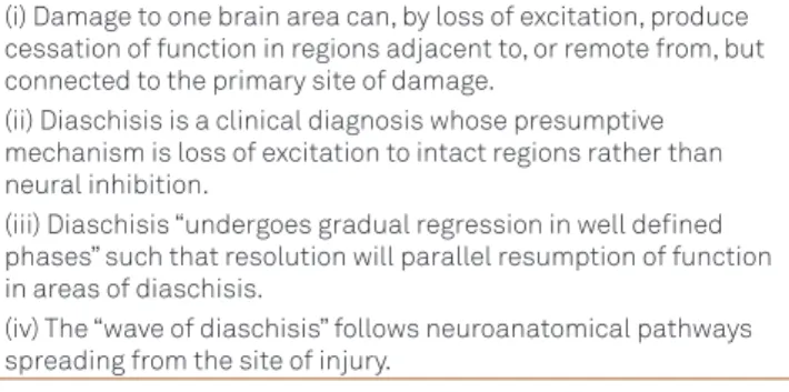
Table 1. Aspects of diaschisis emphasized by von Monakow4,5,7.
(i) Damage to one brain area can, by loss of excitation, produce cessation of function in regions adjacent to, or remote from, but connected to the primary site of damage.
(ii) Diaschisis is a clinical diagnosis whose presumptive mechanism is loss of excitation to intact regions rather than neural inhibition.
(iii) Diaschisis “undergoes gradual regression in well deined phases” such that resolution will parallel resumption of function in areas of diaschisis.
(iv) The “wave of diaschisis” follows neuroanatomical pathways spreading from the site of injury.
Table 2. Types of diaschisis according von Monakow with
updated information4,7-10.
(a) Diaschisis corticospinalis (or cerebrospinalis)
Functional depression from a motor cortex injury to the spinal cord along pyramidal tract ibers.
Later, a cortical-cerebellar diaschisis was also recognized, along cortical-pontine-cerebellar ibers.
(b) Diaschisis associativa
Intrahemispheric: cortical suppression of other cortical areas via corticocortical association ibers.
Later, a thalamic-cortical diaschisis was also described via thalamic-cortical ibers.
Interhemispheric (diaschisis commissuralis or
corticocommissuralis): cortical injury of one hemisphere can produce contralateral functional depression of the other hemisphere via ibers of the corpus collosum.
L1: cortical lesion [inset with magnified view of the cerebral cortex], L2: thalamic lesion, 1: projection fibers [a: corticospinal, b: cortical-pontine-cerebellar], 2: associative intrahemisferic fibers [cortical-cortical], 3: associative interhemispheric or commissural fibers (corpus callosum), 4: thalamic-cortical fibers. The sites of lesion (L1 and L2) and of diaschisis are represented over a coronal magnetic resonance image
(composite and distorted), based on von Monakow’s drawings6,9 and
additional information4,10.
Figure. Types of diaschisis after a focal cortical lesion.
L1
thalamic-cortical diaschisis commissural
diaschisis
crossed cerebellar diaschisis
cerebrospinal diaschisis
489
Eliasz Engelhardt et al. Von Monakow: diaschisis
Von Monakow affirms that: “The concept of diaschisis is the basis of the fundamental distinction, in experimen-tal physiology and in human clinics, between (a) the ini-tial or temporary symptoms (domain of diaschisis prop-er) and (b) the permanent or residual symptoms (domain of the secondary anatomical degenerations proper). The temporary symptoms have a fairly typical character in le-sions of certain parts of the brain and, just as regularly as they come, they will go away again hours or days, gen-erally after a longer time, even when the focus remains stable”7,8.
he basic proposal that emerged from von Monakow’s ideas was that damage to one part of the brain must have disruptive efects on other parts, which may later wear of and be associated with some recovery of function. hus, he established the diferentiation between localization of func-tion and localizafunc-tion of symptoms5,6.
1. Ditunno JF, Little JW, Tessler A, Burns AS. Spinal shock revisited: a four-phase model. Spinal Cord 2004;42:383-395.
2. Hall M. On the diseases and derangements of the nervous system.
London: H. Baillière; 1841. p. 226-239, 247-252.
3. Brown-Séquard CE. Lectures on the physiology and pathology of the
nervous system, and on the treatment of organic nervous affections. Lecture II - Part I. On organic affections and injuries of the spinal cord producing some of the symptoms of spinal hemiplegia. Lancet 1869;2:1-3.
4. Feeney DM, Baron JC. Diaschisis. Stroke 1986;17:817-830.
5. Finger S, Koehler PJ, Jagella C. The Monakow concept of diaschisis: origins and perspectives. Arch Neurol 2004;61:283-288.
6. Jagella EC, Krestel HE. Constantin von Monakow: ein Begründer der
Schweizerischen Neurologischen Gesellschaft. Schweiz Arch Neurol Psychiatr 2008;159:247-251.
7. von Monakow C. Localization of brain functions. In: von Bonin G (ed.) Some papers on the cerebral cortex. Springield: Charles C Thomas; 1960. p. 231-250.
8. von Monakow C, Mourge R. Introduction Biologique a l’Étude de la
Neurologie et de la Psychopatologie. Paris: Librairie Félix Alcan; 1928. p. 27-30,168,390-391.
9. Wiesendanger M. Constantin von Monakow (1853-1930): a pioneer
in interdisciplinary brain research and a humanist. C R Biologies 2006;329:406-418.
10. Mountz JM. Nuclear medicine in the rehabilitative treatment evaluation in stroke recovery. Role of diaschisis resolution and cerebral reorganization. Eur Medicophys 2007;43:221-239.
References
DIASCHISIS NOWADAYS