462
ARTICLE
DOI: 10.1590/0004-282X20130062
Myasthenia gravis and thymus: long-term
follow-up screening of thymectomized
and non-thymectomized patients
M i a s t e n i a g r a v i s e t i m o : i n v e s t i g a ç ã o n o s e g u i m e n t o d e l o n g o
p r a z o e m p a c i e n t e s t i m e c t o m i z a d o s e n ã o t i m e c t o m i z a d o s
Paulo José Lorenzoni, Lucas Pires Augusto, Cláudia Suemi Kamoi Kay, Rosana Herminia Scola, Lineu Cesar Werneck
hymoma is strongly associated with myasthenia gravis (MG), particularly at the onset of the disease1. hymus investiga-tion in MG patients is typically recommended at the onset of MG or when patients present with clinical deterioration or a progres-sive increase of anti-acetylcholine receptor (AChR) antibody2,3.
However, it is unknown if repeated screenings for thymo-ma at ixed intervals, even in the absence of MG deteriora-tion, are necessary when the initial screening is negative. In addition, therapeutic guidelines do not mention the impor-tance of thymus follow-up if the initial screening is negative1,4.
he main objective of this study was to determine the re-currence rate and incidence of new thymoma in a series of MG patients without clinical deterioration who underwent long-term follow-up.
METHODS
We included MG patients who fulilled the following criteria: (1) diagnosis based on abnormal repetitive nerve
Study carried out at Hospital de Cl ínicas, Universidade Federal do Paraná (UFPR), Curitiba PR, Brazil.
Neuromuscular Disorders Service, Neurology Division, Internal Medicine Department, Hospital de Cl ínicas, UFPR, Curitiba PR, Brazil.
C o r r e s p o n d e n c e : Rosana Herminia Scola; Serviço de Doenças Neuromusculares, Hospital de Clínicas da UFPR; Rua General Carneiro 181 / 3º andar; 80060-900 Curitiba PR - Brasil; E-mail: [email protected]
C o n fl i c t o f i n t e r e s t : There is no conflict of interest to declare.
Received 29 November 2012; Received in final form 05 March 2013; Accepted 12 March 2013. ABSTRACT
Thymoma screening is recommended at the onset of myasthenia gravis (MG) or when patients with MG present with clinical deterioration or a progressive increase of anti-acetylcholine receptor antibody. However, it is unknown if it is necessary to repeat the screening of thymoma at fixed intervals, even in the absence of MG deterioration, when the initial screening is negative. We analyzed the recurrence rate and incidence of new thymoma in a series of patients with well-controlled MG. The sample consisted of 53 patients, aged 17 to 72 years, and the follow-up varied between 75 and 472 months. The chest computerized tomography detected thymus abnormalities in eight patients at the initial screening and no abnormalities in all patients at a second screening after five years. The findings of this study support the classical opinion that screening for thymoma should be recommended only if there is clinical deterioration due to the disease.
K e y w o r d s : myasthenia gravis, thymus gland, thymoma, tomography.
RESUMO
A investigação de timoma é recomendada em pacientes com miastenia gravis (MG) no início da doença, em caso de haver piora clínica ou aumento dos níveis do anticorpo antirreceptor de acetilcolina. Contudo, não foi estabelecido se é necessário repetir a investigação de timo-ma em intervalos fixos, na ausência de piora clínica, quando a investigação inicial foi negativa. A taxa de recorrência e a incidência de novo timoma foram analisadas em uma série de pacientes com MG bem controlada. A amostra consiste de 53 pacientes, idade entre 17 e 72 anos, com tempo de acompanhamento variando entre 75 e 472 meses. A primeira tomografia computadorizada de tórax detectou anormalidades no timo em oito pacientes durante a investigação inicial da doença e nenhuma anormalidade no segundo exame, após cinco anos de doença, em todos os pacientes. Os achados desse estudo corroboram a clássica opinião de que a investigação de timoma deveria ser recomendada somente se houver piora clínica da doença.
463 Paulo José Lorenzoni et al. Myasthenia gravis and thymus
stimulation (RNS) and/or presence of anti-AChR antibody; (2) follow-up for more than ive years; (3) well-controlled disease; (4) a irst contrast-enhanced chest computerized tomography (CT) at the beginning of the disease; and (5) a second contrast-enhanced chest CT after ive years. MG was considered well-controlled if the patients had pharmaco-logical remission or minimal manifestations of disease, ac-cording to the Myasthenia Gravis Foundation of America’s (MGFA) clinical classiication standard4.
Relevant data, including age, gender, clinical status, serum anti-AChR antibody levels, RNS indings, thymus histopatholo-gy and treatment, were recorded at the second chest CT screen-ing. To measure the clinical status of the patients, MG compos-ite (MGC) was performed at the second chest CT screening5.
MG patients were classiied according previous thymec-tomy status into the thymectomized group (TG) and the non-thymectomized group (NTG).
All studies were conducted after obtaining patient con-sent in the out-patient clinic or during hospital admission for diagnostic investigation.
RESULTS
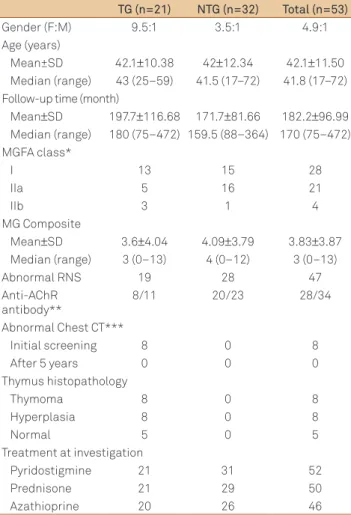
he sample consisted of 53 patients (44 female and 9 male), aged 17 to 72 years (median 41.8 years; mean 42.1±11.50 years). here were 21 TG patients (39.6%) and 32 NTG (60.4%). he duration of follow-up in our hospi-tal varied between 75 and 472 months, with a mean time of 182.2±96.99 months. MGFA classiication revealed 52.8% of patients in class I, 39.6% in class IIa and 7.5% in class IIb. he MGC varied between 0 and 13, with a mean of 3.83±3.87. Serological analysis was performed in 34 patients; serum anti-AChR antibody (binding type) was present in 82.35%.
he RNS test was abnormal in 88.67% and normal in the remaining 11.33%.
he chest CT detected thymus abnormalities in 8 pa-tients (16.3%) at the initial screening and the absence of thymus abnormalities at the second screening in all pa-tients after 5 years. he 8 papa-tients with abnormalities in the initial screening had a mean age of 46.12±9.65 years and a mean follow-up of 203.75±154.86 months. In these patients, the most common chest CT indings detected were thymo-ma at the initial screening (100%) and the absence of thy-mus abnormalities compatible with thymoma at the sec-ond screening (100%).
Twenty-one patients were thymectomized for thymoma or management of disease symptoms. hymus histopathol-ogy revealed thymoma in 8 patients, thymic hyperplasia in 8 and no abnormalities in 5. he suspicion of thymoma in 8 patients based on the initial chest CT screening (16.3%) was conirmed in all 8 by histopathological study.
he drugs used for MG treatment at the second chest CT screening were pyridostigmine in 98.11% of patients, predni-sone in 94.33% and azathioprine in 86.79%.
Table shows the differences between the TG and NTG groups.
DISCUSSION
In the present study, we did not ind any recurrence of thymoma in MG patients with previous thymectomy or new thymoma after at least ive years in MG patients without thy-mectomy. he MGFA classiication and MGC score showed that for our patients MG was well-controlled at the time the second chest CT was performed4,5. his inding suggests that screening for thymoma after ive years of follow-up is not necessary for patients with well-controlled MG.
hymoma recurrences are often associated with the on-set or aggravation of MG1,2,6. In addition, the complete sur-gical resection of thymoma has been reported as the only signiicant independent prognostic factor inluencing thy-moma recurrence in previously thymectomized MG pa-tients1,7. MG in our patients with previous thymoma was well controlled, and they had undergone complete surgical
Table. Characteristics of thymectomized and non-thymectomized groups of myasthenia gravis patients (n=53).
TG (n=21) NTG (n=32) Total (n=53) Gender (F:M) 9.5:1 3.5:1 4.9:1 Age (years)
Mean±SD 42.1±10.38 42±12.34 42.1±11.50 Median (range) 43 (25–59) 41.5 (17–72) 41.8 (17–72) Follow-up time (month)
Mean±SD 197.7±116.68 171.7±81.66 182.2±96.99 Median (range) 180 (75–472) 159.5 (88–364) 170 (75–472) MGFA class*
I 13 15 28
IIa 5 16 21
IIb 3 1 4
MG Composite
Mean±SD 3.6±4.04 4.09±3.79 3.83±3.87 Median (range) 3 (0–13) 4 (0–12) 3 (0–13)
Abnormal RNS 19 28 47
Anti-AChR antibody**
8/11 20/23 28/34
Abnormal Chest CT***
Initial screening 8 0 8
After 5 years 0 0 0
Thymus histopathology
Thymoma 8 0 8
Hyperplasia 8 0 8
Normal 5 0 5
Treatment at investigation
Pyridostigmine 21 31 52
Prednisone 21 29 50
Azathioprine 20 26 46
464 Arq Neuropsiquiatr 2013;71(7):462-464
resection. his could help explain the absence of tumor re-currence in our cases.
Chest CT is currently considered the irst choice for the screening of thymoma. It has been stated that chest CT scans should be conducted yearly for the irst 2, 5 or 10 years fol-lowing the initial thymectomy and every 2 years afterwards, especially in thymomatous patients2,7,8. However, routine screening of thymoma in well-controlled MG patients is rare-ly discussed in the literature3.
High-doses of steroids were previously reported to reduce the size of invasive thymoma, indicating that the growth of a thymoma also depends on steroids9. We did not describe pa-tient steroid doses, but almost all of them used 1 mg/kg/day of prednisone at the beginning of the disease. his could have inluenced our indings; however, we cannot analyze the rela-tionship between steroids and thymoma because we did not observe thymoma in our sample. Despite the absence of new thymoma in our subjects, cases of new thymoma have been reported in patients using steroids3.
Other characteristics of MG patients associated with thy-moma, such as an abnormal RNS or the presence of AChR antibody, could be useful in screening for recurrent or new thymoma. his study and other studies have not established
this correlation. However, some MG patients have other an-tibodies that bind in a cross-striational pattern to skeletal and heart muscle tissue sections (striational antibodies)10. Striational antibodies that react with epitopes on the mus-cle proteins titin, ryanodine receptor (RyR), and voltage-gat-ed potassium receptor (Kv1.4) are expressvoltage-gat-ed in the thymoma tissue of MG patients10. Clinically, the presence of striational antibodies and CT scans of the anterior mediastinum show a similar sensitivity for screening of thymoma in MG patients10. he frequencies of striational antibodies are generally high in MG patients with thymoma, but these antibody assays could not be performed in our patients.
The clinical presentation of MG and its course and out-come are highly dependent on thymus pathology and antibody status. The findings of this study support the classical opinion that screening for thymoma should be recommended only if there is clinical deterioration associ-ated with the disease. However, we also speculate that the detection of striational antibodies, associated with chest CT screening, could provide more specific information about subgroups of well-controlled MG patients, includ-ing the recurrence rate or chance of new thymoma durinclud-ing long-term follow-up.
1. Zielinski M. Management of myasthenic patients with thymoma. Thorac Surg Clin 2011;21:47-57.
2. Evoli A, Minisci C, Di Schino C, et al. Thymoma in patients with MG: characteristics and long-term outcome. Neurology 2002;59: 1844-1850.
3. Sugawara M, Wada C, Okawa S, et al. Long-term follow up of thymus in patients with myasthenia gravis. J Neuroimmunol 2010;221:121-124.
4. Jaretzki 3rd A, Barohn RJ, Ernstoff RM, et al. Myasthenia gravis: recommendations for clinical research standards. Task Force of the Medical Scientific Advisory Board of the Myasthenia Gravis Foundation of America. Ann Thorac Surg 2000;70:327-334.
5. Burns TM, Conaway M, Sanders DB. The MG Composite: a valid and reliable outcome measure for myasthenia gravis. Neurology 2010;74:1434-1440.
6. Kondon K, Monden Y. Thymoma and myasthenia gravis: a clinical study of 1,089 patients from Japan. Ann Thorac Surg 2005;79: 219-224.
7. Margaritora S, Cesario A, Cusumano G, et al. Thirty-five-year follow-up analysis of clinical and pathologic outcomes of thymoma surgery. Ann Thorac Surg 2010;89:245-252.
8. Margaritora S, Cesario A, Cusumano G, et al. Single-centre 40-year results of redo operation for recurrent thymomas. Eur J Cardiothorac Surg 2011;40:894-900.
9. Kirkove C, Berghmans J, Noel H, van de Merckt J. Dramatic response of recurrent invasive thymoma to high doses of corticosteroids. Clin Oncol (R Coll Radiol) 1992;4:64-66.