• Nenhum resultado encontrado

Arq. NeuroPsiquiatr. vol.71 número7

N/A
N/A
Protected

Academic year: 2018

Share "Arq. NeuroPsiquiatr. vol.71 número7"

Copied!
1
0
0

Texto

(1)

496

IMAGES IN NEUROLOGY

DOI:

10.1590/0004-282X20130071

Unilateral abdominal bulging

caused by L1-L2 herniation

Abaulamento abdominal unilateral causado por hérnia L1-L2

Marco A. Lima

1,2

, Pericles Maranhão-Filho

1

1MD PhD; Department of Neurosurgery, Instituto Nacional do Câncer (INCA), Rio de Janeiro RJ, Brazil;

2MD PhD; Instituto de Pesquisa Clínica Evandro Chagas - Fundação Oswaldo Cruz (Ipec/FIOCRUZ), Rio de Janeiro RJ, Brazil.

Correspondence: Marco A. Lima; Avenida Alexandre Ferreira 420 / apto. 403; 22470-220 Rio de Janeiro RJ - Brasil; E-mail: [email protected] Conflict of interest: There is no conlict of interest to declare.

Received 02 April 2012; Received in inal form 22 February 2013; Accepted 01 March 2013.

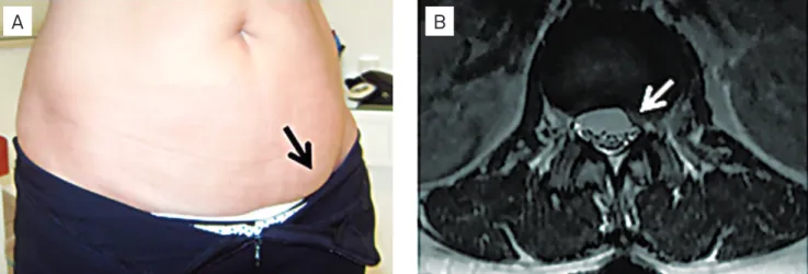
A 62-year-old woman noticed, after a sharp lumbar pain,

a non-painful bulging mass in the left inguinal fossa upon

standing upright, which disappeared in the supine position

(Fig 1A). Neurological examination revealed hypoesthesia

above the left inguinal ligament. Abdominal computerized

tomography (CT) scan showed no evidence of tumor or

in-guinal hernia. Lumbar spine magnetic resonance imaging

(MRI) indicated L1-2 disk herniation compressing the left L1

nerve root (Fig 1B).

A cutaneous sensory recurrent branch from the anterior

branches of L1 of the iliohypogastric and ilioinguinal nerves

innervates a small narrow band-shaped area on the

abdomi-nal wall above the pubis. he clinical picture and the imaging

results were indicative of a lesion at L1 root

1,2

.

1. Billet FP, Ponssen H, Veenhuizen D. Unilateral paresis of the abdominal wall: a radicular syndrome caused by herniation of the Ll-2 disc? J Neurol Neurosurg Psychiatry 1989;52:678-692.

References

2. Mumenthaler M, Schliack H. Patologia de los nervios periféricos. Diagnóstico y tratamiento. Barcelona: Ediciones Toray; 1976.

Fig 1.

(A) Bulging of the left lower part of the abdomen while standing upright. (B) Axial T2-weighted MRI sequence showing a

L1-L2 disk herniation and left side displacement of the dura mater at the L1 level.

Imagem

Fig 1. (A) Bulging of the left lower part of the abdomen while standing upright. (B) Axial T2-weighted MRI sequence showing a L1- L1-L2 disk herniation and left side displacement of the dura mater at the L1 level.

Referências

Documentos relacionados

Na hepatite B, as enzimas hepáticas têm valores menores tanto para quem toma quanto para os que não tomam café comparados ao vírus C, porém os dados foram estatisticamente

vegetative branches from Quitéria outcrop: (a) medial portion of a branch, demonstrating the leafless principal branch (I), the distribution of first order branches (II) and of

The exam revealed well- defined cortical waveforms following excitation of all sensory nerves, except the one arising from the lateral cutaneous branch of the right

he iliohypogastric, ilioinguinal, lateral cutaneous nerve of the thigh and femoral nerves emerge laterally to the lateral edge of this muscle, while the gen- itofemoral and

The first mentions of the nerve branches of the elbow capsule date from 1844, in descriptions of a branch of the cutaneous nerve perforating the brachial muscle and reaching the

The following arteries supply the knee joint: the genicular branches of the femoral, popliteal and anterior tibial recurrent arteries and the descending branch of the lateral

The probability of attending school four our group of interest in this region increased by 6.5 percentage points after the expansion of the Bolsa Família program in 2007 and