Endorectal Magnetic Resonance Imaging in Persistent
Hemospermia
Adilson Prando
Department of Radiology and Diagnostic Imaging, Vera Cruz Hospital, Campinas, Sao Paulo, Brazil
ABSTRACT
Objective:To present the spectrum of abnormalities found at endorectal magnetic resonance imaging (E-MRI), in patients with persistent hemospermia.
Materials and Methods: $UHYLHZRI(05,¿QGLQJVREVHUYHGLQSDWLHQWVZLWKSHUVLVWHQWKHPRVSHUPLDZDVSHUIRUPHG
DQGUHVXOWVFRPSDUHGZLWKWKRVHUHSRUWHGLQWKHOLWHUDWXUH)ROORZXSZDVSRVVLEOHLQRISDWLHQWVZLWKKHPR -spermia.
Results:(05,VKRZHGDEQRUPDO¿QGLQJVLQRISDWLHQWVZLWKKHPRVSHUPLD7KHVH¿QGLQJVZHUHDKHPRU
-UKDJLFVHPLQDOYHVLFOHDQGHMDFXODWRU\GXFWLVRODWHGQ RURUDVVRFLDWHGZLWKFRPSOLFDWHGPLGOLQHSURVWDWLFF\VW Q RUEKHPRUUKDJLFFKURQLFVHPLQDOYHVLFXOLWLVLVRODWHGQ RURUDVVRFLDWHGZLWKFDOFXOLZLWKLQ GLODWHGHMDFXODWRU\GXFWVQ RUFKHPRUUKDJLFVHPLQDOYHVLFOHDVVRFLDWHGZLWKFDOFXOLZLWKLQGLODWHGHMDFXODWRU\ GXFWQ RURUZLWKLQVHPLQDOYHVLFOHQ RUGQRQFRPSOLFDWHGPLGOLQHSURVWDWLFF\VWQ RU DQGHSURVWDWHFDQFHUQ RU6XFFHVVIXOWUHDWPHQWZDVPRUHIUHTXHQWLQSDWLHQWVZLWKFKURQLFLQÀDPPDWRU\DQGRU
obstructive abnormalities.
Conclusion: E-MRI should be considered the modality of choice, for the evaluation of patients with persistent hemosper-mia.
Key words: hemospermia; diagnostic imaging; magnetic resonance imaging
Int Braz J Urol. 2008; 34: 171-9
INTRODUCTION
Hemospermia or hematospermia is not an uncommon clinical urological problem among adult men, but its exact prevalence remains unknown. He-mospermia is prevalent in young males with a mean
DJHRI\HDUV8URJHQLWDOLQÀDPPDWLRQDQG
infection are usually considered the most common cause of hemospermia in this group of patients. In young males often only simple, tailored
investiga-WLRQVDQGDSSURSULDWHWUHDWPHQWDUHUHTXLUHG,QROGHU
SDWLHQWVDERYH\HDUVRIDJHRUWKRVHZLWKUHFXUUHQW
hemospermia or associated symptoms, other benign
FDXVHV DQG UDUHO\ PDOLJQDQF\ FDQ EH IRXQG
Imaging evaluation of patients with recurrent hemospermia is usually performed by transrectal
XOWUDVRXQG7586,QFRQWUDVWWR7586HQ
-dorectal magnetic resonance imaging (E-MRI) has the ability to identify hemorrhage within the reproductive structures, but despite its superior diagnostic capabil-ity there are only few reports describing its utilcapabil-ity in
Our aim was to illustrate the spectrum of ab-normalities found at E-MRI in patients with persistent hemospermia.
MATERIALS AND METHODS
%HWZHHQ 0DUFK DQG 0D\
consecutive patients with persistent hemospermia
RI DQ DYHUDJH GXUDWLRQ RI PRQWKV UDQJH PRQWKV XQGHUZHQW (MRI at our institution.
0HDQSDWLHQWDJHZDV\HDUVUDQJHyears).
6L[W\SDWLHQWVZHUHDV\PSWRPDWLFH[FHSWIRU
hemospermia. One or more associated symptoms,
ODERUDWRULDORUFOLQLFDO¿QGLQJVZHUHREWDLQHGLQWKH UHPDLQLQJSDWLHQWVIUHTXHQF\RUXUJHQF\
Q SHULQHDOGLVFRPIRUWRUSDLQQ HMDFX
-ODWRU\SDLQQ DUWHULDOK\SHUWHQVLRQQ DQG KHPDWXULDQ $IWHUWUHDWPHQWIROORZXSZDV
obtained in 37 patients. Conventional MR imaging
ZDVSHUIRUPHGZLWKD705LPDJHU6LJQD*( 0HGLFDO6\VWHPV0LOZDXNHH:,3DWLHQWVZHUH
examined by usingWKHERG\FRLOIRUVLJQDODFTXLVL -tion and a combina-tion of a pelvic phased-array coil
*(0HGLFDO6\VWHPV7RUVR3$ZLWKDFRPPHUFLDO -ly available balloon-covered endorectal coil (Endo
$7'0HGUDG3LWWVEXUJK3$IRUVLJQDOUHFHSWLRQ
The balloon-covered endorectal coil wasLQÀDWHG
ZLWKP/RIOLTXLGSHUÀXRURFDUERQ2Q05
images, the prostate was evaluated with transverse spin-echo T1-weighted MR images by using the followingSDUDPHWHUV UHSHWLWLRQ WLPH PVHFHFKR
WLPHPVHFPLQLPXPVHFWLRQ WKLFNQHVVPP
PDWUL[[WZRVLJQDOVDFTXLUHG¿HOGRIview,
FPLQWHUVHFWLRQJDSPPEDQGZLGWK
kHz. TransverseDQGWUDQVYHUVHREOLTXH7ZHLJKWHG images were obtained with theIROORZLQJSDUDPHWHUV
VHFWLRQWKLFNQHVVPPPDWUL[[ WKUHHVLJQDOVDFTXLUHG¿HOGRIYLHZFP LQWHUVHFWLRQJDSPPEDQGZLGWKN+])RU
the transverse images, phase encoding was in the
ULJKWWROHIWGLUHFWLRQ7ZHLJKWHGVDJLWWDO05LP
-DJHVZHUHREWDLQHGZLWKWKHIROORZLQJSDUDPHWHUV VHFWLRQWKLFNQHVVPPPDWUL[[ WZR VLJQDOV DFTXLUHG ¿HOG RI YLHZ FP LQWHUVHFWLRQJDSPPEDQGZLGWKN+])RU WKH7ZHLJKWHGVDJLWWDO05LPDJHVSKDVHHQFRG
-ing was in the superior-to-inferior direction. After
WUHDWPHQWIROORZXSZDVSRVVLEOHLQRI
patients.
RESULTS
In patients with hemospermia, E-MRI
VKRZHGDEQRUPDO¿QGLQJVLQRXWRIWKHSDWLHQWV +HPRUUKDJHZLWKLQWKHVHPLQDOYHVLFOHRUWKH HMDFXODWRU\GXFWZDVUHFRJQL]HGLQRISDWLHQWV %ORRGZLWKLQVHPLQDOYHVLFOHRUHMDFXODWRU\
duct appears as areas of high signal intensity on T1-weighted spin-echo images representing the presence of metahemoglobin due to subacute hemorrhage
The imaging criteria used to characterize
VHPLQDO YHVLFXOLWLV ZHUH GLIIXVH ZDOO WKLFNHQLQJ RIWKHVHPLQDOYHVLFOHZLWKORZ7ZHLJKWHGVLJQDO
intensity, loss of convolutions and proteinaceous or
KHPRUUKDJLFÀXLGFRQWHQWZLWKYDULDEOHVLJQDOLQWHQ
-VLW\RQ7ZHLJKWHGDQG7ZHLJKWHGLPDJHV 7KXVVLJQL¿FDQWDEQRUPDO(05,¿QGLQJVREVHUYHG LQ WKLV JURXS RI SDWLHQWV ZHUH D KHPRUUKDJLF
seminal vesicle and ejaculatory duct, isolated (n =
RURUDVVRFLDWHGZLWKFRPSOLFDWHGPLGOLQH
SURVWDWLFF\VWQ RU)LJXUHEKHP
-RUUKDJLFFKURQLFVHPLQDOYHVLFXOLWLVLVRODWHGQ RU)LJXUHRUDVVRFLDWHGZLWKFDOFXOLZLWKLQ
GLODWHGHMDFXODWRU\GXFWVQ RUFKHPRU
-rhagic seminal vesicle associated with calculi within
GLODWHGHMDFXODWRU\GXFWQ RURUZLWKLQ VHPLQDOYHVLFOHQ RU)LJXUHGQRQ FRPSOLFDWHGPLGOLQHSURVWDWLFF\VWQ RU DQGHSURVWDWHFDQFHUQ RU)LJXUH
Thirteen patients with hemospermia
under-ZHQWWUDQVXUHWKUDOHQGRVFRSLFWUHDWPHQWXQURR¿QJ
of the midline cysts or ductal obstruction and resec-tion, fulguraresec-tion, and dilatation of ejaculatory duct
REVWUXFWLRQ 7KLV DSSURDFK ZDV VXFFHVVIXO LQ
patients with dilated hemorrhagic seminal vesicle(s) and ejaculatory duct associated with complicated
PLGOLQHSURVWDWLFF\VWLQSDWLHQWVZLWKKHPRUUKDJLF
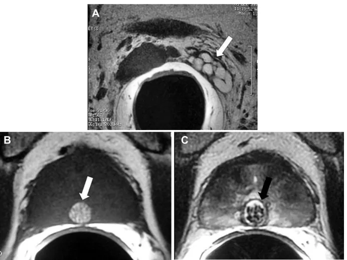
Figure 1 – Hemorrhagic seminal vesicle associated with a complicated midline prostatic cyst (utricular cyst). A 54-year-old man, with history of persistent hemospermia. A) E-MRI, axial plane, T1-weighted image, showing a hemorrhagic normal-walled left seminal vesicle (arrow). Hemorrhage is recognized due the presence of high signal intensity on T1-weighted images. B) and C) E-MRI, axial T1 and T2-weighted images respectively, showing a complicated midline prostatic cyst (arrow) containing blood and several small calculi.
A
B
C
midline prostatic cyst. Hemospermia disappeared
FRPSOHWHO\ LQ RXW RI SDWLHQWV IROORZLQJ DQ
E-MRI diagnosis of hemorrhagic chronic seminal
YHVLFXOLWLVDQGVXEVHTXHQWDQWLPLFURELDODQGRUDQWL LQÀDPPDWRU\GUXJV6SRQWDQHRXVHOLPLQDWLRQRID
seminal vesicle calculus was reported by one patient with complete remittance of the hemospermia. Two patients suspected to have prostate cancer due to the
SUHVHQFHRIIRFDOK\SRLQWHQVHDUHDRQ7ZHLJKWHG
images, in the peripheral zone of the prostate, were
IXUWKHUHYDOXDWHGZLWK7586JXLGHGELRSV\JXLGHG E\PDJQHWLFUHVRQDQFHLPDJLQJ¿QGLQJV7KLV WHFKQLTXHDOORZHGWKHGLDJQRVLVRIFDQFHULQRQO\
one of these patients.
COMMENTS
Although hemospermia is usually a benign and self-limiting condition, it provokes great concern and anxiety in sexually active patients. Hemospermia
PD\EHVHFRQGDU\WRLQÀDPPDWLRQLQIHFWLRQGXFWDO
obstruction or cysts, benign neoplasm, vascular ab-normalities, systemic or iatrogenic factors and rarely malignant tumors. History and physical examination are often unrevealing (1). In patients younger than
\HDUVDQLQIHFWLYHFDXVHLQWKHXURJHQLWDOWUDFWLV WKHPRVWFRPPRQHWLRORJLFDOIDFWRU)DFWRUVWKDW
duration of hemospermia, whether it is persistent and the presence of associated symptoms or signs such as weight loss, local or bony pain, fever, lower urinary tract symptom and hematuria. It is widely accepted that persistent hemospermia or hemospermia with an associated symptom and hemospermia in older
SDWLHQWVUHTXLUHVPRUHH[WHQVLYHLQYHVWLJDWLRQ
In our small series of patients, laboratorial
RU FOLQLFDO ¿QGLQJV ZHUH SUHVHQW LQ RXW SD
-WLHQWVIUHTXHQF\RUXUJHQF\Q SHULQHDO GLVFRPIRUWRUSDLQQ HMDFXODWRU\SDLQQ DUWHULDOK\SHUWHQVLRQQ DQGKHPDWXULDQ
Both patients with hematuria with normal E-MRI
¿QGLQJVZHUHVXEPLWWHGWRGLUHFWULJLGDQGÀH[LEOH F\VWRVFRS\ 3DSLOODU\ XUHWKULWLV ZDV IRXQG LQ RQH
patient.
7586FDQEHFRQVLGHUHGDVDIHQRQLQYDVLYH
and relatively inexpensive method, which allows clear images of the reproductive system structures.
7586 KDV DQ DFFXUDWH GLDJQRVWLF UDWH RI EHWZHHQ DQGIRUWKHHYDOXDWLRQRIKHPRVSHUPLD
E-MRI has superior imaging capability since offers higher spatial resolution for the visualization of the whole seminal tract. E-MRI allows the
demonstra-WLRQRIQRUPDOYDULDWLRQVSUHVHQFHRIKHPRUUKDJH
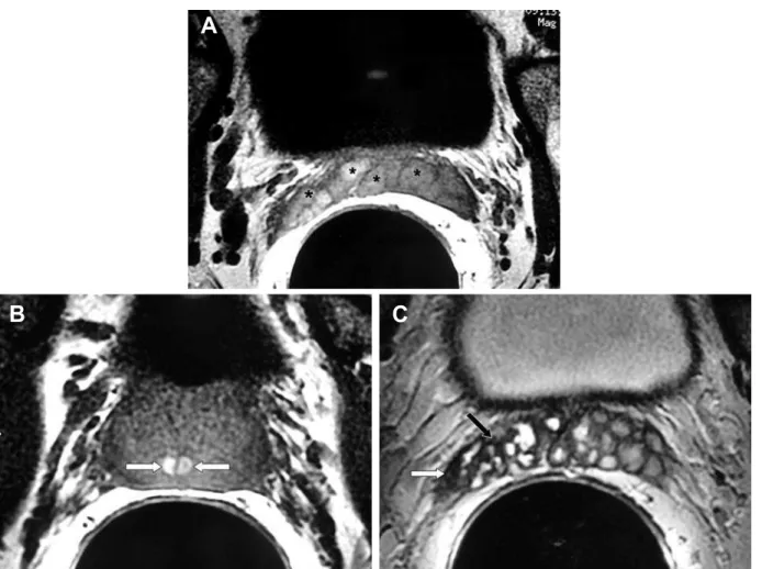
and evident signs of chronic infection, obstruction Figure 2 – Hemorrhagic chronic seminal vesiculitis. A 62-year-old man, with history of persistent hemospermia and perineal discom-fort. A) and B) E-MRI ,axial plane, T1-weighted images. Note high-signal intensity hemorrhage in both seminal vesicles (asterisk) and in both ejaculatory ducts (white arrows). C) and D) E-MRI, axial plane, T2-weighted images ,showing imaging features consistent with chronic seminal vesiculitis: diffuse thickening of the of the seminal vesicles with low T2-weighted signal intensity (dark arrow) and loss of convolutions( white arrow).These abnormalities are more evident in the right seminal vesicle which appeared contracted in comparison with the left seminal vesicle.
A
DQGPDOLJQDQFLHV&RQWUDU\WR758605,KDVWKH
ability to accurately identify hemorrhage within the seminal tract due to its characteristic signal behavior (high signal intensity on T1-weighted images).
Imaging studies have considered a wide range
RI HWLRORJLFDO IDFWRUV IRU KHPRVSHUPLD SURVWDWLF FDOFL¿FDWLRQSURVWDWLFK\SHUWURSK\SURVWDWLWLVPLG -line prostatic cyst (utricular), mid-line extra-prostatic cyst, seminal vesicle cyst or calculi, dilatation of the seminal vesicles or the ejaculatory ducts, ejaculatory duct cyst, blood within normal or thick-walled seminal vesicle (seminal vesiculitis) or the ejaculatory duct,
seminal vesicle amyloidosis, periprostatic varicosities
DQGSURVWDWLFFDUFLQRPD
6RPHRIWKHVHDEQRUPDOLWLHVVXFKDVSURV
-tatic hypertrophy, dilatation of the seminal vesicle(s),
SURVWDWLFFDOFL¿FDWLRQDQGQRQFRPSOLFDWHGPLGOLQH
prostatic cyst, can be found in asymptomatic patients.
6HPLQDOYHVLFOHVGLODWDWLRQIRUH[DPSOHKDVEHHQ
described as a very common cause of hemospermia
EXWLWLVNQRZQWKDWYDULRXV¿OOLQJVWDWHV RI WKH VHPLQDO YHVLFOHV DUH TXLWH QRUPDO )RU WKLV
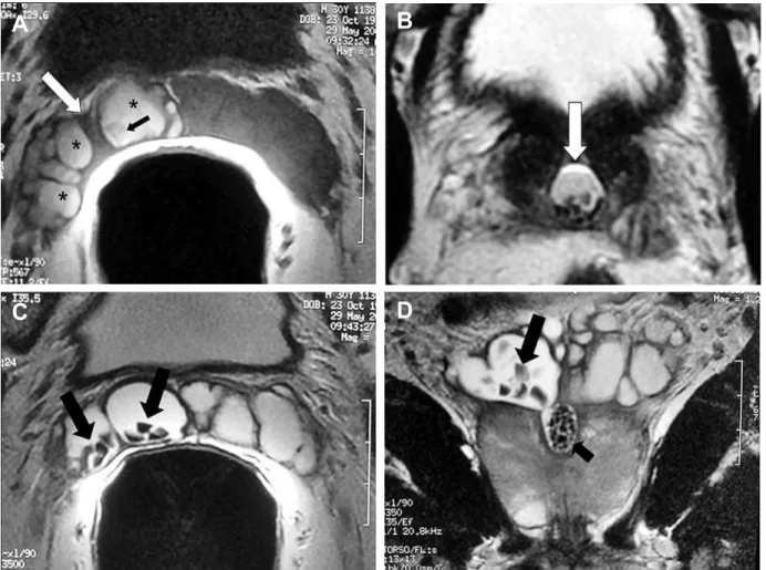
reason, we are speculating that perhaps there is a tendency to consider many incidental and common Figure 3 – Hemorrhagic seminal vesicle associated with calculi within the right seminal vesicle and dilated ejaculatory duct. A 30-year-old man, with history of persistent hemospermia and ejaculatory pain. A) E-MRI, axial plane, T1-weighted image, showing a hemorrhagic dilated ejaculatory duct (white arrow), containing blood (asterisk) and calculi (black arrow). B) E-MRI, axial,T2-weighted image ,better shows the calculi within the dilated ejaculatory duct (arrow). C) and D) E-MRI, axial and coronal T2-weighted images respectively, nicely demonstrates the presence of several stones within the right seminal vesicle (arrows) and within the dilated right ejaculatory duct (small arrow). Note the contiguity of the dilated seminal vesicle with the dilated right ejaculatory duct. This is an
HVVHQWLDO¿QGLQJIRUWKHGLIIHUHQWLDWLRQEHWZHHQGLODWHGHMDFXODWRU\F\VWIURPPLGOLQHSURVWDWLFF\VW
A
B
urological abnormalities as the etiological factor of
KHPRVSHUPLD7KLVFRXOGSRVVLEO\H[SODLQ
why the success rate of the treatment was variable in our small series of patients. Transurethral endoscopic treatment was more effective in patients with clear
REVWUXFWLYH¿QGLQJVDQGIDLOHGLQSDWLHQWVZLWKQRQ
complicated, non obstructive, midline prostatic cyst. This mechanism could also explain why therapy with
DQWLPLFURELDO DQG RU DQWLLQÀDPPDWRU\ GUXJV ZDV
more effective in patients with evident manifestation of seminal vesiculitis and failed in the majority of pa-tients with hemorrhagic seminal vesicle. Although the
ODFNRIKLVWRORJLFDOFRQ¿UPDWLRQRIFKURQLFVHPLQDO
vesiculitis (no seminal vesicle biopsy) is a limitation of our study, we may assume that our imaging criteria for chronic seminal vesiculitis is correct since in most
RIWKHSDWLHQWVZLWKWKLV05,¿QGLQJVKHPRVSHUPLD GLVDSSHDUHGDIWHUDGHTXDWHDQWLPLFURELDODQWLLQÀDP -matory treatment.
In conclusion, E-MRI should be considered the modality of choice for the evaluation of patients with persistent hemospermia. In our series, the most
VLJQL¿FDQW(05,¿QGLQJVZHUHKHPRUUKDJLFVHPL -nal vesicle and ejaculatory duct, isolated or associated
ZLWKFRPSOLFDWHGPLGOLQHSURVWDWLFF\VWKHPRUUKDJLF
chronic seminal vesiculitis, isolated or associated with calculi within dilated ejaculatory ducts, hemorrhagic seminal vesicle associated with calculi within dilated
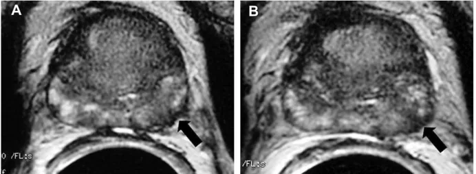
Figure 4 – Prostate cancer. A) and B) E-MRI, axial plane, T2-weighted images showing focal area of low signal intensity in the lat-eral aspect of the left periphlat-eral zone (arrow) associated with irregular thickening of the capsule of the prostate. TRUS-guided biopsy
GLUHFWHGE\WKHVH¿QGLQJVDOORZHGWKHGLDJQRVLVRISURVWDWHFDQFHU*OHDVRQVFRUH
ejaculatory duct or within seminal vesicle, non-com-plicated midline prostatic cyst and prostate cancer.
6XFFHVVIXOWUHDWPHQWZDVLQIDFWPRUHIUHTXHQWLQ SDWLHQWVZLWKFKURQLFLQÀDPPDWRU\DQGRUREVWUXFWLYH
abnormalities.
CONFLICT OF INTEREST
None declared.
REFERENCES
0XQNHO ZLW] 5 .UDVQRNXWVN\ 6 /LH - 6KDK 60 %D\VKWRN-.KDQ6$&XUUHQWSHUVSHFWLYHVRQKHPD
-WRVSHUPLDDUHYLHZ-$QGURO $PHXU$7RXLWL'-LUD+HO$ODPL0%RXPGLQ+
$EEDU0+HPRVSHUPLDGLDJQRVLVDQGWKHUDSHXWLF DVSHFWV6HYHQFDVHUHSRUWV$QQ8URO3DULV
)OHWFKHU06+HU]EHUJ=3U\RU-37KHDHWLRORJ\DQG LQYHVWLJDWLRQRIKDHPRVSHUPLD%U-8URO
0XOKDOO-3$OEHUWVHQ3&+HPRVSHUPLDGLDJQRVLV DQGPDQDJHPHQW8URORJ\
$KPDG,.ULVKQD16+HPRVSHUPLD-8URO
+DQ0%UDQQLJDQ5($QWHQRU-$5RHKO.$&DWD
-ORQD:-$VVRFLDWLRQRIKHPRVSHUPLDZLWKSURVWDWH FDQFHU-8URO
:RULVFKHFN-+3DUUD52&KURQLFKHPDWRVSHUPLD DVVHVVPHQWE\WUDQVUHFWDOXOWUDVRXQG8URORJ\
(WKHULQJWRQ 5- &OHPHQWV 5 *ULI¿WKV *- 3HHOLQJ :%7UDQVUHFWDO XOWUDVRXQG LQ WKH LQYHVWLJDWLRQ RI KDHPRVSHUPLD&OLQ5DGLRO <DJFL&.XSHOL67RN&)LWR]6%DOWDFL6*RJXV
2 (IILFDF\ RI WUDQVUHFWDO XOWUDVRQRJUDSK\ LQ WKH HYDOXDWLRQRIKHPDWRVSHUPLD&OLQ,PDJLQJ
7RULJLDQ'$5DPFKDQGDQL3+HPDWRVSHUPLDLPDJ
-LQJ¿QGLQJV$EGRP,PDJLQJ :HLQWUDXE03'H0RX\(+HOOVWURP:-1HZHU
modalities in the diagnosis and treatment of ejaculatory
GXFWREVWUXFWLRQ-8URO
/HQFLRQL52UWRUL6&LRQL'0RUHOOL*&HUHWWL( &RVRWWLQL0HWDO(QGRUHFWDOFRLO05LPDJLQJ¿QG
-LQJVLQKHPRVSHUPLD0$*0$ 3UDQGR$.XUKDQHZLF]-%RUJHV$32OLYHLUD(0-U
)LJXHLUHGR(3URVWDWLFELRSV\GLUHFWHGZLWKHQGRUHFWDO 05VSHFWURVFRSLFLPDJLQJ¿QGLQJVLQSDWLHQWVZLWK
HOHYDWHG SURVWDWH VSHFL¿F DQWLJHQ OHYHOV DQG SULRU QHJDWLYHELRSV\¿QGLQJVHDUO\H[SHULHQFH5DGLRORJ\
0DHGD+7R\RRND1.LQXNDZD7+DWWRUL5)XUXND
-ZD70DJQHWLFUHVRQDQFHLPDJHVRIKHPDWRVSHUPLD 8URORJ\
*DUFLD15*R]DOHV,)0DWHR&3&DVWUR*(7HOOR $0+HPDWRVSHUPLD\TXLVWHGHOFRQGXFWRPOOHULDQR $UFK(VS8URO
)XVH+6XPL\D+,VKLL+6KLPD]DNL-7UHDWPHQW
of hemospermia caused by dilated seminal vesicles
E\GLUHFWGUXJLQMHFWLRQJXLGHGE\XOWUDVRQRJUDSK\-8URO
)XUX\D6.DWR+$FOLQLFDOHQWLW\RIF\VWLFGLODWDWLRQ RIWKHXWULFOHDVVRFLDWHGZLWKKHPRVSHUPLD-8URO
,VKLNDZD02NDEH+2\D7+LUDQR07DQDND0 2QR0HWDO0LGOLQHSURVWDWLFF\VWVLQKHDOWK\PHQ LQFLGHQFHDQGWUDQVDEGRPLQDOVRQRJUDSKLF¿QGLQJV $-5$P-5RHQWJHQRO
&XUUDQ6$NLQ2$JLOGHUH$0=KDQJ-+ULFDN+ 5DGHPDNHU-(QGRUHFWDO05,RISURVWDWLFDQGSHUL
- SURVWDWLFF\VWLFOHVLRQVDQGWKHLUPLPLFV$-5$P-5RHQWJHQRO
Accepted after revision: February 25, 2008
Correspondence address: 'U$GLOVRQ3UDQGR $Y$QGUDGH1HYHV
&DPSLQDV63%UD]LO )D[
(PDLODSUDQGR#PSFFRPEU
EDITORIAL COMMENT
Hemospermia can be considered a chal-lenging situation for both urologists and radiolo-gists, given its relatively high prevalence and poor understanding. Transrectal ultrasound, despite being a good modality for prostate evaluation and guided-intervention, has limited applications for dedicated
seminal vesicles imaging, especially regarding
identi-¿FDWLRQRIEORRGZLWKLQWKHGXFWV7KHDUWLFOHIURP 'U3UDQGRFRQ¿UPVWKHHYROYLQJUROHRI0DJQHWLF
contrast resolution (the ability to characterize different structures and components, like blood). Endorectal MRI is now considered the modality of choice for local staging of prostate cancer, including seminal
YHVLFOHVLQYDVLRQ7KHGHYHORSPHQWDQGLQFUHDV -ing availability of 3 Tesla MR scanners can further improve the application of this imaging modality in the evaluation of the seminal vesicles, since its intrinsic high signal intensity might exempt the need for an endorectal coil (3). Nowadays, regardless these
VSHFL¿F WHFKQRORJLFDO DVSHFWV ZH FDQ FRQ¿GHQWO\
state that MRI is the imaging modality of choice for evaluation of hemospermia and other seminal vesicles diseases.
REFERENCES
&KR,5/HH065KD.++RQJ6-3DUN66.LP 0-0DJQHWLFUHVRQDQFHLPDJLQJLQKHPRVSHUPLD-8URO
:DQJ/+ULFDN+.DWWDQ0:&KHQ+1.XURLZD .(LVHQEHUJ+)HWDO3UHGLFWLRQRIVHPLQDOYHVLFOH LQYDVLRQLQSURVWDWHFDQFHULQFUHPHQWDOYDOXHRIDGG -ing endorectal MR imag-ing to the Kattan nomogram.
5DGLRORJ\
6RVQD-3HGURVD,'HZROI:&0DKDOODWL+/HQNLQ
-VNL5(5RIVN\1005LPDJLQJRIWKHSURVWDWHDW 7HVODFRPSDULVRQRIDQH[WHUQDOSKDVHGDUUD\FRLO WRLPDJLQJZLWKDQHQGRUHFWDOFRLODW7HVOD$FDG 5DGLRO
Dr. Ronaldo Hueb Baroni
Radiologist, Body Imaging Department Institute of Radiology, USP and Albert Einstein Israelita Hospital São Paulo, SP, Brazil E-mail: [email protected]
EDITORIAL COMMENT
In his paper published in this issue of the
,QWHUQDWLRQDO%UD]LOLDQ-RXUQDORI8URORJ\3UDQGR
presents an overview of abnormalities found at en-dorectal coil magnetic resonance imaging (MRI) in patients with persistent hemospermia.
The use of MRI instead of imaging modali-ties such as transrectal ultrasonography or computed
WRPRJUDSK\VHHPVTXLWHHYLGHQW0DJQHWLFUHVRQDQFH LPDJLQJDOORZVGLUHFWPXOWLSODQDULPDJHDFTXLVLWLRQ
and offers superb soft tissue contrast, enabling ac-curate depiction and characterization of soft tissues within the pelvis and facilitating the demonstration of blood products within the male reproductive system.
:KHQFRPELQHGZLWKDQHQGRUHFWDOFRLOWKHLPDJH
resolution can be further increased, providing unsur-passed image detail of the prostate gland, ejaculatory
GXFWVDQGVHPLQDOYHVLFOHV+HQFH3UDQGRIRXQGDE -normalities that were directly related to hemospermia
LQDERXWRIFDVHV
On the other hand, uncomplicated
hemosper-PLDXVXDOO\KDVRQO\PLQRUFOLQLFDOVLJQL¿FDQFHDQG
needs no immediate imaging evaluation, especially in
\RXQJHUSDWLHQWVOHVVWKDQ\HDUVRIDJH+RZHYHU
in cases of persistent or complicated hemospermia, it
FDQEHYHU\GLVTXLHWLQJIRUSDWLHQWVDQGIUXVWUDWLQJIRU
urologists to have no information about the location or
WKHHWLRORJ\RIWKHEOHHGLQJ6RIDUWUDQVUHFWDOXOWUDVR
-QRJUDSK\KDVEHHQWKHH[DPLQDWLRQRI¿UVWFKRLFHLQ
these patients. It is a relatively inexpensive and readily
DYDLODEOHWHFKQLTXHWKDWDOORZVWKHLGHQWL¿FDWLRQRI
benign prostatic hyperplasia, dilated ejaculatory ducts or seminal vesicles, and obvious lithiasis, cystic lesions, or tumors. On the other hand, transrectal ultrasound cannot directly prove the presence of blood products within the ejaculatory ducts or seminal vesicles and will fail to disclose more subtle abnormalities. Although computed tomography (CT) can readily demonstrate
WKHSUHVHQFHRIFDOFL¿FDWLRQVDQGKLJKGHQVLW\EORRG
in the younger patient group. MRI does not suffer from the abovementioned inconveniences.
:H VWURQJO\ EHOLHYH KRZHYHU WKDW LQ WKH
majority of patients with hemospermia no immediate
LPDJLQJHYDOXDWLRQLVUHTXLUHG)XUWKHUPRUHWUDQVUHF -tal sonography remains a valid and readily accessible
SULPDU\WHFKQLTXHWRGLVFORVHPRUHREYLRXVDEQRU -malities of the prostatovesicular complex. However, in complicated or persistent hemospermia, certainly
LQSDWLHQWVDERYH\HDUVRIDJH05,PD\KDYHWKH
potential to disclose more subtle abnormalities that remain obscure on transrectal ultrasonographic exami-nation. Although we are currently not aware of any study having directly compared the diagnostic value of transrectal ultrasound with that of MRI, it is not unreasonable to expect that MRI might become the
LPDJLQJPRGDOLW\RI¿UVWFKRLFHWRHYDOXDWHSDWLHQWV
with persistent or complicated hemospermia.
Dr. Geert M. Villeirs & Dr. Willem OosterlinckWillem Oosterlinck
'HSWRI5DGLRORJ\*09DQG8URORJ\:2 *KHQW8QLYHUVLW\+RVSLWDO *HQW%HOJLXP
E-mail: [email protected]
EDITORIAL COMMENT
I read with great interest the article by Dr.
3UDQGRLQZKLFKKHHYDOXDWHV¿QGLQJVRIHQGRUHFWDO
magnetic resonance (E-MRI) in patients with persistent hemospermia. Although in the majority hemospermia
LVDEHQLJQDQGVHOIOLPLWLQJFRQGLWLRQWKHTXHVWLRQ
lies in how to investigate these patients. To date, a small number of studies using an endorectal coil for evaluation of patients with hematospermia have been published. Magnetic resonance is the current gold standard for imaging the accessory sex glands and their ducts and, E-MRI promotes an excellent multiplanar anatomic evaluation of the prostate gland, seminal vesicles and ejaculatory ducts. However, we know
WKDWWUDQVUHFWDOXOWUDVRQRJUDSK\7586LVDQHIIHF
-WLYHDQGZLGHO\XVHGWHFKQLTXHDVDSULPDU\PRGDOLW\ IRUSDWLHQWVZLWKKHPRVSHUPLD7586FDQDOVRGHWHFW
dilatation, cysts and stones in the seminal vesicles, prostate and ejaculatory ducts. The greatest advantage
RI(05,RYHU7586LVLWVDELOLW\WRUHYHDOKHPRUUKDJH
in the seminal vesicles or prostate. I also agree that Dr.
3UDQGRLPDJLQJFULWHULDIRUFKURQLFVHPLQDOYHVLFXOLWLV
is by far superior to those that we can infer by using
75863UREDEO\WKHDGGLWLRQRIFRQWUDVWJDGROLQLXP
further improves resolution of magnetic resonance for
LQÀDPPDWRU\VLJQV
Infective cause in the urogenital tract is the
PRVWFRPPRQHWLRORJLFDOIDFWRU'U3UDQGRFRQ¿UPV ¿QGLQJVRILQÀDPPDWRU\FRQGLWLRQVDVDFRPPRQDV -sociation with hemospermia and this is demonstrated
LQUHFHQWVWXGLHVZKHUHFXUUHQWODERUDWRU\WHFKQLTXHV GHWHFWHGDSDWKRJHQLQRIFDVHVRIKHPRVSHUPLD
(1).
In summary, current evidence suggests that, for patients with persistent hemospermia, endorectal
FRLO05,VKRXOGEHSHUIRUPHGZKHQ7586LVXQVDW -isfactory or nondiagnostic.
REFERENCE
%DPEHUJHU(0DGHE56WHLQEHUJ-3D]$6DWLQJHU ,.UD2]=HWDO'HWHFWLRQRIVH[XDOO\WUDQVPLWWHG
pathogens in patients with hematospermia. Isr Med