Arq Neuropsiquiatr 2001;59(1):112-115
A RARE CASE OF INTRAMEDULLARY LIPOMA
ASSOCIATED WITH CYST
Asdrubal Falavigna
1, Ana Claudia Segatto
2, Karine Salgado
3ABSTRACT - Intramedullary lipomas are benign tumours of the spinal cord corresponding to 1% of all primitive intramedullary tumours. We report a rare case of true intramedullary lipoma associated with cyst. The patient underwent subtotal resection and the diagnosis was made by histopathological examination. There was postoperative neurological improvement.
KEY WORDS: lipoma, spinal cord, magnetic resonance imaging, dermoid cyst.
Caso raro de lipoma intramedular cístico
RESUMO Os lipomas intramedulares são tumores benignos da medula espinhal correspondendo a 1% dos tumores primitivos intramedulares. Relatamos um caso raro de lipoma intramedular verdadeiro associado com cisto . O paciente foi submetido a ressecção cirúrgica parcial com melhora neurológica no pós-operatório, sendo o diagnóstico histopatológico de lipoma.
PALAVRAS-CHAVE: lipoma, medula espinhal, ressonância magnética, cisto dermóide.
Disciplina de Neurologia da Faculdade de Medicina da Universidade de Caxias do Sul (UCS): 1Professor Assistente da Disciplina de
Neurologia da Faculdade de Medicina da Universidade de Caxias do Sul, Mestrando em Neurocirurgia da UNIFESP - Escola Paulista de Medicina de São Paulo; 2Médica Residente de Clínica Médica do Hospital Saúde; 3Neuropatologista.
Received 3 June 2000, received in final form 4 September 2000. Accepted 11 September 2000.
Dr. Asdrubal Falavigna - Rua Dr. Moreira César 2712/1 - 95034-000 Caxias do Sul RS - Brasil. E-mail: [email protected]
Lipomas of the spinal cord are uncommon1-4. The
majority of reports found an incidence of 1% of all spinal cord tumours5. Intramedullary lipomas are
even more rare, their incidence being estimated as 0.45 to 0.6% when they are not associated with spi-nal cord dysrhaphism6. The origin of these lesions is
unknown and the clinical presentation may mimic any of a variety of other spinal cord processes7.
CASE
A 48-year-old woman presented with a 10-year his-tory of progressive bladder dysfunction. Five years after the onset of bladder dysfunction, she has developed numb-ness and a cramp in both lower limbs. At this time, she had a clinical examination and was investigated with elec-tromyography and a lumbar computed tomography (CT) that were normal. There was a gradual worsening of leg numbness with distal anesthesia in the last three months and she was unable to walk unaided in the last month prior to admission. At admission the general physical ex-amination revealed that she was 120 Kg overweight. The cardiovascular and respiratory systems were normal. There were no motor, reflex or sensory changes in the upper extremities. Both lower limbs were spastic with the knee
and ankle jerks exaggerated. There were grade IV spastic paraparesis, bilateral. Babinskis sign was positive bilater-ally. There was depression of spinothalamic and posterior column sensibility up to the level of D10. Muscle spasms were present in both lower limbs. There were no signs of spinal dysraphism.
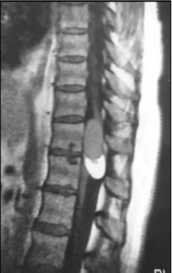
Routine blood tests were normal. Urine culture revealed the growth of E. coli that was treated with antibiotic therapy. Plain radiographs of the dorsal-lumbar spine were unremarkable. Dorsal MRI (Philips Gyroscan T5-NT) re-vealed an intradural lesion extending from D11 to D12 displacing the spinal cord anteriorly. The inferior part of the lesion was high-signal intensity on T1-weighted im-ages (TR 450 / TE 15) which was characteristic of a fat component (Fig. 1). At the upper border of the lesion there was a cyst that had the same MRI characteristics as the cerebrospinal fluid on T2-weighted images (TR 3700 / TE 130) with a fat component in the lower portion that ap-pear hyperintense to normal neural parenchyma. There was indefinition of the thoracic spinal cord at the level of the cyst without any gadolinium enhancement (Fig. 2).
Arq Neuropsiquiatr 2001;59(1) 113
Fig 2. Axial section in T2-weighted dor-sal resonance images (TR 5600 / TE 140) showing the cyst with high-signal inten-sity occuping the whole spinal canal at D11 level (upper images) and the lipoma expelling the spinal cord in the antero-lateral part of the spinal canal at D12 level (lower images).
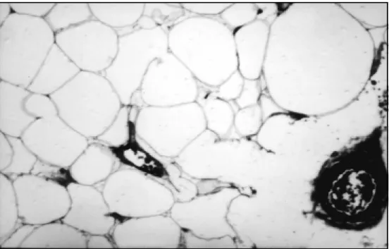
was performed in D11 and D12. It revealed a pale, spindle-shaped, non-pulsatile and bulging dural sac . The micro-scope was introduced into the field. The dura was opened and a yellowish tumor was seen with a cyst on the upper margin. After opening the cyst a plane of cleavage be-tween the tumor and the neural tissue could be defined. In the remaining areas the tumor was continuous to the spinal cord and the nerve roots by a fine, whitish, fibrous capsule. Subtotal removal was, therefore carried out. A watertight dural closure was performed followed by a muscle and skin closure. The pathological examination revealed that the tumor was composed by mature adipocytes with fibrous tissue, smooth tissue, scattered strands of normal nervous tissue and a few vascular chan-nels (Fig 3).
After surgery there was recovery of sensation in the lower limbs and bladder function. Motor power improved allowing her to climb stairs and walk without help. The sensation of cramps disappeared.
DISCUSSION
Intradural lipomas are often continuous with dorsal subcutaneous fat through dysraphic defects in the posterior neural arch8. Spinal lipomas not
as-sociated with spinal dysrhaphism in adults are re-garded as tumoral and so termed true lipomas8.
Their incidence was estimated to be 0.45 to 0.6%6.
There is an insignificant sex difference6, 9.
Intramedul-lary lipomas not associated with spinal dysrhaphism and cutaneous abnormality usually appear neurologi-cally in the third decade10,11. They generally have a
ten-dency to extend longitudinally, occupying several seg-ments of the spinal cord12. The thoracic area appeares
to be most frequently involved1.
114 Arq Neuropsiquiatr 2001;59(1)
The exact pathogenesis of spinal lipoma is still unclear8. They are considered embryonic
malforma-tions of the neural tube10. Bostroem13 believed that
the lipoma was an inclusion of misplaced primitive cells within the neural tube at the time of its clo-sure. Ehni and Love2 postulated the hypothesis that
these lipomas are of mesenchymal rather than neu-ral origin and they appear because of local failure control over the formation of fat from the pericapilla-ry mesenchymal cells. As suggested by McClone et al.14, spinal cord lipomas are thought to arise from
premature disjunction of the cutaneus ectoderm from the forming neural tube.
The neurological abnormalities are variable depend-ing upon the size and location of the lipoma15. The
clinical course is typically one of slow progression, with occasional remission, which could be related to changes in the systemic metabolism of fat with loss of weight3,5,16. Deterioration in the neurological status
has been noted during periods of rapid weight gain and in treatment with steroids, which apparently re-sults in the swelling of the lipomatous tissue17. Acute
spinal cord compression syndromes do not occur, per-haps as a result of the soft texture of the lipoma16.
Because of the wide variety of clinical manifestations, the differential diagnosis includes both mass lesions and degenerative disorders that may affect the spinal
cord12,18. The presence of a subcutaneous lipoma
over-lying the suspect clinical level in a patient with spinal cord compression, may lead to the clinical diagnosis of an associated intraspinal lipoma in 10% of cases1.
Before the days of CT and MRI, the diagnosis of spinal lipoma could only be suggested by aspecific X-ray features and myelography1,2,5,8,12,15,17,19.
Wors-ened clinical signs were observed after lumbar punc-ture for myelography1,12. CT scanning not only readily
identifies the fatty nature of the tumour but also defines its extent. MRI visualizes the entire spinal cord and the spinal canal in multiple planes16,17,20. In
the evaluation of spinal lipomas MRI has proved su-perior to CT in the sagittal plane while CT has got the edge over MRI in the axial plane15,20,21. In many
instances the contrast-enhanced MRI may be the only investigation required prior to surgical intervention11.
Relaxation times of fat on T2-weighted images are variable and can appear hyperintense, isointense, or hypointense when compared to normal neural pa-renchyma. However high signal intensity due to the very short relaxation times of fat on T1-weighted images is characteristic of lipomas22. There were
sev-eral reports of intramedullary lipomas associated with syringomyelia and hydromyelia, but its combi-nation with a cyst is rare1, 3-5,7,10,15-20,23-26.
The main purpose of surgery for lipoma is not total removal but decompression of the adjacent neural structures, since both types, extramedullary and intramedullary lipomas, are closely involved with the surrounding parenchyma and adjacent nerve roots2,3,9,27. Subtotal removal carries no higher risk
of recurrence than total removal and total removal has resulted in postoperative neurological dys-function due to disruption of spinal cord
Arq Neuropsiquiatr 2001;59(1) 115
ments11,17,28. The success of surgery depends strictly
on the severity of the preoperative neurological defi-cit and an early intervention is advocated to avoid later irreparable damage. Patients who present with severe neurological involvement are unlikely to im-prove with surgical resection although pain syndromes have responded well1,5,17,19. It should be emphasized
that if the spinal cord lipomas are asymptomatic they should be left alone4. The multiplanar capacity of MRI
makes it possible to follow the growth of residual or asymptomatic lipomas.
Histologically, lipomas are an mixture of highly vascularized lobulated fatty tissue separated by deli-cate connective tissue and interposed in the neural tissue10. All intraspinal lipoma cells also have the
same metabolic properties as normal adipocytes suggesting that intraspinal lipomas are not lipoma-tous tumors but hamartomalipoma-tous lesions, capable of growth and regression and can be influenced by diet and weight gain17,18.
In conclusion, pre-operative differentiation between intradural dermoid cyst and lipoma is often unfeasible unless the presence of a dermoid cyst is revealed by the appearance of a characteristic mixed-signal intensity. Particularly in this case, the unusual association with a cyst brings more difficulties to their differential diagnosis.
REFERENCES
1. Caram PC, Scarcella G, Carton CC. Intradural lipomas of the spinal cord with particular emphasis on the intramedullary lipomas.J Neurosurg 1957;14:28-42.
2. Ehni G, Love JG. Intraspinal lipomas: report of cases, review of literature and clinical and pat hological study.Arch Neurol Psychiatry 1945;53:1-28. 3. Johnson RE, Roberson GH. Subpial lipoma of the spinal cord.
Radiol-ogy 1974;111:121-125.
4. Razack N, Jimenez OF, Aldana P, Ragheb J. Intramedullary holocord lipoma in an athlete:case rep ort.Neurosurgery 1990;42:394-397.
5. Rogers HM, Long DM, Chou SN, French LA. Lipomas of the spinal cord and cauda equina.J Neurosurg 1987;34:349-354.
6. McCormick PC, Stein BM. Intramedullary tumors in adults.Neurosurg Clin North Am 1990;1:609-630.
7. Dick P. Intramedullary lipoma: diagnosis and treatment.Spine 1992;17:979-981.
8. Love JG, Daly DD, Harris LE. Tight filum terminale: report of condi-tion in three siblings.JAMA 1961;31:176.
9. McLone DG, Naidich TP. Laser resection of fifty spinal lipomas. Neu-rosurgery 1986;18:611-615.
10. Medjek L, Adjmi M, Hammoum S. Lipomes rachidiens intraduraux: à propos de 2 cas.J Radiol 1992;73:653-656.
11. Fujiwara F, Tamaki N, Nagashima T, Nakamura M. Intradural spinal lipomas not associated with spinal dysraphism: a report of four cases. Neurosurgery 1995;37:1212-1215.
12. Foster JJ. Spinal intradural lipomas.Intern Surg 1966;46:480-486. 13. Bostroem E. Üeber die pialen epidermoid Dermoid und Lipome.Path
1897;8:1.
14. McClone DG, Mutluer S, Naidich TP. Lipomeningoceles of the conus medullaris.Concep Pediat Neurosurg 1982;3:170-177.
15. Goyal M, Mishra NK, Gaikwad S, Jayasundar R. Cervical intramedul-lary lipoma with unusual MRI features: case report.Neuroradiology 1996;38:117-119.
16. Lunardi P, Missori P, Ferrante L, Fortuna A. Long-term results od sur-gical treatment of spinal lipomas: report of 18 cases.Acta Neurochir 1990;104:64-68.
17. Khamlichi A, Ouahabi A, Amrani F, Agdach R, Bellakhdar F. Lipomes intramédullaires.Neurochirurgie 1989;35:366-370.
18. McGillucuddy GT, Shucart W, Kwan ESK. Intradural spinal lipomas. Neurosurgery 1987;21:343 - 346.
19. Fornari M, Pluchino F, Solero CL, et al. Microsurgical treatment of intramed-ullary spinal cord tumours.Acta Neurochir 1988;43:(Suppl):3-8. 20. Lantos G, Epstein F, Kay L. Magnetic resonance imaging of intradural
spinal lipoma.Neurosurgery 1987;20:469-472.
21. Behari S, Banerji D, Gupta RK, Agarwal P, Chhabra DK. Problems in differentiating intradural lipoma from dermoid on magnetic resonance imaging.Australas Radiol 1997;41:196-198.
22. Wilson JT, Shapiro RH, Wald SL. Multiple intradural spinal lipomata with intracranial extension.Pediatr Neurosurg 1996;24:5-7. 23. Amendola MA, Garfinkle WB, Ostrum BJ, Katz R, Katz RI.
Preopera-tive diagnosis of a rupture intracranial dermoid cyst by computerized tomography.J Neurosurg 1978;48:1035-1037.
24. Stookey B. Intradural spinal lipoma.Arch Neurol Psychiatry 1927;18:16-42. 25. Joubert J, Durrheim DN, Copley IB. Cervical intraspinal lipoma in a
pregnant patient.Br J Neurosurg 1993;7:437-441.
26. Swanson HS, Barnett JC. Intradural lipomas in children.Pediatrics 1962;29:911-926.
27. Timmer FA, Van Rooij WJJ, Beute GN, Teepen JLJM. Intramedullary lipoma.Neuroradiology 1996;38:159-160.