Totally implantable venous catheters: history, implantation
technique and complications
Cateteres venosos totalmente implantáveis: histórico, técnica de implante e
complicações
Antonio Eduardo Zerati1
*
, Nelson Wolosker1, Nelson de Luccia1, Pedro Puech-Leão1
Abstract
Access to the venous system is of vital importance for diagnosis and treatment of patients with the most varied range of clinical conditions, whether for taking blood samples or for infusion of solutions. In 1616, Harvey described the circulatory system on the basis of studies in animals and 4 decades later Sir Christopher Wren conducted the irst intravenous infusions in living beings. Since then there has been constant evolution in access technique and infusion devices. Of particular note is the creation of long-term catheters in the 1970s, particularly totally implantable devices, which revolutionized cancer treatment, increasing both safety and comfort for oncology patients. he objectives of this article are to review historical data on vascular access and discuss the implantation technique and the main complications associated with procedures for placement and use of totally implantable venous access devices.
Keywords: historical article; catheters; vascular access devices; infection; ultrasonography, interventional.
Resumo
O acesso ao sistema venoso, seja para coleta de amostras de sangue ou para infusão de soluções, é de vital importância para o diagnóstico e tratamento de pacientes com as mais variadas condições clínicas. Desde que Harvey, em 1616, descreveu o sistema circulatório a partir de estudos em animais e que Sir Christopher Wren, 4 décadas depois, realizou a primeira infusão endovenosa em seres vivos, a evolução na técnica de acesso e nos dispositivos para infusão tem sido constante. Merece destaque a criação dos cateteres de longa duração na década de 1970, em especial os totalmente implantáveis, que revolucionaram o tratamento do câncer, aumentando a segurança e o conforto dos pacientes oncológicos. Este artigo tem como objetivo a revisão de dados históricos relativos ao acesso vascular e a discussão da técnica de implante e das principais complicações associadas ao procedimento de colocação e ao uso dos cateteres totalmente implantáveis.
Palavras-chave: artigo histórico; cateteres; dispositivos de acesso vascular; infecção; ultrassonograia de intervenção.
1Universidade de São Paulo – USP, Faculdade de Medicina, Hospital das Clínicas, São Paulo, SP, Brazil.
Financial support: None.
Conlicts of interest: No conlicts of interest declared concerning the publication of this article. Submitted: November 17, 2016. Accepted: April 04, 2017.
HISTORY
The physiology of blood vessels began to be revealed in the seventeenth century when Harvey, who conducted experiments with animals, published a description of the circulatory system in the 1616 work Excercitatio Anatomica de Moto Cordis et Sanguinus in Animalibus.1 This knowledge made it possible, a few decades later, to conduct interventions in the blood vessels of living beings, as Folly did in
1654, when he conducted the irst blood transfusion
between two animals using a silver tube inserted into an artery in the donor and a bone cannula inserted into a vein in the recipient.2
In 1656, Sir Christopher Wren, best known as the architect responsible for St Paul’s Cathedral,
performed the irst infusion into the venous system
of living beings, administering, opium, beer, and wine into the veins of dogs, for which he employed a goose quill connected to a pig’s bladder.3
In 1663, Robert Boyle and in 1667, Richard Lower described blood transfusions from animals to humans.4
The irst blood transfusion between human beings did
not happen until 1818, when Blundell5 transfused to a patient in postpartum hemorrhagic shock blood that had been extracted from a different person.
In 1831, O’Shaughnessy,6 and the following year Latta,7 successfully treated cholera patients with intravenous infusions of saline solution, and the same principle was described for treatment of people in shock.8
The irst polyethylene catheter introduced by
puncture via the lumen of a needle was created in 1945 and was then released commercially under the name Intracath (BD Worldwide, Franklin Lakes, New Jersey).9
Access to the venous system by puncture was pioneered by the French military surgeon Robert Aubaniac, who described the technique in 1952.10 The method he used of puncturing the subclavian
vein enabled infusion of greater volumes of luids
more rapidly for treating people in hypovolemic
shock on the battleield. The technique described
by Aubaniac involved a medial access, guiding the puncture laterally and inferiorly in the direction of the fossa adjacent to the sternum. Postmortem dissections showed that the point at which the catheters entered the subclavian vein was close to the junction with the internal jugular vein.10
In 1952, Seldinger11 described intravascular insertion
of catheters, advancing them along a lexible guidewire
introduced by puncture. This technique remains the basis for procedures used for endovascular access today.
Insertion of central catheters via peripheral veins in the limbs was described in 1960 by Wilson, with the objective of monitoring the central venous pressure of critical patients.12
Percutaneous supraclavicular access to the subclavian vein was described in 1965 by Yoffa.13 At the time, other techniques for percutaneous catheterization of the internal and external jugular veins were already in use.3
Evolution to long-term access routes began in 1973, when Broviac created a silicone catheter that exited via the anterior wall of the thorax after subcutaneous tunneling from the puncture site. The device was synthesized in silicone and included a polyester cuff
that provoked an inlammatory reaction, offering better ixation of the catheter by adhesion of the cuff
to subcutaneous tissue.14
In 1979, Hickman adapted Broviac’s device, creating a new, larger-caliber, model that could be used for plasmapheresis and bone marrow transplantation (BMT).15
Another major step in the evolution of vascular accesses was the creation of totally implantable catheters. This technique emerged during the 1970s, after Belin et al.16 described implantation of a central venous catheter (CVC) with a subcutaneous chamber for infusion of parenteral nutrition, in 1972. In 1982, Niederhuber et al.17 released the results of experiments with 30 totally implantable devices used to treat patients with cancer, 20 of which were placed with the tip in a central venous position, while the remainder were in arterial positions. Such totally implantable catheters are widely used today, primarily for cancer treatment, and are the subject of this article.
VASCULAR ACCESSES IN CANCER/ ONCOLOGY PATIENTS
To ensure that it is capable of offering patients both safety and comfort, the choice of vascular access should take a variety of factors into account, such as
deinition of which drugs will be administered, the
predicted duration of treatment, the frequency with which the access will be employed, the possibility that it will be needed for transfusion of blood products, and the condition of the patient’s peripheral venous network.
TYPES OF CATHETERS
Different types of venous access can be classiied
Short-duration peripheral catheters are manufactured
from telon or silicone, are around 35 to 52 mm long,
and are inserted via puncture of peripheral veins, in a low-risk procedure. They are inexpensive, offer short durability, and are most often used in clinical practice with hospitalized patients.
Short-duration central venous catheters are polyurethane devices of 20 to 30 cm in length and with calibers of up to 8 Fr, that are implanted via puncture of a central vein (internal jugular, subclavian, axillary, or femoral), with the tip positioned close to the cavoatrial junction. There are versions with single or multiple lumens, and they are always for continuous use, exclusively in patients who have been admitted to hospital. Home use is not recommended, because the risks of infection and/or of displacement of the device are greater, because they are not tunneled, and are held
ixed in place by a non-absorbable suture to the skin at the entry oriice. The largest-caliber model (12 Fr), known as the Shilley catheter, offers the high low
rates needed for hemodialysis sessions or apheresis, with the drawback that they are short-duration.
Peripherally inserted central catheters (PICCs)
are also inserted by puncture of a supericial vein,
generally in an upper limb (antecubital, basilic, cephalic), or guided by ultrasonography (US), also by puncture of the brachial vein. These catheters are not tunneled, but they offer long duration and the tip is maintained in a central position. They can be used continuously or intermittently, for treatment at home or in hospital settings. The insertion procedure is low-risk and can be performed at the bedside, foregoing the convenience of controlling advancement of the catheter with imaging. Since these catheters are long
(50 to 65 cm in length) and of ine caliber (up to 5 Fr),
they are not appropriate for infusion of large volumes in short periods of time. They offer the advantage of ease of removal, but there are disadvantages related to issues of esthetics and comfort.
Tunneled catheters offer greater durability, because the subcutaneous path is a protective factor against infections,18 in addition to providing
better ixation for the device.19 Semi-implantable
catheters are introduced via an entry oriice in the
skin (generally in the anterior wall of the thorax) and passed via a subcutaneous route to the site of entry into a central vein, from which point they continue in the intravascular space until the tip reaches its position close to the cavoatrial junction. There are two main types of semi-implantable catheters: a more malleable model with symmetrical tips to the lumens (generally two), known as a Hickman line, and a more rigid
type that can pass mean lows of 350 to 450 mL/min
and has tips designed to minimize recirculation of blood (lumens with symmetrical tips – for example, Palindrome™ –, Covidien, asymmetrical tips – for example, Mahurkar™, Covidien –, or separate tips – Splitcath, Medcomp), in general known as permcaths. Both types have a Dacron cuff that is placed within the subcutaneous tunnel, ideally 2 cm
from the catheter entry oriice. This cuff provokes an inlammatory reaction, leading to adherence, resulting in better ixation of the device around 1 month after
implantation.
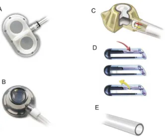
Another type of long-stay catheter is the totally implantable version, known as a portacath. These catheters have a diameter less than 10 Fr and can be implanted via a peripheral or central vein and, after taking a subcutaneous path, are connected to a reservoir (port) that is generally implanted over the muscular fascia of the site chosen for construction of the pocket that will accommodate the port. No part of the assembly is outside of the body and so this type of catheter has a lower risk of infection and greater durability than the semi-implantable type.18 Ports are fabricated from titanium or plastic and may have single or double chambers (Figure 1). Devices are available with and without valves and in some valved models the valve is positioned in the port and in others it is at the catheter tip (Figure 1). The theoretical advantage of valved catheters is to reduce the occurrence of malfunction caused by intracatheter thrombi, by preventing
inadvertent relux of blood. However, the superiority of valved catheters has not been conirmed.20,21
Some newer catheter models are more resistant and allow infusion of fluids at higher pressures
Table 1. Classiication of the most widely used types of catheter.
Catheter Duration Insertion/position of tip Frequency of use
Jelco Up to 4 days P/P Continuous
Short-term CVC Up to 3 weeks C/C Continuous
PICC Up to 12 months P/C Continuous/Intermittent
Semi-implantable Months to years C/C Continuous/Intermittent
Totally implantable Years C, P/C Intermittent
(up to 5 mL/s, 300 psi), such as injection via the catheter using a contrast pump for imaging exams (for example, Dignity - Medcomp, PowerPort - Bard).
Long-term catheters (PICC, semi-implantable and totally implantable) are manufactured from silicone or polyurethane, and each has different characteristics. While silicone offers better biocompatibility and lower risk of provoking thrombosis,22 a polyurethane catheter has thinner walls, allowing a larger diameter internal lumen in relation to a silicone catheter with the same external diameter, resulting in a lower risk of obstruction.19
INDICATIONS FOR USE
Peripheral accesses are preferred for short-term infusion of solutions (a few days) in patients with a preserved venous network and for infusion of solutions that are not vesicant. If vesicant solutions leak, they cause intense irritation, formation of boils (vesicles) and tissue necrosis. Patients being treated with non-vesicant chemotherapy for shorter periods
can beneit from this type of access.
Central venous access becomes more appropriate than peripheral when the solution to be infused has a pH < 5.0 or > 9.0, osmolarity > 500 mOsm/L, or vesicant characteristics.23,24 Other indications include a need to monitor central venous pressure and factors making peripheral access impossible, which are relatively common in oncology patients. Short-term central venous access should only be used with inpatients and for periods of less than 3 weeks.19
In cases for which central access will be needed for longer periods (some months) or the patient must be cared for at home, a PICC is an alternative option.
Nowadays, PICCs are increasingly itted for patients
on outpatient chemotherapy, because they allow intermittent use. Since part of the catheter remains outside of the body, exiting via the puncture site, they can cause discomfort.
High low semi-implantable catheters (permcath)
are indicated for patients who require hemodialysis for longer periods and for individuals on apheresis programs, which consists of a process for collecting peripheral stem cells mobilized into the blood circulation after treatment with granulocyte colony stimulating factor (G-CSF), preparatory to bone marrow transplantation. Hickman catheters offer the possibility of simultaneous infusion of different solutions, including blood products, in addition to their use for BMT. They also enable blood samples to be drawn for analysis, thereby offering increased comfort by avoiding frequent vein punctures, and can also be used for administration of prolonged intravenous parenteral nutrition.25
The main indications for totally implantable catheters are a need for frequent venous access, use of vesicant drugs, and a peripheral venous system that cannot be used for access. These catheters require percutaneous puncture to access the port, which is why they are more indicated for intermittent use, allowing the skin to recover during intervals in treatment. They are almost exclusively used for chemotherapy treatment of cancer patients.26
TECHNIQUES FOR PLACEMENT OF TOTALLY IMPLANTABLE CATHETERS
The operation to implant one of these catheters is performed in an appropriate setting, in which the patient’s vital signs can be monitored and imaging
support is available, particularly luoroscopy equipment.
In general, this infrastructure is found in operating theaters and radiology suites.
The type of anesthesia depends on the patient’s clinical status and the surgical team’s preferences. Generally, local anesthesia combined with sedation
is suficient. Since this is a clean operation, antibiotic
prophylaxis is not required.
Choice of the implantation site is based on which vein will be used to insert the catheter and the site in which the port pocket will be created. The preference is for insertion into veins that drain to the superior vena cava system. An anterior chest wall that does not offer adequate conditions is a relative indication for choosing veins of the inferior vena cava system, since the port can be placed in a number of alternative sites, such as the upper limbs.27 However, thrombosis of the superior vena cava is an absolute indication for insertion via internal saphenous or femoral veins.28 In exceptional situations, alternative options are translumbar puncture of the inferior vena cava, trans-hepatic percutaneous access, cannulation of collateral veins and recanalization of obstructed veins.29-31
The access technique is dependent of the vessel
chosen. In general, supericial veins (external jugular,
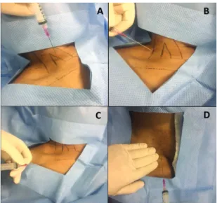
cephalic, basilic, and saphenous) are accessed by dissection, whereas deep veins (internal jugular, subclavian, and femoral) are reached by puncture26,32 (Figure 2). Reinements in materials (needles, guidewires) have resulted in puncture of deep veins becoming the procedure of choice in the majority of centers. Utilization of ultrasonography in the operating room makes it possible to assess the vein chosen for puncture, allowing diagnosis of asymptomatic thrombosis before the operation is started. This resource also enables puncture to be guided by ultrasound, reducing the risk of accidents, such as arterial puncture and pneumothorax (Figure 3).33,34
When the option chosen is dissection of a supericial
vein, a venotomy is performed to allow the catheter to be inserted and advanced until the tip reaches the central position. The vessel is ligated distally and a proximal ligature is placed around the catheter, taking care not to constrict it. In the case of larger caliber veins, a suture around the incision, rather than
ligature, allows maintenance of blood low, avoiding thrombophlebitis (Figure 4).
The venous path to the atrium is straighter on the right, which is why this side is preferred for insertion. In cases in which there are tumors in the thorax area (for example, breast cancer), even if there is no impediment to the catheter’s passage on that side,
Figure 2. Deep vein puncture techniques most commonly used for insertion of venous catheters. (A) Puncture anterior of the internal jugular vein (IJV). Entry between the bellies of the sternocleidomastoid muscle, with the needle angled at 45º in the direction of the ipsilateral nipple; (B) Puncture posterior to the IJV. Needle inserted in the medial direction, below the clavicular branch of the sternocleidomastoid muscle; (C) Infraclavicular puncture of the subclavian vein, with entry between the medial and lateral thirds of the clavicle; (D) Puncture of the femoral vein medial of the site where the femoral arterial pulse is palpated.
the procedure is generally conducted contralateral to the tumor.
The proximal extremity of the catheter is placed at the cavoatrial junction, carefully monitoring for possible arrhythmia provoked by the device. In many cases, the tip of the catheter may enter the right atrium, without harming the patient.
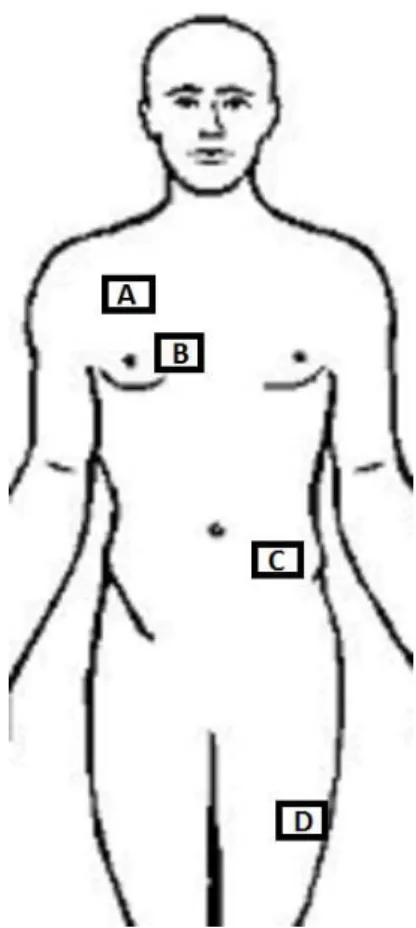
The port pocket should be created in site that is irm
and is distant from areas in which the skin has lost integrity, such as result from stoma, radiodermatits, or ulcerous tumoral lesions. Whenever possible, the port is implanted in the anterior thorax wall, just above the fascia of the pectoral muscle (Figure 5). In obese patients, very deep subcutaneous tissue could cause
dificulties with puncturing the port, if it were placed
directly against the muscle fascia. In such cases, the
port pocket can be created more supericially, within
the adipose plane, leaving subcutaneous tissue a minimum of 2 cm deep over the device.
When access is achieved via the internal saphenous or femoral veins, the port pocket can be constructed in the abdomen, medial of the anterior superior iliac crest, or on the anterolateral surface of the thigh (Figure 5).
After the port pocket has been correctly prepared, using rigorous hemostasis to reduce the risks of infection, the catheter is advanced along its subcutaneous path from the vein insertion site to the port pocket. After this, radiology is used once more to ascertain that the tip of the device is still in the
correct position and another low/backlow test is
performed. The port is then connected to the catheter
and positioned in the pocket, where it is ixed with
two non-absorbable sutures to the muscle fascia. Before closure of the subcutaneous tissue and the
skin, and another low/backlow test is performed,
this time by puncture of the port, rinsing the catheter with at least 20 mL of saline and infusing a heparin solution before removing the needle.
COMPLICATIONS ASSOCIATED WITH TOTALLY IMPLANTABLE CATHETERS RELATED TO THE IMPLANTATION PROCEDURE
Intercurrent conditions caused by the operation to implant the device are related to accidents that occur during puncture to access a central vein, such as pneumothorax, hemothorax, and inadvertent arterial puncture, or to navigation of endovascular devices (guidewire, introducer, catheter), which include venous drilling and myocardial injury.35,36
Hematoma and early infections at the pocket or along the catheter path are also adverse events that
Figure 4. Dissection of the external jugular vein (EJV) to insert a long-term catheter. (A) Proximal and distal repair of the EJV; (B) Venorrhaphy, avoiding distal ligature of the EJV, maintaining low through the vessel.
can be associated with the operation to place totally implantable catheters.26
When this procedure is conducted using anatomic parameters as a guide, the risk of pneumothorax caused during subclavian and jugular punctures can be as high as 3%,37,38 while accidental punctures of arteries occur in 5 to 10% of cases.38 In some studies, guiding the procedure with US eliminated occurrence of hemothorax and pneumothorax and was associated with an inadvertent arterial puncture rate of less than 1%.39 However, in contrast with the present study, these studies did not compare rates with punctures conducted using anatomic parameters.
At our service, in a total of 1,255 procedures to implant totally implantable catheters, there were 18 (1.4%) complications related to the procedure, including one (0.1%) case of pneumothorax and 14 (1.1%) inadvertent arterial punctures, nine of which occurred in US-guided procedures (0.9% of the total number of procedures).40 Our data show that not using US to guide venous puncture is a risk factor for iatrogenic arterial puncture, but not for other complications, such as hemothorax and pneumothorax.40
COMPLICATIONS ASSOCIATED WITH TOTALLY IMPLANTABLE CATHETERS RELATED TO USE OF THE IMPLANT
Infectious complications
Infectious complications are most frequently related with long-term catheters and are the principal cause for early removal (before the end of the treatment) of the catheter.26 Infection may occur in the pocket or in the bloodstream.
Infection of the port pocket
Diagnosis is by clinical examination when there are phlogistic signs (pain, hyperemia, increased local temperature) in the area of the port. There may be pus build up in the pocket, sometimes accompanied by dehiscence with drainage of purulent secretions. Conservative treatment does not generally achieve good results, and in the majority of cases the catheter has to be removed and systemic antibiotic therapy given.
Bloodstream infections
Diagnosis of bloodstream infections (BSI) in patients with long-term catheters is still a serious challenge. Fever and shivering are generally associated with
BSI, but these are nonspeciic symptoms. When a
BSI is suspected, paired blood cultures (BC) should be conducted (aerobic and anaerobic) of samples from the central catheter and from the peripheral
vascular access. A diagnosis of BSI is conirmed in
the following situations:
- The same infectious agent grows in both the catheter and the peripheral BCs.
- Central and peripheral BCs are positive:
- Time difference before positive result: central BC grows a microorganism at least 2 hours before the peripheral BC.
- Quantitative BC: central BC has at least three times greater growth of the infectious agent than the peripheral BC.
- Central BC positive and peripheral BC negative.
- There is sepsis with no other presumable focus of infection.
While waiting for the BC results, empirical treatment should cover both Gram-positive and Gram-negative
agents. After identiication of the infectious agent,
treatment should be adjusted to match the culture results,41 maintaining systemic antibiotics, combined with lock therapy for 7 to 14 days. After 72 hours of effective antibiotic treatment combined with lock therapy, a repeat pair of BCs should be conducted on samples collected via the catheter, irrespective of the clinical response observed. If there are still positive results for the same infectious agent, then the catheter should be removed.
Patients with bacteremia or fungemia that persists for 72 hours after removal of the catheter should be given antibiotic therapy for 4 to 6 weeks.
Table 2 lists situations that demand immediate removal of the catheter, with no attempt to save it.
In addition to taking precautions with antisepsis and asepsis during the implantation procedure, there is evidence that insertion by puncture is associated with a lower risk of infection than insertion by venous dissection.42 It is also advisable to use double-chamber portals only when strictly necessary and reserve the catheter exclusively for chemotherapy treatments.
At our institution, infectious complications were the most frequent type, with a prevalence of 13%, 0.35/1,000 days of use.40 The greater part (66%) of
Table 2. Indications for removal of long-term catheters.
Hemodynamic instability
Blood culture positive for Staphylococcus aureus, Candida spp Sepsis or bacteremia that remain after 48 hours of appropriate antibiotic therapy
these intercurrent conditions were late and therefore were associated with using the device and not with the implantation procedure.40
Non-infectious complications
Deep venous thrombosis
In addition to presence of factors associated with cancer that increase the risks of deep venous thrombosis, such as hypercoagulability, endothelial injury from the chemotherapy agents, and venous compression by the tumor, the presence of a catheter can itself be considered a risk factor.
Deep venous thrombosis (DVT) can cause signs and symptoms such as pain along the path of the vein, edema of a limb, facial edema, and presence of collateral venous circulation in the chest wall. Diagnosis is achieved using imaging exams, such as venous duplex scan of the cervical and abdominal regions and of limbs. If thrombosis is suspected in the venous brachiocephalic trunk or the superior vena cava, then computed tomography angiography or magnetic resonance angiography are more appropriate. However, patients are very often asymptomatic and diagnosis an incidental result of routine tests conducted during cancer treatment.43
Once a diagnosis of DVT has been made, full anticoagulation is initiated (as long as there are no clinical contraindications). Highly symptomatic patients, with extensive thromboses, such as cases of superior vena cava syndrome, may be candidates
for ibrinolytic treatment, weighing up the risks of
hemorrhagic complications.44
If the catheter is still functioning correctly, it
should be left in place, since there is no beneit from
removing it and there is a risk of provoking additional venous thromboses by placing another catheter at a different site. Removal is restricted to cases in which the catheter is no longer patent, which happens when the DVT involves the tip of the device.45
The likelihood of occurrence of catheter-related DVT is reduced by maintaining the tip of the catheter close to or within the right atrium, even in cases in which the device is implanted via a femoral or saphenous access.
Among the non-infectious complications recorded at our institution, there were 27 (2.2%) cases of DVT, equating to 0.06/1,000 days of use.40 This rate is compatible with what is reported in other studies, in which the frequency of DVT varies from 0.03 to 1.2/1,000 days of use.46 Some authors report that the risk of DVT is greater when a subclavian vein access is used, when compared with insertion
via the internal jugular,47-50 whereas others state that the subclavian access is better than other accesses in terms of DVT incidence.49 Femoral access has also
been identiied as involving higher risk of DVT in
some studies.47,48 However, our data do not show a relationship between site of insertion and occurrence of DVT.40
Malfunctions
Malfunctions may involve dysfunctions preventing blood drawing only or of both blood drawing and infusion of medications. Malfunction may be the result of technical failure during implantation, such as incorrect positioning of the tip of the catheter, excessive angulation, or pinching of the catheter (Figure 6). The last of these three is most common when the catheter is inserted via puncture of the
subclavian vein, since the space between the irst rib
and the clavicle is narrow. Malfunction immediately
after the catheter is irst punctured is indicative of
technical failure of the implantation procedure.26 The catheter’s presence in the intravascular space can
provoke ibrin formation around it, impeding relux by
acting as a valve mechanism when negative pressure is created during aspiration (Figure 7). With catheters
that have a slit-shaped valve at the tip, a ibrin layer
may not only prevent blood from being drawn, but
also infusion of luids.45,51
Another condition that can impact on functioning is formation of thrombi in the catheter lumen, caused
by relux of blood that may occur, for example, when
negative pressure is created by removal of the puncture needle from the port.52
Investigation of a malfunctioning catheter begins by checking the puncture. Very often failure to achieve
relux is the result of incorrect puncture of the port.
The next step is a simple chest X-ray to analyze the
position of the catheter. The tip could be misplaced because of technical failure during implantation, or as the result of migration after a successful implantation. If the catheter is correctly positioned, without excessive angulation and with no signs of fracture or pinching,
ibrinolysis can be attempted and often produces good
results for dysfunctions occurring less than 15 days previously.26
A DVT may cause loss of function if it involves the tip of the catheter.26
Embolization of the catheter
This can occur if the catheter becomes detached from the port or is fractured, which is more frequent in cases in which the device is implanted by puncture of the subclavian vein.45,51
Suspicion is aroused if the catheter will not allow blood to be drawn and the patient complains of pain on infusion of medications. A simple X-ray may show the catheter detached from the port or completely fractured and possible embolization of the catheter. Partially damaged catheters do not provoke embolization and are diagnosed by an examination with contrast in which contrast leakage will be seen.
In these cases, removal of the device is mandatory. If a total fracture with migration has taken place, it may be possible to remove the catheter with endovascular techniques.
Port rotation
If the port becomes rotated, the puncture area will be against the chest wall and the base will be facing out, preventing puncture.26
A profile chest X-ray can show rotation of metallic portals. However, if the port is made from a radiotransparent material (plastic), palpation should
be suficient for diagnosis, since the port will not be
visible on the imaging exam.
Treatment requires a surgical operation to reposition
and ix the port.
Extrusion of the port
Dehiscence of the skin with exposure of the port can be a result of an infection, but may also be caused by necrosis of skin, which can adhere to the port if there
is insuficient subcutaneous tissue over the device.53 To avoid this complication, the best available site should be chosen for construction of the port pocket, avoiding areas with too little adipose tissue, such as close to the manubrium of the sternum. In patients
with cachexia, low-proile portals should be preferred.
Materials failures
Nowadays, primary failures of devices are rare, but can still be observed at high-volume centers.
Chemotherapy is an option for a large proportion of cancer patients and since it is based on the infusion of intravenous drugs intermittently and for prolonged periods, totally implantable catheters are often chosen.26,28 These devices increase comfort and safety of infusion treatments, since many of the drugs used are vesicant and it is not uncommon for cancer patients to have problems that make peripheral access
dificult. As long as they are accessed at specialized
centers and by nursing teams who have been trained to use these devices, totally implantable catheters also enable intravenous infusion of other medications and drawing of blood samples for laboratory analysis. These functions are especially useful when the patient has been admitted to hospital for treatment of cancer or an intercurrent clinical condition. More recent models (for example, Dignity - Medcomp, PowerPort - Bard) allow infusion of solutions using injection pumps, accepting pressures up to 300 psi and infusion rates up to 5 mL/s, making it possible to perform examinations such as computed tomography with contrast injected via the catheter.
Maintenance of the device is of great importance for these patients, considering the long duration of antineoplastic treatment. Therefore, determination of the risk factors for complications related to long-term catheters is essential to maintaining them until treatment is complete.54
Despite the advances that have been achieved with relation to construction of catheters and operating techniques,26,28 the complications related
to implantation procedures and use of the device that have been described above remain a challenge to the multidisciplinary team responsible for treating these patients.
Variations in implantation technique and differences related to occurrence of complications and their management may be related to institutional issues, which should motivate every oncology center to monitor the progress of their patients who have totally implantable catheters.
REFERENCES
1. Harvey W. Exercitatio anatomica de motu cordis et sanguinis in animalibus. Florence: R. Lier & Co.; 1628.
2. Barsoum N, Kleeman C. Now and then, the history of parenteral fluid administration. Am J Nephrol. 2002;22(2-3):284-9. PMid:12097754. http://dx.doi.org/10.1159/000063775.
3. Dudrick SJ. History of vascular access. JPEN J Parenter Enteral Nutr. 2006;30(1, Supl):S47-56. PMid:16387910. http://dx.doi.org/10.11 77/01486071060300S1S47.
4. Gordon HE. Historical development of vascular access procedures. In: Wilson SE, editor. Vascular access surgery. 2. ed. Chicago: Year Book; 1988. p. 1.
5. Blundell J. Successful case of transfusion. Lancet. 1829;I:431-2.
6. O’Shaughnessy WB. Experiments on the blood in cholera. Lancet. 1832;40:1831-2.
7. Latta T. Injections in cholera. Lond Med Gazz. 1832;379-82.
8. Matas RM. A clinical report on intravenous saline infusion in the Charity Hospital, New Orleans (1881 to 1891). New Orleans Med Surg J. 1891;181(14):83-8.
9. Zimmermann B. Intravenous tubing for parenteral therapy. Science. 1945;101(2631):567-8. PMid:17780134. http://dx.doi.org/10.1126/ science.101.2631.567.
10. Aubaniac R. L’injection intraveineuse sous-claviculaire: advantages et technique. Presse Med. 1952;60(68):1456. PMid:13027062.
11. Seldinger SI. Catheter replacement of the needle in percutaneous arteriography; a new technique. Acta Radiol. 1953;39(5):368-76. PMid:13057644. http://dx.doi.org/10.3109/00016925309136722.
12. Wilson JN, Owens JC. Continuous monitoring of venous pressure in optimal blood volume maintenance. Surg Forum. 1961;12:94-6. PMid:14007384.
13. Yoffa D. Supraclavicular subclavian venepuncture and catheterisation. Lancet. 1965;2(7413):614-7. PMid:4157504. http://dx.doi.org/10.1016/ S0140-6736(65)90519-2.
14. Broviac JW, Cole JJ, Scribner BH. A silicone rubber atrial catheter for prolonged parenteral alimentation. Surg Gynecol Obstet. 1973;136(4):602-6. PMid:4632149.
15. Hickman RO, Buckner CD, Clift RA, Sanders JE, Stewart P, Thomas ED. A modified right atrial catheter for access to the venous system in marrow transplant recipients. Surg Gynecol Obstet. 1979;148(6):871-5. PMid:109934.
16. Belin RP, Koster JK Jr, Bryant LJ, Griffen WO Jr. Implantable subcutaneous feeding chamber for noncontinuous central venous alimentation. Surg Gynecol Obstet. 1972;134(3):491-3. PMid:4621851.
17. Niederhuber JE, Ensminger W, Gyves JW, Liepman M, Doan K, Cozzi E. Totally implanted venous and arterial access system to replace external catheters in cancer treatment. Surgery. 1982;92(4):706-12. PMid:7123491.
18. Maki DG, Kluger DM, Crnich CJ. The risk of bloodstream infection in adults with different intravascular devices: a systematic review of 200 published prospective studies. Mayo Clin Proc. 2006;81(9):1159-71. PMid:16970212. http://dx.doi.org/10.4065/81.9.1159.
19. Gallieni M, Pittiruti M, Biffi R. Vascular access in oncology patients. CA Cancer J Clin. 2008;58(6):323-46. PMid:18971486. http://dx.doi. org/10.3322/CA.2008.0015.
20. Biffi R, De Braud F, Orsi F, et al. A randomized, prospective trial of central venous ports connected to standard open-ended or Groshong catheters in adult oncology patients. Cancer. 2001;92(5):1204-12. PMid:11571734. http://dx.doi.org/10.1002/1097-0142(20010901)92:5<1204::AID-CNCR1439>3.0.CO;2-9.
21. Zottele Bomfim GA, Wolosker N, Yazbek G, et al. Comparative study of valved and nonvalved fully implantable catheters inserted via ultrasound-guided puncture for chemotherapy. Ann Vasc Surg. 2014;28(2):351-7. PMid:24094470. http://dx.doi.org/10.1016/j. avsg.2013.01.025.
22. Galloway S, Bodenham A. Long-term central venous access. Br J Anaesth. 2004;92(5):722-34. PMid:15003979. http://dx.doi. org/10.1093/bja/aeh109.
23. Registered Nurses’ Association of Ontario RNAO [site na Internet]. Ontario: Assessment and Device Selection for Vascular Access; 2004. [atualizado 2008; citado 2016 set 08]. http://rnao.ca/bpg/ guidelines/assessment-and-device-selection-vascular-access
24. Bishop L, Dougherty L, Bodenham A, et al. Guidelines on the insertion and management of central venous access devices in adults. Int J Lab Hematol. 2007;29(4):261-78. PMid:17617077. http://dx.doi.org/10.1111/j.1751-553X.2007.00931.x.
25. Silveira RCCP. O cuidado de enfermagem e o cateter de Hickman: a busca de evidências [tese]. Ribeirão Preto: Universidade de São Paulo; 2005.
26. Wolosker N, Yazbek G, Nishinari K, et al. Totally implantable venous catheters for chemotherapy: experience in 500 patients. Sao Paulo Med J. 2004;122(4):147-51. PMid:15543368. http:// dx.doi.org/10.1590/S1516-31802004000400003.
27. Zerati AE, Wolosker N, Motta-Leal-Filho JM, Nabuco PH, Puech-Leão P. Totally implantable venous catheters: insertion via internal jugular vein with pocket implantation in the arm is an alternative for diseased thoracic walls. J Vasc Access. 2012;13(1):71-4. PMid:21748723. http://dx.doi.org/10.5301/JVA.2011.8486.
28. Wolosker N, Yazbek G, Munia MA, Zerati AE, Langer M, Nishinari K. Totally implantable femoral vein catheters in cancer patients. Eur J Surg Oncol. 2004;30(7):771-5. PMid:15296992. http://dx.doi. org/10.1016/j.ejso.2004.05.019.
29. Power A, Singh S, Ashby D, et al. Translumbar central venous catheters for long-term haemodialysis. Nephrol Dial Transplant. 2010;25(5):1588-95. PMid:20023114. http://dx.doi.org/10.1093/ ndt/gfp683.
30. Powell S, Belfield J. Complex central venous catheter insertion for hemodialysis. J Vasc Access. 2014;15(Supl 7):S136-9. PMid:24817471. http://dx.doi.org/10.5301/jva.5000250.
32. Yazbek G, Zerati AE, Malavolta LC, Nishinari K, Wolosker N. Endovascular techniques for placement of long-term chemotherapy catheters. Rev Hosp Clin Fac Med Sao Paulo. 2003;58(4):215-8. PMid:14534674. http://dx.doi.org/10.1590/S0041-87812003000400005.
33. Cavanna L, Civardi G, Vallisa D, et al. Ultrasound-guided central venous catheterization in cancer patients improves the success rate of cannulation and reduces mechanical complications: a prospective observational study of 1,978 consecutive catheterizations. World J Surg Oncol. 2010;8(1):91. PMid:20958986. http://dx.doi. org/10.1186/1477-7819-8-91.
34. Peris A, Zagli G, Bonizzoli M, et al. Implantation of 3951 long-term central venous catheters: performances, risk analysis, and patient comfort after ultrasound-guidance introduction. Anesth Analg. 2010;111(5):1194-201. PMid:20829559. http://dx.doi.org/10.1213/ ANE.0b013e3181f333c1.
35. Dariushnia SR, Wallace MJ, Siddiqi NH, et al. Quality improvement guidelines for central venous access. J Vasc Interv Radiol. 2010;21(7):976-81. PMid:20610180. http://dx.doi.org/10.1016/j. jvir.2010.03.006.
36. Teichgräber UK, Gebauer B, Benter T, Wagner HJ. Central venous access catheters: radiological management of complications. Cardiovasc Intervent Radiol. 2003;26(4):321-33. PMid:14667113.
37. Dariushnia SR, Wallace MJ, Siddiqi NH, et al. Quality improvement guidelines for central venous access. J Vasc Interv Radiol. 2010;21(7):976-81. PMid:20610180. http://dx.doi.org/10.1016/j. jvir.2010.03.006.
38. Teichgräber UK, Gebauer B, Benter T, Wagner HJ. Central venous access catheters: radiological management of complications. Cardiovasc Intervent Radiol. 2003;26(4):321-33. PMid:14667113.
39. Fankhauser GT, Fowl RJ, Stone WM, Money SR. Elimination of pneumothorax and hemothorax during placement of implantable venous access ports using ultrasound and fluoroscopic guidance. Vascular. 2013;21(6):345-8. PMid:23493277. http://dx.doi. org/10.1177/1708538112472279.
40. Zerati AE, Figueredo TR, de Moraes RD, et al. Risk factors for infectious and noninfectious complications of totally implantable venous catheters in cancer patients. J Vasc Surg Venous Lymphat Disord. 2016;4(2):200-5. PMid:26993868. http://dx.doi.org/10.1016/j. jvsv.2015.10.008.
41. Mermel LA, Allon M, Bouza E, et al. Clinical practice guidelines for the diagnosis and management of intravascular catheter-related infection: 2009 Update by the Infectious Diseases Society of America. Clin Infect Dis. 2009;49(1):1-45. PMid:19489710. http:// dx.doi.org/10.1086/599376.
42. O’Grady NP, Alexander M, Dellinger EP, et al. Guidelines for the prevention of intravascular catheter-related infections. Centers for Disease Control and Prevention. MMWR Recomm Rep. 2002;51(RR-10):1-29. PMid:12233868.
43. Frykholm P, Pikwer A, Hammarskjöld F, et al. Clinical guidelines on central venous catheterisation. Swedish Society of Anaesthesiology
and Intensive Care Medicine. Acta Anaesthesiol Scand. 2014;58(5):508-24. PMid:24593804. http://dx.doi.org/10.1111/aas.12295.
44. Debourdeau P, Farge D, Beckers M, et al. International clinical practice guidelines for the treatment and prophylaxis of thrombosis associated with central venous catheters in patients with cancer. J Thromb Haemost. 2013;11(1):71-80. PMid:23217208. http:// dx.doi.org/10.1111/jth.12071.
45. Nishinari K, Wolosker N, Bernardi CV, Yazbek G. Totally implantable ports connected to valved catheters for chemotherapy: experience from 350 Groshong devices. J Vasc Access. 2010;11(1):17-22. PMid:20119917.
46. Wolosker N, Yazbek G, Nishinari K, et al. Totally implantable venous catheters for chemotherapy: experience in 500 patients. Sao Paulo Med J. 2004;122(4):147-51. PMid:15543368. http:// dx.doi.org/10.1590/S1516-31802004000400003.
47. Frykholm P, Pikwer A, Hammarskjöld F, et al. Clinical guidelines on central venous catheterisation. Swedish Society of Anaesthesiology and Intensive Care Medicine. Acta Anaesthesiol Scand. 2014;58(5):508-24. PMid:24593804. http://dx.doi.org/10.1111/aas.12295.
48. Debourdeau P, Farge D, Beckers M, et al. International clinical practice guidelines for the treatment and prophylaxis of thrombosis associated with central venous catheters in patients with cancer. J Thromb Haemost. 2013;11(1):71-80. PMid:23217208. http:// dx.doi.org/10.1111/jth.12071.
49. Araújo C, Silva JP, Antunes P, et al. A comparative study between two central veins for the introduction of totally implantable venous access devices in 1201 cancer patients. Eur J Surg Oncol. 2008;34(2):222-6. PMid:17566692. http://dx.doi.org/10.1016/j. ejso.2007.04.003.
50. Verso M, Agnelli G. Venous thromboembolism associated with long-term use of central venous catheters in cancer patients. J Clin Oncol. 2003;21(19):3665-75. PMid:14512399. http://dx.doi. org/10.1200/JCO.2003.08.008.
51. Pignataro BS, Nishinari K, Wolosker N, Bomfim GA. Fracture and migration into the coronary sinus of a totally implantable catheter introduced via the right internal jugular vein. BMJ Case Rep. 2014;1:2014. PMid:25452299.
52. Araújo C, Silva JP, Antunes P, et al. A comparative study between two central veins for the introduction of totally implantable venous access devices in 1201 cancer patients. Eur J Surg Oncol. 2008;34(2):222-6. PMid:17566692. http://dx.doi.org/10.1016/j. ejso.2007.04.003.
53. Nishinari K, Bernardi CV, Wolosker N, Yazbek G. Retained catheter: a rare complication associated with totally implantable venous ports. J Vasc Access. 2010;11(2):159-61. PMid:20119912.
*
Correspondence
Antonio Eduardo Zerati Universidade de São Paulo – USP, Faculdade de Medicina, Hospital das Clínicas Rua Joaquim Floriano, 820, cj. 61 – Itaim Bibi CEP 04534-003 - São Paulo (SP), Brazil Tel.: +55 (11) 3071-1464 E-mail: [email protected]
Author information
AEZ - Tenured Professor, Hospital das Clínicas, Faculdade de Medicina, Universidade de São Paulo (USP). NW - Associate Professor, Faculdade de Medicina, Universidade de São Paulo (USP). NL - Full Professor of Vascular and Endovascular Surgery, Faculdade de Medicina, Universidade de São Paulo (USP). PPL - Full Professor of Vascular and Endovascular Surgery, Faculdade de Medicina, Universidade de São Paulo (USP).
Author contributions
Conception and design: AEZ, NW, NL, PPL Analysis and interpretation: AEZ, NW Data collection: AEZ Writing the article: AEZ Critical revision of the article: AEZ, NW, NL, PPL Final approval of the article*: AEZ, NW, NL, PPL Statistical analysis: AEZ Overall responsibility: AEZ