ABSTRACT
Objective: Many patients with proportional reductions in FVC and FEV1 on spirometry show no reduction in TLC. The aim of this study was to evaluate the role that measuring lung volumes and airway resistance plays in the correct classiication of patients with a possible restrictive pattern on spirometry. Methods: This was a prospective study involving adults with reduced FVC and FEV1, as well as an FEV1/FV(C) ratio within the predicted range. Restrictive lung disease (RLD) was characterized by TLC below the 5th percentile, as determined by plethysmography.
Obstructive lung disease (OLD) was characterized by high speciic airway resistance, signiicant changes in post-bronchodilator FEV1, or an FEF25-75% < 50% of predicted, together with a high RV/TLC ratio. Nonspeciic lung disease (NLD) was characterized by TLC within the predicted range and no obstruction. Combined lung disease (CLD) was characterized by reduced TLC and indings indicative of airlow obstruction. Clinical diagnoses were based on clinical suspicion, a respiratory questionnaire, and the review of tests of interest. Results: We included 300 patients in the study, of whom 108 (36%) were diagnosed with RLD. In addition, 120 (40%) and 72 (24%) were diagnosed with OLD/CLD and NLD, respectively. Among the latter, 24 (33%) were clinically diagnosed with OLD. In this sample, 151 patients (50.3%) were obese, and obesity was associated with all patterns of lung disease. Conclusions: Measuring lung volumes and airway resistance is often necessary in order to provide an appropriate characterization of the pattern of lung disease in patients presenting with a spirometry pattern suggestive of restriction. Airlow obstruction is common in such cases.
Keywords: Spirometry; Airway resistance; Lung volume measurements.
Lung volumes and airway resistance in
patients with a possible restrictive pattern
on spirometry
Kenia Schultz1,2, Luiz Carlos D’Aquino3, Maria Raquel Soares4, Andrea Gimenez5, Carlos Alberto de Castro Pereira4,5
Correspondence to:
Kenia Schultz. Avenida dos Imigrantes, 519, apto. 301, Noêmia Vitali, CEP 29707-040, Colatina, ES, Brasil. Tel.: 55 27 99959-5877. Fax: 55 27 3711-1366. E-mail: [email protected]
Financial support: None. INTRODUCTION
The American Thoracic Society (ATS)/European
Respiratory Society (ERS) task force proposed deinitions
for the various patterns of lung disease.(1) Restrictive
lung disease (RLD) was deined as a reduction in TLC
below the 5th percentile of the predicted value and a normal FEV1/VC ratio. Obstructive lung disease (OLD)
was deined as an FEV1/VC ratio below the 5th percentile
of the predicted value. Mixed or combined lung disease
(CLD) was characterized by FEV1/VC and TLC below the
5th percentile of the predicted values.
A combination of reduced VC and preserved FEV1/
(F)VC is used in order to infer the presence of RLD; however, in approximately 40% of such cases, TLC is not
reduced.(2,3) According to the ATS/ERS task force, OLD is
characterized by a combination of reduced (F)VC, FEV1/
(F)VC above the lower limit of normal, and TLC within
the predicted range.(1) This functional abnormality was
later designated nonspeciic lung disease (NLD).(4) In a
sample of 100 patients presenting with reduced (F)VC,
FEV1/(F)VC above the lower limit of normal, and TLC within the predicted range, 68 had evidence of airway disease, whereas the remaining 32 had signs of restriction.(4)
The proportional reduction in FVC and FEV1 in patients
with OLD can be explained by airway closure with air
trapping.(5) Obesity reduces (F)VC more than it does FEV
1
(6)
and can therefore result in a preserved FEV1/(F)VC ratio
in the presence of OLD. As occurs with diseases affecting
respiratory mechanics or respiratory muscle strength,
obesity can, in and of itself, result in NLD.(4) Although
COPD and asthma account for most OLDs, a wide range
of other diseases, including bronchiolar diseases and
some interstitial lung diseases, are associated with airlow
obstruction and can result in proportional reductions in
FVC and FEV1.
(7) In addition, smoking (either current or
past) is associated with various lung diseases and can contribute to an obstructive component.
Spirometry is considered the method of choice for
detecting airlow limitation caused by OLD. However, airlow limitation is multifactorial. One such factor is high airway
resistance (Raw).(8) In many patients with spirometry
results suggestive of RLD, Raw measurements can reveal
airlow obstruction. It is commonly believed that Raw is a parameter that is not suficiently sensitive in cases of peripheral airway disease; however, a classic study
showed a close correlation between airway conductance
1. Programa de Pós-Graduação em Ciências da Saúde, Instituto de Assistência Médica ao Servidor Público Estadual, São Paulo (SP) Brasil.
2. Centro Universitário do Espírito Santo, Colatina (ES) Brasil.
3. Faculdade de Medicina, Universidade da Região de Joinville, Joinville (SC) Brasil.
4. Universidade Federal de São Paulo, São Paulo (SP) Brasil.
5. Centro de Diagnósticos Brasil, São Paulo (SP) Brasil.
Submitted: 27 March 2016.
Accepted: 31 July 2016.
(Gaw) and bronchiolar diameter.(9) It is possible that
Gaw alone is abnormal in patients with bronchiolitis. (10)
In 2012, reference values for speciic Raw were derived
from a large sample of healthy adults.(11)
The objective of the present study was to evaluate the role that measuring lung volumes and Raw plays
in the inal functional classiication of patients with
spirometry results suggestive of RLD.
METHODS
Data collection was performed in the pulmonary function laboratories of Centro Diagnóstico Brasil
(n = 217) and the São Paulo Hospital for State Civil
Servants (n = 83) in the period between December of
2011 and December of 2013. Pulmonologists certiied in pulmonary function testing by the Brazilian Thoracic
Association (BTA) and the lead author of the present study prospectively selected all spirometry results
suggestive of RLD. Clinical diagnosis was established by
the pulmonologist requesting the test, by administering
a standardized respiratory questionnaire adapted from a
previously published questionnaire (Appendix 1: http:// www.jornaldepneumologia.com.br/detalhe_anexo. asp?id=46)(12) and by reviewing ancillary test results
or analyzing the results of additional tests, including chest X-rays, chest CT scans, and echocardiograms,
requested on the basis of clinical suspicion. All pul-monary function tests were performed in accordance with the BTA guidelines.(13) All patients gave written
informed consent.
Inclusion criteria
The inclusion criteria were as follows: 1) being an adult whose age and height were within the reference range(14); 2) having FVC below the lower limit of
normal, i.e., below the 5th percentile of the reference population(14); 3) having FEV
1/FVC and FEV1/VC equal
to or above the lower limit of normal, i.e., above the 5th percentile of the reference population(14);
4) having a deinitive clinical diagnosis (for asthma,
physician-diagnosed asthma and a patient report of two
or more episodes of wheezing, which were alleviated by bronchodilator use; for COPD, physician-diagnosed COPD, chronic cough/dyspnea—a Medical Research Council scale score ≥ 2—and past or current smoking;
patients diagnosed with obesity were in most cases referred for preoperative evaluation for bariatric surgery, including those with a complaint of dyspnea
without meeting criteria for diseases such as asthma);
and 5) having performed pulmonary function tests in accordance with the BTA/ATS/ERS acceptability and reproducibility criteria.(13,15-17)
Patients whose tests were not in accordance with
the aforementioned criteria were excluded, as were
those without a deinitive diagnosis by the end of the
analysis period.
All pulmonary function tests were performed with
a Sensor Medics 6200 Bodybox system and a Collins system (Ferraris Respiratory, Louisville, CO, USA).
Lung volumes were determined by whole-body ple-thysmography. For lung volumes, the predicted values
were those proposed by Crapo et al.(18) Reduced TLC
was characterized by values below the 5th percentile. RV and the RV/TLC ratio were considered high when
they were above the 95th percentile of the reference values.(18) Spirometry was repeated after administration
of a bronchodilator (400 µg of albuterol aerosol). A
signiicant bronchodilator response was characterized
by FEV1 ≥ 0.20 L and 7% of predicted, in accordance with Soares et al.(19)
Raw was measured by mean linear intercept values, as recommended by Matthys et al., after analysis of
at least ive pressure-low loops.(20) Only satisfactory,
reproducible loops were accepted. The predicted values
used for calculation were those proposed by Piatti
et al.(11) Values above 8.0 cmH
2O/s in females and
8.6 cmH2O/s in males were considered high (mean
± 1.64 SD).
Satisfactory single-breath DLCO measurements were
obtained in 260 patients. The reference values were based on those proposed by Miller et al.(21)
After data collection, the patterns of lung disease were divided into four groups:
RLD—characterized by TLC below the lower limit of
normal and no obstruction(1)
OLD—characterized by one or more of the following: high speciic Raw corrected for lung volume (Raw × Lv); a signiicant change in FEV1 after bronchodilator
administration (ΔFEV1 > 0.20 L and 7% of predicted);
and FEF25-75% < 50% of predicted with a high RV/TLC ratio (see the Results section)
CLD—characterized by reduced TLC and indings indicative of airlow obstruction, including high Raw × Lv; FEF25-75% < 50% with a high RV/TLC ratio; and
a signiicant bronchodilator response
NLD—characterized by TLC within the predicted range and no functional indings indicative of obstruction
All values were expressed as mean ± standard deviation. The groups were compared by means of the
Student’s t-test and ANOVA (for continuous independent
variables), and the chi-square test (for nominal variables).
Correlations between Raw × Lv and functional parameters
were determined by Spearman’s test. The distribution
of Raw × Lv was lognormal, and Raw × Lv values were transformed for comparison. ROC curve analysis was
used in order to correlate functional parameters and
the RV/TLC ratio with speciic Raw. Statistical analysis was performed with the IBM SPSS Statistics software package, version 20 (IBM Corp., Armonk, NY, USA). The level of signiicance was set at α = 0.05.
RESULTS
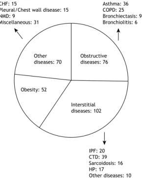
Clinical diagnoses were divided into four groups:
obstructive diseases, interstitial diseases, obesity, and other diseases (Figure 1).
Of the 300 patients included in the present study,
151 (50.3%) were obese, but only 52 (17.3%) had a
inal diagnosis of obesity without other conditions. In addition, 172 (57.3%) had TLC below the lower limit of normal (RLD), and 128 (42.7%) had TLC within the predicted range (n = 127) or high TLC (n = 1).
RV and the RV/TLC ratio were above the upper limit
of normal in 46 (15.3%) and 126 (42.0%), respectively.
High Raw × Lv was observed in 97 patients (32.3%). Raw × Lv (and Gaw/Lv) correlated more strongly with
FEF25-75% (rs = 0.55) than with FEV1/FVC (rs = 0.50) or percent predicted FEV1 (rs = 0.27; p < 0.01 for all).
Raw × Lv also correlated signiicantly with the RV/ TLC ratio (rs = 0.46; p < 0.001). ROC curve analysis
showed that the area under the ROC curve was higher
for FEF25-75% than for FEV1/FVC or percent predicted FEV1 (i.e., 0.75; p < 0.001) for differentiating between
patients with and without high Raw × Lv. An FEF25-75% of
less than 50% had a sensitivity of 40% and a speciicity of 89% for detecting high speciic Raw. With regard to lung volume measurements, the RV/TLC ratio had the highest area under the curve for characterizing airlow obstruction (0.75; p < 0.01). Given that a high RV/TLC ratio and FEF25-75% < 50% can each be found
in patients with RLD or NLD, they were combined in order to characterize airlow obstruction. A combination of high RV/TLC and FEF25-75% < 50% was found in 46
patients. In 14 of those, the aforementioned parameters constituted the only evidence of obstruction.
A signiicant bronchodilator response was observed in
23 patients (7.7%). The most common clinical diagnoses in those patients were obstructive diseases (n = 12)
and obesity (n = 5). Of the 14 patients diagnosed with congestive heart failure (CHF), only 1 (7.0%) had a signiicant bronchodilator response.
On the basis of one or more of the aforementioned criteria, 120 patients (40.0%) had airlow obstruction. Of the 120 patients with OLD, 64 (53.3%) had TLC
below the lower limit of normal and were therefore
considered to have CLD. Of the 128 patients with TLC within the predicted range, 72 (56.2%) had no airlow obstruction and were therefore classiied as having NLD.
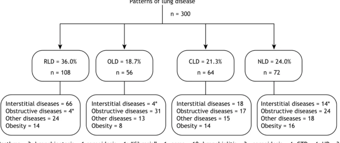
All patterns of lung disease found in the present study and the respective clinical diagnoses are shown in Figure 2. In the four groups of lung diseases there were patients diagnosed with obesity and patients diagnosed with other diseases. Four of the patients who were diagnosed with asthma had RLD, and 17
of the patients in the CLD group had a diagnosis of OLD, asthma being the most common obstructive
disease (n = 10).
Of the 72 patients who were diagnosed with NLD,
24 (33.3%) had a clinical diagnosis of obstructive
disease: asthma, in 11; COPD, in 8; bronchiectasis, in 3; and bronchiolitis, in 3. Therefore, of the 300
patients included in the present study, 144 (i.e., the aforementioned 24 plus the 120 who were diagnosed
with OLD or CLD, accounting for 48.0% of the sample)
had obstructive disease.
Table 1. General characteristics of patients with spirometry results suggestive of restrictive lung disease (n = 300).a
General characteristics Results
Age, years 56.2 ± 14.4
Males/females, n/n 117/183
Nonsmokers/smokers/ former smokers, n/n/n
187/34/79
Body mass index, (kg/m2) 31.0 ± 7.9 aValues expressed as mean ± SD, except where otherwise indicated.
Table 2. Functional characteristics of patients with spirometry
results suggestive of restrictive lung disease (N = 300).a
Functional characteristics Results
VC% 66.2 ± 11.0
FVC% 65.0 ± 10.6
FEV1% 64.5 ± 10.8
FEV1/FVC 0.81 ± 0.06
FEF25-75% 74.6 ± 28.8
RV% 99.5 ± 32.0
RV/TLC 0.44 ± 0.10
TLC% 78.6 ± 14.2
Raw× Lv 8.44 (4.17-9.09)
Raw × Lv: speciic airway resistance corrected for lung
volume. aValues expressed as mean ± SD or median (interquartile range).
CHF: 15
Pleural/Chest wall disease: 15 NMD: 9
Miscellaneous: 31
Asthma: 36 COPD: 25 Bronchiectasis: 9 Bronchiolitis: 6
Other diseases: 70
Obstructive diseases: 76
Obesity: 52
Interstitial diseases: 102
IPF: 20 CTD: 39 Sarcoidosis: 16 HP: 17
Other diseases: 10
Figure 1. Clinical diagnoses in patients with spirometry
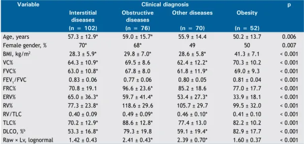
Several variables, including age, gender, and functional results, were compared among the four major clinical
diagnostic groups (Table 3). The patients with a inal
diagnosis of obesity were younger and had a body mass index above 35 kg/m2 (79%); therefore, most
were referred for preoperative evaluation for bariatric surgery. Expiratory reserve volume (ERV) was lowest in the group of patients who were diagnosed with obesity, who, in comparison with the group of patients diagnosed with interstitial diseases, had higher percent
predicted VC, FVC, RV, and TLC; more preserved DLCO; and similar Raw × Lv.
DISCUSSION
The present study conirms the indings of previous
studies(2-4) showing that spirometric indings of reduced
FVC and FEV1 associated with a preserved FEV1/FVC
ratio are of limited value in establishing a functional diagnosis. In addition, the study shows that lung volume and Raw measurements provide a more consistent
functional characterization.
It is widely recognized that adequate expiratory effort and time are required in order to characterize reduced FVC on spirometry. Incomplete exhalation
often results in a restrictive pattern on spirometry. In the present study, all tests were carefully performed and reviewed.
The ATS/ERS task force deined RLD as a reduction in TLC below the 5th percentile of the predicted value
and a normal FEV1/FVC ratio.(1) The reference values for
TLC are therefore of great importance. In the present study, we used the values proposed by Crapo et al.(18)
Although one study derived reference values for lung
volumes in a sample of healthy adults in Brazil,(22) the
number of individuals included in that study was small.
We cannot exclude the possibility that patients with obstructive diseases that were classiied as having RLD
or CLD in the present study would have been better classiied had there been a more suitable equation for calculating predicted lung volumes. However, cases of asthma with true restriction (reduced TLC)
not associated with obesity have been described in the literature, including those with changes in lung function.(23) Such cases are sometimes encountered
in clinical practice.
RLD can be due to interstitial diseases, such as
pulmonary ibrosis; non-respiratory conditions that
secondarily impede lung expansion, such as muscle
weakness, pleural diseases, obesity, and kyphoscoliosis;
and conditions that directly affect lung function, such
as CHF. Several studies have found the prevalence of spirometry-deined RLD to be 7-14%.(24-26) The
prevalence of RLD is higher in males, heavy smokers, elderly individuals, individuals with a lower level of education, individuals with diabetes, individuals with
CHF, and individuals with a very low or very high body mass index. Heavy smokers commonly have
smoking-related interstitial lung disease, which can
result in RLD or CLD.(27)
The obesity epidemic does not spare developing countries. For several reasons, obesity introduces confounding factors in the interpretation of lung
function. Obesity affects lung volume measurements
and spirometric values, particularly by reducing ERV and, consequently, functional residual capacity.(28) In
the present study, ERV was signiicantly lower in the group of patients diagnosed with obesity. Proportional reductions in FVC and FEV1 resulting in a preserved or
slightly increased FEV1/FVC ratio have been reported
in obese individuals. However, although statistically signiicant, reductions in FVC and FEV1 are typically
small, and FEV1, FVC, and TLC usually remain within the range of predicted values.(6,28)
Figure 2. Functional diagnoses (based on lung volume and airway resistance measurements) and corresponding clinical
diagnoses in 300 patients with spirometry results suggestive of restrictive lung disease. RLD: restrictive lung disease; OLD: obstructive lung disease; CLD: combined lung disease; NLD: nonspeciic lung disease; HP: hypersensitivity pneumonia; CTD: connective tissue disease; and IPF: idiopathic pulmonary ibrosis.
Interstitial diseases = 66 Obstructive diseases = 4* Other diseases = 24 Obesity = 14
Interstitial diseases = 4* Obstructive diseases = 31 Other diseases = 13 Obesity = 8
Interstitial diseases = 18 Obstructive diseases = 17 Other diseases = 15 Obesity = 14
Interstitial diseases = 14* Obstructive diseases = 24 Other diseases = 18 Obesity = 16
*asthma = 3; bronchiectasis = 1 sarcoidosis = 1; “fibrosis” = 1 HP = 1; scleroderma = 1
asma = 10; bronchiolitis = 3 COPD = 2; bronchiectasis = 2
sarcoidosis = 4; CTD = 4; HP = 3; “fibrosis”= 2; IPF = 1
Patterns of lung disease
n = 300
n = 108 n = 56 n = 64 n = 72
In our study, obese individuals constituted half of the
sample. Obesity was found in patients with RLD, OLD, CLD, and NLD. The interaction among obesity, lung function, asthma, and COPD has been the subject of
several studies and excellent reviews.(6,29-31) Obesity
is associated with an increased risk of asthma.(30,31)
In obese individuals, dyspnea can be attributed to obesity itself or asthma, resulting in overdiagnosis and underdiagnosis.(32) Methacholine challenge testing is
useful in such cases.(4)
Lung volume measurements can aid in differentiating
between RLD and OLD in patients with spirometry
results suggestive of restriction.(1) One study showed
that the level of agreement between clinical diagnosis and diagnosis based on pulmonary function test results (including lung volume measurements) is low and therefore does not allow differentiation between
RLD and OLD.(33)
In the present study, Raw measurements and the combination of FEF25-75% and a high RV/TLC ratio
allowed the diagnosis of airlow obstruction in several
cases, the level of agreement between that diagnosis
and clinical diagnosis being signiicant. Given that the
total cross-sectional area of the airways decreases dramatically from the periphery to the central regions of the lung, Raw measurements are theoretically less
sensitive to peripheral changes. However, measurements of speciic Raw can be useful. One study showed
that Gaw measurements were more sensitive than
spirometry for detecting airlow obstruction in patients
with bronchiolitis obliterans syndrome.(10)
In patients with COPD, in whom obstruction is
peripheral and mostly mild, the FEV1/FVC ratio is
low and Raw or speciic Gaw is within the predicted range; however, in the pulmonary function laboratory
setting, the opposite has also been observed. A study conducted nearly 30 years ago showed that a com-bination of clinical and whole-body plethysmography
data detected 18% of airlow obstruction cases.(34) In
a classic study of 26 postmortem lungs from sudden death victims, a nearly perfect hyperbolic correlation was found between mean bronchiolar diameter and Raw (r = 0.89), whereas the correlation between mean segmental bronchial diameter and Raw was
not signiicant.(9) In the present study, high Raw ×
Lv was signiicantly associated with reduced FEF25-75%
and increased RV/TLC, suggesting a correlation with
peripheral airway obstruction. All patients had an FEV1/
FVC ratio within the predicted range.
As occurred with obesity, a inal diagnosis of “other diseases” was made in patients with RLD, OLD, CLD, and NLD. Diseases included CHF, pleural disease,
chest wall disease (particularly kyphoscoliosis), and
neuromuscular disease. Of the 102 patients with
interstitial lung disease, only 66 (65.0%) had RLD alone,
as conirmed by TLC measurements. The remaining 36 had OLD alone (n = 4), CLD (n = 18), or NLD (n = 14). Combined pulmonary ibrosis and emphysema
is a relatively common condition, given that both are smoking-related diseases.(35) In patients with connective
tissue disease, bronchiolitis and emphysema associated with interstitial disease and muscle weakness can
result in OLD and NLD, respectively.(36,37) In patients
with hypersensitivity pneumonia or sarcoidosis, airway
involvement is common and can result in OLD.(38,39)
In a study by Hyatt et al.,(4) 68% of the patients
with NLD had a inal diagnosis of OLD, which is in
accordance with the ATS/ERS guidelines stating that
proportional reductions in FVC and FEV1 with TLC within
the predicted range are indicative of OLD. However,
Table 3. Anthropometric and functional variables in patients with spirometry results suggestive of restrictive lung
disease (N = 300), by clinical diagnosis.a
Variable Clinical diagnosis p
Interstitial diseases
Obstructive diseases
Other diseases Obesity
(n = 102) (n = 76) (n = 70) (n = 52)
Age, years 57.3 ± 12.9* 59.0 ± 15.7* 55.9 ± 14.4 50.2 ± 13.7 0.006
Female gender, % 70* 68* 49 50 0.007
BMI, kg/m2 28.3 ± 5.9* 29.8 ± 7.0* 28.6 ± 5.8* 41.3 ± 7.1 < 0.001
VC% 64.3 ± 10.9* 69.5 ± 8.6 62.4 ± 12.2* 70.3 ± 10.2 < 0.001
FVC% 63.0 ± 10.8* 67.8 ± 8.0 61.8 ± 11.9* 69.0 ± 9.3 < 0.001
FEV1/FVC 0.83 ± 0.06 0.77 ± 0.06 0.80 ± 0.05 0.81 ± 0.04 < 0.001 FRC% 70.8 ± 19.1 96.6 ± 23.6* 85.2 ± 18.6 77.0 ± 17.7 < 0.001 ERV% 65.0 ± 36.3* 59.7 ± 41.4* 53.4 ± 27.3* 33.9 ± 18.1 < 0.001 RV% 77.3 ± 23.8* 118.6 ± 29.6 105.7 ± 29.7 99.5 ± 32.0 < 0.001 RV/TLC 0.40 ± 0.09 0.49 ± 0.09* 0.46 ± 0.10* 0.41 ± 0.10 < 0.001 TLC% 70.2 ± 12.9* 88.6 ± 12.8* 77.4 ± 13.0 82.2 ± 10.2 < 0.001 DLCO, %b 53.3 ± 16.8* 79.3 ± 19.8 59.1 ± 19.4* 82.9 ± 17.7 < 0.001
Raw × Lv, lognormal 1.42 ± 0.43 2.41 ± 0.43* 2.39 ± 0.70* 1.60 ± 0.37 < 0.001
FRC: functional residual capacity; ERV: expiratory reserve volume; and Raw × Lv: speciic airway resistance
corrected for lung volume. aValues expressed as mean ± SD, except where otherwise indicated. bn = 260. *p <
many of the patients with airway disease had a reduced FEV1/slow VC ratio. In the present study, such cases were excluded, and, as a result, only one third of all
NLD patients were clinically diagnosed with OLD. Our sample selection strategy limits generalizability
of results. Because of the large number of patients routinely treated at the study facilities, selected patients were not consecutive. It is possible that there was discrepancy between functional and clinical diagnoses,
given that not all tests for other causes of RLD were
performed. However, we believe that the objective of
the present study was achieved.
In conclusion, lung volume and Raw measurements are often necessary in order to provide an appropriate
characterization of the pattern of lung disease in patients
with spirometry results suggestive of restriction.
Diseases accompanied by airlow obstruction can result
in a restrictive pattern on spirometry.
REFERENCES
1. Pellegrino R, Viegi G, Brusasco V, Crapo RO, Burgos F, Casaburi R, et al. Interpretative strategies for lung function tests. Eur Respir J. 2005;26(5):948-68. http://dx.doi.org/10.1183/09031936.05.0003520 5
2. Aaron SD, Dales RE, Cardinal P. How accurate is spirometry at predicting restrictive pulmonary impairment? Chest. 1999;115(3):869-73. http://dx.doi.org/10.1378/chest.115.3.869
3. Venkateshiah SB, Ioachimescu OC, McCarthy K, Stoller JK. The utility of spirometry in diagnosing pulmonary restriction. Lung. 2008;186(1):19-25. http://dx.doi.org/10.1007/s00408-007-9052-8
4. Hyatt RE, Cowl CT, Bjoraker JA, Scanlon PD. Conditions associated with an abnormal nonspeciic pattern of pulmonary function tests. Chest. 2009;135(2):419-24. http://dx.doi.org/10.1378/chest.08-1235
5. Stănescu D, Veriter C. A normal FEV1/VC ratio does not exclude airway obstruction. Respiration. 2004;71(4):348-52. http://dx.doi. org/10.1159/000079638
6. Salome MC, King GG, Berend N. Effects of obesity on lung function. In: Dixon AE, Clerisme-Beaty EM, editors. Obesity and lung disease: a guide to management. New York: Springer Science; 2013. p. 1-20. http://dx.doi.org/10.1007/978-1-62703-053-3_1
7. Ryu JH, Scanlon PD. Obstructive lung diseases: COPD, asthma, and many imitators. Mayo Clin Proc. 2001;76(11):1144-53. http://dx.doi. org/10.4065/76.11.1144
8. Kaminsky DA. What does airway resistance tell us about lung function? Respir Care. 2012;57(1):85-96. http://dx.doi.org/10.4187/ respcare.01411
9. Niewoehner DE, Kleinerman J. Morphologic basis of pulmonary resistance in the human lung and effects of aging. J Appl Physiol. 1974;36(4):412-8.
10. Bassiri AG, Girgis RE, Doyle RL, Theodore J. Detection of small airway dysfunction using speciic airway conductance. Chest. 1997;111(6):1533-5. http://dx.doi.org/10.1378/chest.111.6.1533
11. Piatti G, Fasano V, Cantarella G, Tarantola C. Body plethysmographic study of speciic airway resistance in a sample of healthy adults. Respirology. 2012;17(6):976-83. http://dx.doi.org/10.1111/j.1440-1843.2012.02206.x
12. Aguiar VA, Beppu OS, Romaldini H, Ratto OR, Nakatani J. Validity of a respiratory modiied questionnaire (ATS-DLS-78) as a tool of an epidemiologic study in Brazil [Article in Portuguese]. J Pneumol. 1988;14(3):111-6.
13. Sociedade Brasileira de Pneumologia e Tisiologia. Diretrizes para testes de função pulmonar. J Pneumol. 2002;28(Suppl 3):S1-S238.
14. Pereira CA, Sato T, Rodrigues SC. New reference values for forced spirometry in white adults in Brazil. J Bras Pneumol. 2007;33(4):397-406. http://dx.doi.org/10.1590/S1806-37132007000400008
15. Pereira CA. Espirometria. J Pneumol. 2002;28(Suppl 3)S1-S82.
16. Standardization of Spirometry, 1994 Update. American Thoracic Society. Am J Respir Crit Care Med. 1995;152(3):1107-36. http:// dx.doi.org/10.1164/ajrccm.152.3.7663792
17. Wanger J, Clausen JL, Coates A, Pedersen OF, Brusasco V, Burgos F, et al. Standardisation of the measurement of lung volumes. Eur Respir J. 2005;26(3):511-22. http://dx.doi.org/10.1183/09031936.05 .00035005
18. Crapo RO, Morris AH, Clayton PD, Nixon CR. Lung volumes in healthy nonsmoking adults. Bull Eur Physiopathol Respir. 1982;18(3):419-25.
19. Soares AL, Pereira CA, Rodrigues SC. Spirometric changes in obstructive disease: after all, how much is signiicant? J Bras Pneumol. 2013;39(1):56-62. http://dx.doi.org/10.1590/S1806-37132013000100008
20. Matthys H, Orth U. Comparative measurements of airway resistance. Respiration.1975;32(2):121-34. http://dx.doi.org/10.1159/000193642
21. Miller A, Thornton JC, Warshaw R, Anderson H, Teirstein AS, Selikoff IJ. Single breath diffusing capacity in a representative sample of the population of Michigan, a large industrial state. Predicted values, lower limits of normal, and frequencies of abnormality by smoking history. Am Rev Respir Dis. 1983;127(3):270-7.
22. Neder JA, Andreoni S, Castelo-Filho A, Nery LE. Reference values for lung function tests. I. Static volumes. Braz J Med Biol Res. 1999;32(6):703-17. http://dx.doi.org/10.1590/s0100-879x1999000600006
23. Miller A, Palecki A. Restrictive impairment in patients with asthma. Respir Med. 2007; 101(2):272-6. http://dx.doi.org/10.1016/j. rmed.2006.05.008
24. Mannino DM, Ford ES, Redd SC. Obstructive and restrictive lung disease and functional limitation: data from the Third National Health and Nutrition Examination. J Intern Med. 2003;254(6),540-7. http:// dx.doi.org/10.1111/j.1365-2796.2003.01211.x
25. Soriano JB, Miravitlles M, García-Río F, Muñoz L. Sánchez G, Sobradillo V, et al. Spirometrically-deined restrictive ventilatory defect: population variability and individual determinants. Prim Care Respir J. 2012;21(2):187-93. http://dx.doi.org/10.4104/ pcrj.2012.00027
26. Wan ES, Hokanson JE, Murphy JR, Regan EA, Make BJ, Lynch DA, et al. Clinical and radiographic predictors of GOLD-unclassiied smokers in the COPDGene study. Am J Respir Crit Care Med. 2011;184(1):57-63. http://dx.doi.org/10.1164/rccm.201101-0021OC
27. Sverzellati N, Guerci L, Randi G, Calabrò E, La Vecchia C, Marchianò A, et al. Interstitial lung diseases in a lung cancer screening trial. Eur Respir J. 2011;38(2):392-400. http://dx.doi. org/10.1183/09031936.00201809
28. Jones RL, Nzekwu MM. The effects of body mass index on lung volumes. Chest. 2006;130(3):827-33. http://dx.doi.org/10.1378/ chest.130.3.827
29. Nicolacakis K, Skowronski ME, Coreno AJ, West E, Nader NZ, Smith RL, et al. Observations on the physiological interactions between obesity and asthma. J Appl Physiol (1985). 2008;105(5):1533-41. http://dx.doi.org/10.1152/japplphysiol.01260.2007
30. Brashier B, Salvi S. Obesity and asthma: physiological perspective. J Allergy (Cairo). 2013;2013:198068. http://dx.doi. org/10.1155/2013/198068
31. Brazzale DJ, Pretto JJ, Schachter LM. Optimizing respiratory function assessments to elucidate the impact of obesity on respiratory health. Respirology. 2015;20(5):715-21. http://dx.doi.org/10.1111/ resp.12563
32. van Huisstede A, Castro Cabezas M, van de Geijn GJ, Mannaerts GH, Njo TL, Taube C, et al. Underdiagnosis and overdiagnosis of asthma in the morbidly obese. Respir Med. 2013;107(9):1356-64. http:// dx.doi.org/10.1016/j.rmed.2013.05.007
33. Hong Y, Ra SW, Shim TS, Lim CM, Koh Y, Lee SD, et al. Poor interpretation of pulmonary function tests in patients with concomitant decreases in FEV1 and FVC. Respirology. 2008;13(4):569-74. http:// dx.doi.org/10.1111/j.1440-1843.2008.01274.x
34. Gilbert R, Auchincloss JH Jr. The interpretation of the spirogram. How accurate is it for ‘obstruction’? Arch Intern Med. 1985;145(9):1635-9. http://dx.doi.org/10.1001/archinte.1985.00360090103016
35. Jankowich MD, Rounds SI. Combined pulmonary ibrosis and emphysema syndrome: a review. Chest. 2012;141(1):222-31. http:// dx.doi.org/10.1378/chest.11-1062
in connective tissue disease. Curr Opin Pulm Med. 2012;18(5):418-27. http://dx.doi.org/10.1097/MCP.0b013e328356803b
37. Wells AU. Pulmonary function tests in connective tissue disease. Semin Respir Crit Care Med. 2007;28(4):379-88. http://dx.doi. org/10.1055/s-2007-985610
38. Bourke SJ, Carter R, Anderson K, Boyd J, King S, Douglas B, Boyd
G. Obstructive airways disease in non-smoking subjects with pigeon fanciers’ lung. Clin Exp Allergy. 1989;19(6):629-32. http://dx.doi. org/10.1111/j.1365-2222.1989.tb02758.x