w w w . r b o . o r g . b r
Case
Report
Combined
anterior
and
posterior
cruciate
ligaments
avulsion
from
the
tibial
side
in
adult
patient:
case
report
夽
Marcos
George
de
Souza
Leão
∗,
Erika
Santos
Santoro,
Rafael
Lima
Avelino,
Ronan
Campos
Granjeiro,
Nilton
Orlando
Junior
OrthopedicsandTraumatologyService,Fundac¸ãoHospitalAdrianoJorge,Manaus,AM,Brazil
a
r
t
i
c
l
e
i
n
f
o
Articlehistory:
Received27February2013
Accepted22March2013
Keywords:
Fracturesbone
Anteriorcruciateligament
Posteriorcruciateligament
Knee/surgery Therapeutics
a
b
s
t
r
a
c
t
Theauthorsdescribeararecaseofa28-year-oldmalepatient,victimofmotorcyclecrash,
withdirectimpactontherightknee,whosustainedabicruciateligamentfractureavulsion
fromthetibialside,dislocatedandwithlargedimensions,withoutassociatedligamentary
lesions;hehasundergonesurgicaltreatment–openreductionandinternalfixation,ofthe
avulsions,andthefollowupwasatleastsixmonths,presentinggoodoutcomeusingthe
Tegner–Lysholmscale.
©2013SociedadeBrasileiradeOrtopediaeTraumatologia.PublishedbyElsevierEditora
Ltda.Allrightsreserved.
Fratura
avulsão
simultânea
das
inserc¸ões
tibiais
dos
ligamentos
cruzados
anterior
e
posterior
em
adulto
Palavras-chave:
Fraturasósseas
Ligamentocruzadoanterior
Ligamentocruzadoposterior
Joelho/cirurgia Terapia
r
e
s
u
m
o
Osautoresrelatamorarocasodeumpacientede28anos,vítimadeacidentedemoto,com
traumadiretonojoelhodireito,queapresentoufraturaavulsãodasinserc¸õestibiaisdos
ligamentoscruzadosanterioreposterior,desviadasedegrandesdimensões,semoutras
lesõesligamentaresassociadas,semsimilarnaliteratura.Opacientefoisubmetidoa
trata-mentocirúrgicocomfixac¸ãodasavulsões.Comseguimentoambulatorialdeseismeses,
evoluiucombomresultado.
©2013SociedadeBrasileiradeOrtopediaeTraumatologia.PublicadoporElsevierEditora
Ltda.Todososdireitosreservados.
Introduction
Injuriestothe cruciate ligaments oftheknee are typically
ofintrasubstance nature,withtears tocollagenfibers.Less
夽
WorkperformedatFundac¸ãoHospitalAdrianoJorge,Manaus,AM,Brazil.
∗ Correspondingauthor.
E-mail:[email protected](M.G.deSouzaLeão).
frequently,theyimplyavulsionfracturesattheinsertion
loca-tion,generallyonthetibialsurface.Avulsionsofthecruciate
ligamentsofthekneecanbeseenwellonroutineradiographs.
Thus,theyenablediagnosisofthisspecifictypeofinjuryand,
dependingontheclassificationofthefracture;theymaybe
2255-4971/$–seefrontmatter©2013SociedadeBrasileiradeOrtopediaeTraumatologia.PublishedbyElsevierEditoraLtda.Allrightsreserved.
treatedeitherconservativelyorsurgically,whichwillboth
pro-ducegoodresults.Overthelastdecade,arthroscopicfixation
ofavulsionsofthecruciateligaments hasbecomepopular,
along withopen fixation. Thechoice ofsurgical technique
andfixationmaterial,aswellastheresults,dependonthe
typeoffracture and particularlyonthe size,displacement,
comminutionandorientationoftheavulsedfragment.1
Wereportararecase,withoutanysimilarcasesinthe
liter-ature,ofapatientwhowasavictiminamotorcycleaccident,
withdisplacedavulsionfracturesofbothcruciateligaments
oftheknee,attheirtibialinsertions,whichweretreated
sur-gically.
Case
report
Thepatientwas a28-year-old manwho wasa victimin a
motorcycleaccident,withdirecttraumatohisrightknee.He
wasinitiallyattendedattheemergencyservice,wherethe
ini-tialradiographswereproduced(Figs.1and2),whichshowed
atibialavulsionfracture ofthe anteriorand posterior
cru-ciateligaments.Hislegwas immobilizedfrom theinguinal
regiontothefootandhewassenttoouroutpatientservice.
WeexaminedhimandappliedtheTegner–Lysholm
question-naire(35points).Hepresentedapainfulknee,effusion++/4,
Lachmann++,anteriordrawer+andposteriordrawer++,and
wasnegativeforvarusandvalgusstress.Computed
tomogra-phy(Fig.3)andmagneticresonance(Fig.4)wererequested.
Thediagnosiswasconfirmedandtheavulsionswereclassified
asMeyersandMcKeeverIII-Bfortheanteriortibialspineand
Fig.1–APradiographoftheknee(arrow).
Fig.2–Lateralradiographoftheknee(arrow).
IIfortheavulsionoftheposteriorcruciateligament.Because
ofthemagnitudeofthe fragmentdisplacement(the
poste-riorfragmentextendedtothetibialplateau)andthetimethat
hadelapsedsincethetrauma,itwasdecidedtoperformopen
reductionofbothavulsions.
Thepatientunderwentthesurgicalprocedure21daysafter
the trauma. He was initially positioned in horizontal
ven-traldecubitus,whichisthepositionenablingposterioraccess
to the knee as recommended byBurks and Schaffer.1 The
reductionwasperformedandinternalfixationoftheposterior
fracturewasachievedusingtwo3.5mmspongyscrewsand
washers.Followingthis,thepatientwasrepositionedin
hor-izontaldorsaldecubitusandthesurgicalfieldswerechanged
so as to enable anterior access. Limited medial
parapatel-lararthrotomy(minimid-vastus)wasperformed,followedby
reductionandinternalfixationoftheanteriorspine,alsowith
two3.5mmspongyscrewswithwashers(Fig.5).Therewasno
meniscalinterpositionatthefocusofthefracture.
Thepatientwasdischargedfromhospitaltwodaysafter
thesurgery,withanimmobilizer,prophylacticantibioticsand
prophylaxis for deep vein thrombosis(enoxaparin sodium,
40mg,for15days),and wasinstructednottoputhisbody
weightontheoperatedlimb.
Fifteendaysaftertheoperation,physiotherapyconsisting
ofisotonicandisometricexerciseswasstarted.Onthe30th
dayaftertheoperation,thepatientattainedrangeofmotion
of0–90◦.Onthe60thdayoffollow-up,thepatientpresented
range of motion of 0–100◦, with radiographs that showed
Fig.3–Computedtomographysliceshowingdisplaced posteriorfragment.
Fig.4–Magneticresonanceimageshowingwrenchingof tibialspine(arrow).
reductionofthespines.Hewasthenallowedtopartiallybear
weightonthelimb,withcrutches.After10weeksoffollow-up,
hewasreleasedforfullweight-bearing,whilecontinuingwith
therehabilitationprotocol.
Fromthenon,thepatientwasfollowedupevery month
atthekneediseaseoutpatientclinicofourinstitutionuntil
thesixthmonth,whennewradiographswereproduced.At
thatconsultation,thepatientwasreassessedbyanotherknee
Fig.5–Duringtheoperation,showinglargefragmentof thetibialspine(arrow).LFC–lateralfemoralcondyle;MFC– medialfemoralcondyle.
specialist surgeon who had not participated in the
surgi-calprocedure.Thisassessmentshowedthatthepatientwas
freefromsymptoms. Hehadalreadyreturnedtohis
habit-ualworkingactivities.TheTegner–Lysholmkneeevaluation
scorewasmeasuredas94points(goodresult);hisrangeof
motionwas0–115◦;hewasnegativeforLachmannmaneuvers,
negativeforanteriorandposteriordrawers,negativeforpivot
shiftandnegativeforvarusandvalgusstress;andhis
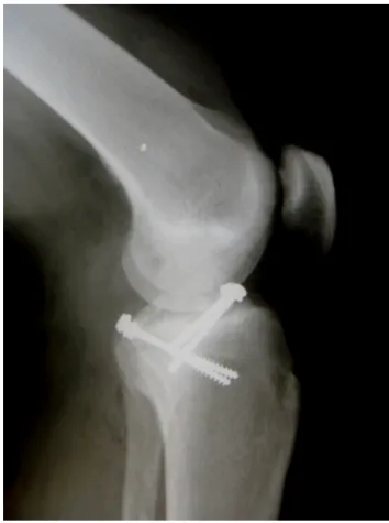
radio-graphsshowedconsolidatedtibialspines(Fig.6).Outpatient
dischargewasthereforegiven.
Discussion
Kneeligamentinjuriesareafrequenttopicinlargenumberof
publishedscientificpapers,particularlyinjuriesofthe
ante-riorcruciateligament(ACL).However,overthelastfewyears,
injuriesoftheposteriorcruciateligament(PCL)havereceived
specialattention,asconfirmedbytheincreasingnumberof
articlesdealingwiththisligament.Fromananatomicalpoint
ofview,the ACLoriginatesfrom theanterior intercondylar
areaofthetibia,immediatelybehindthefixationofthemedial
meniscus.Itsinsertionisintheposteriorpartofthemedial
faceofthelateralcondyleofthefemur,anditsmain
func-tionistoblockanteriordisplacementofthetibiainrelation
tothefemur.ThePCLisfixedtotheanteriorhalfofthelateral
faceofthemedialfemoralcondyle,anditprojectscaudally
andmediallythroughtheintercondylarnotch,towarditstibial
insertion,whichislocatedposteriorly,inferiorlyand
juxtalat-erallytothemedialline ofthetibialplateau. Itactsasthe
mainposteriorstabilizerofthekneeandrestrictsposterior
tibialtranslationinrelationtothefemur.2
AvulsionfracturesoftheACLarerareinjuriesinadultsand
occurin1–5%oftheinjuriestothisligament.3Fracturesofthe
intercondylareminencearebetterdescribedinthepediatric
orthopedicliteratureandoccuratlowerfrequencyinadults.
Thus,thebibliographyonthissubjectisverylimited.4Evenin
children,theseinjuriesareuncommon,affectingonlythreein
Fig.6–Radiographofthekneesixmonthsafterthe operation,showinganatomicalreductionand consolidation.
morefrequentlythantheposteriortibialinsertion.5Whenan
avulsedfragmentisdisplaced,primaryfixationisindicated
inordertopreventanteriorimpactinextension,residual
lax-ityandnon-consolidationoffragmentsandpreservationof
thenative ACL.Several surgicaltreatmentshavebeen
pro-posedfortheseinjuries,goingfrom the conventionalopen
proceduretoinclusionofarthroscopicmethods,whichwere
first described byMcLennan inOchiai et al.in1982,6 with
anumberoffixationmethods:Kirschnerwires,cannulated
screws, sutureswith steel or polyesterwires, anchors and
EndoButton®.Whiletheresultsfromprimaryfixationin
skele-tallyimmaturepatientsare good, thetreatments inadults
presentvariableresults,andsomeauthorshavereportedhigh
ratesofincidenceofpostoperativecomplications.3
In1970,MeyersandMcKeever7 proposedaclassification
systemforfractures oftheanteriortibialspineinchildren,
basedon the degreeofdisplacement ofthe fragment. The
injuriesweredivided into threetypes,but noclassification
foravulsedfracturesofthePCLwasreported.Subsequently,
this classification was modified by Zaricznyj,8 who added
afurthersubtype.Thisclassificationsystemmadeit
possi-bletodefine thebest treatmentinrelationtoeach typeof
fracture:TypeI–withoutdisplacementorwithminimal
dis-placementofthefragment;TypeII–angularelevationofthe
anteriorportionwithfullposteriorhinging;TypeIII–complete
displacement withor withoutrotation; Type IV–
commin-uted.Griffithetal.9modifiedtheclassificationofMeyersand
McKeeverandexpandedtheseconceptstoavulsionfractures
ofthePCL.
There is still some controversy regarding the surgical
indications fortreating PCLinjuries but, foravulsion
frac-tures, surgicalreinsertionofthefragmentisthe procedure
indicated.9 Tibialavulsionfractures ofthe PCLare a small
subgroupthatdiffersfromotherinjuriestothisligamentin
twoways:firstly,earlydiagnosis isgenerallypossibleusing
standard radiographs in which the bone fragment can be
viewed; and secondly, there is no simplified standardized
treatmentprotocolforposteriorapproachestotheknee.10
Severalauthorshaveemphasizedthatsurgicalreinsertion
ofthePCLfragmentproducesbetterresultsthan
conserva-tivetreatment.Surgerymakesitpossibletoperformmeasures
suchasdeepening ofthesite oftibialinsertionofthePCL
andrigidfixationofthebonefragment,whichcanbedone
usingscrewsand washersornon-absorbablethread.These
measuresmayassistinretensioningtheligamentand,
conse-quently,inimprovingtheclinicalevolution.9Furthermore,the
timethatelapsesbetweentheinjuryandthesurgical
proce-dureisanimportantfactortobeconsidered.Overthelastfew
years,newoptionsforfixationofbonefragmentshavebeen
evaluated,alongwithnewproposalsforsurgicalapproaches
towardtheseinjuries,suchascontrollingthereductionunder
arthroscopicviewing.
To treat ACL or PCL avulsions, it is recommended that
fractureswithoutdisplacement(TypeI)shouldbetreated
con-servatively; moderatelydisplaced fractures (TypeII) canbe
managedconservativelyorsurgically;anddisplacedfractures
(Type III) and comminuted fractures (Type IV) are surgical
indications.9Thetypeofsurgicaltreatmentdependsonthe
sizeanddegreeofcomminutionoftheavulsedfracture.
Fix-ation oftheavulsedtibialinsertionofthePCLcanbedone
bymeansofaconventionalopenroute,orarthroscopically.
Thelatterislessaggressivebutitrequiresequipmentandan
experiencedsurgeon.Trickeydescribedasurgicaltechnique
withaposterioraccessrouteintheknee,openreductionand
fixation oftheavulsedfragment. Burks and Schaffer1 used
a simplified accessroute forthe posteriorapproachto the
knee. Arthroscopic reduction and fixation are difficult and
requirealongerlearningcurve.Therefore,reductionand
fix-ationcanbeachievedbymeansofasimplifiedopenaccess
route,particularlyaposteriorroute,whichcanbeusedinany
center. In2011, Shelbourneet al.11 reported inareviewof
thecurrentliteraturethatthecommonestformsoftreatment
forthistypeofinjurymightequallybeopenorarthroscopic
reduction, although controversyremained regarding which
treatmentmethodwasbest.11In2012,Hapaetal.12conducted
abiomechanical studyon sheepandaffirmed thatfixation
usingEndoButton® forfracturesofthetibialeminence
pro-duced initial fixation strength that was greater than with
fixationusinganchorsorothertypesofsuture.Recently,Gui
etal.13contraindicatedarthroscopicfixationforavulsionsof
thePCLpresentinglargefragmentswithaneffectgoingasfar
asthetibialplateau.Insuchsituations,becauseofthe
dif-ficultyinachievingthenecessaryelevationandexposureof
thefocusofthefracture,thebestoptionisopenfixationusing
screws.
For functional evaluation, Lysholm and Gillquist
basicaspectsofthe Larsonscale,but introducesthe
crite-rionofinstabilityand correlatesit withactivity.Thisscale
was subsequently modified byTegner and Lysholm.These
authors recognized the difficulty in havinga score for
lig-ament injuriesand decided atthat juncture toinvestigate
clinicalfindingsandevaluatesymptomsandfunctions.This
scale or questionnaire by Lysholm is composed on eight
questions,withoptions forclosed responses,inwhich the
finalresultisexpressedinnominal andordinalform,such
that “excellent” is 95–100 points, “good” is 84–94 points,
“fair”is65–83pointsand“poor”islessthan orequalto64
points.14
Theinterestinpresentingthiscasearisesbecausethisisa
rareepisodeofsimultaneousavulsionfracturesofthecruciate
ligamentsattheirtibialsites,forwhichnosimilarpublished
papersareavailableintheliterature.Forthiscase,wechoseto
performfixationofthetwofracturesasiftheywereseparate
injuries.Despitethe gravityofthe traumaandthesurgical
complexity,thepatientevolvedsatisfactorily,bothfromthe
functionalandfromthemechanicalpointofview,usingthe
Lysholmquestionnaireandtheusualmaneuverstoverify
lig-amentstability.
Conflicts
of
interest
Theauthorsdeclarenoconflictsofinterest.
r
e
f
e
r
e
n
c
e
s
1. BurksRT,SchafferJJ.Asimplifiedapproachtothetibial attachmentoftheposteriorcruciateligament.ClinOrthop RelatRes.1990;254:216–9.
2. PiedadeSR,MischanMM.Surgicaltreatmentofavulsion fracturesofthekneePCLtibialinsertion:experiencewith21 cases.ActaOrtopBras.2007;15(5):272–5.
3.MontgomeryKD,CavanaughJ,CohenS,WickiewiczTL, RussellF,WarrenRF,etal.Motioncomplicationsafter arthroscopicrepairofanteriorcruciateligamentavulsion fracturesintheadult.Arthroscopy.2002;18(2):171–6.
4.ToyeLR,CummingsDP,ArmendarizG.Adulttibial intercondylareminencefracture:evaluationwithMR imaging.SkeletalRadiol.2002;31(1):46–8.
5.ponsellerPD,StanistiCL.Fraturaseluxac¸õesnaregiãodo joelho.In:BeatyJ,KasserJ,editors.RockwoodeWilkins: fraturasemcrianc¸as.5a.ed.Manole:SãoPaulo;2004.p. 1038–44.
6.OchiaiS,HaginoT,WatanabeY,SengaS,HaroH.Onestrategy forarthroscopicsuturefixationoftibialintercondylar eminencefracturesusingtheMeniscalViperRepairSystem. SportsMedArthroscRehabilTherTechnol.2011;3(1):17.
7.MeyersMH,McKeeverFM.Fractureoftheintercondylar eminenceofthetibia.JBoneJointSurgAm.
1970;52(8):1677–84.
8.ZaricznyjB.Avulsionfractureofthetibialeminence: treatmentbyopenreductionandpinning.JBoneJointSurg Am.1977;59(8):1111–4.
9.GriffithJF,AntonioGE,TongCWC,MingCK.Cruciateligament avulsionfractures.Arthroscopy.2004;20(8):803–12.
10.DhillonMS,SinghHP,NagiOP.Posteriorcruciateligament avulsionfromthetibia:fixationbyaposteromedialapproach. ActaOrthopBelg.2003;69(2):162–7.
11.ShelbourneKD,UrchSE,FreemanH.Outcomesafter arthroscopicexcisionofthebonyprominenceinthe treatmentoftibialspineavulsionfractures.Arthroscopy. 2011;27(6):784–9.
12.HapaO,BarberFA,SünerG,ÖzdenR,DavulS,Bozda ˘gE,etal. Biomechanicalcomparisonoftibialeminencefracture fixationwithhigh-strengthsuture.EndoButton,andsuture anchor.Arthroscopy.2012;28(5):681–7.
13.GuiJ,WangL,JiangY,WangQ,YuZ,GuQ.Single-tunnel suturefixationofposteriorcruciateligamentavulsion fracture.Arthroscopy.2009;25(1):78–85.