1. Department of Rheumatology and Clinical Immunology, Poznan University of Medical Sciences
2. Department of Dermatology, Poznan University of Medical Sciences
3. Department of Pulmonology, Allergology and Pulmonary Oncology, Poznan University of Medical Sciences
4. Department of General Radiology and Neuroradiology, Poznan University of Medical Sciences
Circulating adipokines and organ involvement
in patients with systemic sclerosis
ACTA REUMATOL PORT. 2015;40:156-162
Olewicz-Gawlik A1, Danczak-Pazdrowska A2, Kuznar-Kaminska B3, Batura-Gabryel H3, Katulska K4, Silny W2, Trzybulska D1, Hrycaj P1
digital ulcers (p=0.03) and serum concentrations of le -ptin were associated with the duration of SSc symptoms other than Raynaud’s phenomenon (p<0.01)
Conclusions: Serum adiponectin should be further
in-vestigated as a candidate for SSc activity marker and re-sistin may play a role in ulcer development in SSc patients.
Keywords: Scleroderma; Adipokines; Pulmonary
fi-brosis.
IntroductIon
Systemic sclerosis (SSc) is a chronic, complex disease of unknown etiology characterized by excessive fibrosis, vascular damage and inflammation1. A leading cause of
morbidity and mortality in SSc patients is lung involve-ment2presented as two major pulmonary syndromes:
pulmonary arterial hypertension (PAH) and interstitial lung disease (ILD). White adipose tissue is known to be an active organ that secretes proteins called adipokines. Adipokines take part in regulation of many pathophy -siological processes, both metabolic and inflammatory, but their significance remains obscure3.
Resistin is a 12.5 kDa polypeptide which in humans is highly expressed in bone marrow and lung4and is
se-creted mainly by immune cells5. It is a cysteine-rich
protein which is robustly induced in response to va -rious proinflammatory cytokines, such as tumor necro-sis factor a (TNF-a), interleukin (IL)-6, IL-1b, and re-sistin itself, and has been shown to up-regulate the ex-pression of proinflammatory cytokines such as TNF--a, IL-6 and IL-126-8. The co-culture of T cells and
den-dritic cells treated with resistin enhances the expansion of regulatory T cells, an important source of trans-forming growth factor b (TGF-b), a key profibrotic cy-tokine9.
AbstrAct
Background: In recent years, mediators synthesized in
the adipose tissue, the so-called adipokines, have been reported to play important roles in the pathogenesis of autoimmune rheumatic diseases.
Objective: To compare serum leptin, adiponectin and
resistin levels in patients with systemic sclerosis (SSc) and healthy controls. To find possible relationship be-tween serum levels of adipokines and organ involve-ment with focus on interstitial lung disease in SSc pa-tients.
Patients and Methods: Lung involvement was
as-sessed functionally (body plethysmography, diffusing capacity of the lung for carbon monoxide (DLCO) and six-minute walk test) and radiologically (using average disease extent on high resolution computed tomogra-phy (HRCT) of the lungs, according to the percentage of interstitial changes) in 29 SSc patients. Quantitative sandwich ELISA was used to measure resistin, leptin and adiponectin concentrations in sera of patients and 30 healthy controls.
Results: We found no statistically significant diffe rences
in serum resistin, leptin and adiponectin levels between SSc patients and the controls. However, serum adi ponectin concentrations were significantly lower in acti ve than in inactive patients. They also correlated po siti -vely with vital capacity (VC) (p=0.04) and negati-vely with Valentini disease activity index (p=0.04). Serum re sistin levels were significantly elevated in patients with
Adiponectin is a 30 kDa plasma protein mainly pro-duced in white adipose tissue by mature adipocytes and non-fat cells, although it can also be found in skeletal muscle cells, cardiac myocytes and endothe-lial cells10. Its secretion is suppressed by TNF-a and its
production is also regulated by other proinflammato-ry cytokines, like IL-611.
The third cytokine-like hormone, leptin, is a circu-lating non-glycosylated peptide of 16 kDa12. Leptin
exerts effect on the different cell populations of both innate and adaptive immune responses. It up-regulates the secretion of proinflammatory cytokines like TNF-a, IL-6, IL-12, increases the expression of adhesion molecules, induces the expression of activation mar -kers of monocytes, NK cells and lymphocytes13.
Leptin and adiponectin, and their respective receptors are also expressed in the human lung14,15.
A growing body of evidence indicates that these three adipokines play important roles in autoimmune diseases. Resistin is associated with disease activity and laboratory findings of systemic lupus erythematosus (SLE)16, rheumatoid arthritis (RA)17 and psoriatic
arthritis18, and it was also described as a useful mar ker
of pulmonary vascular involvement in SSc19. In
pa-tients with diffuse SSc, tissue levels of adiponectin have been shown to inversely correlate with skin scores20.
The study by Fang et al. demonstrated the antifibro -tic effects of adiponectin in normal and scleroderma fi-broblasts21. Leptin was found to have opposing,
profi-brotic effects, as it promotes the development of bleomycin-induced lung fibrosis in mice by augmen-tation of TGF-b signaling22. As literature data on serum
adipocytokines levels in SSc patients are sparse and controversial, this study aims to investigate the rela-tionship and possible interactions between resistin, leptin, adiponectin and markers of disease activity and organ involvement in patients with SSc, with particu-lar regard to lung involvement.
mAterIAl And methods PAtIents
We recruited to the pilot study 29 consecutive Cau-casian patients with SSc, according to the American College of Rheumatology (ACR) classification crite-ria23. All patients were also analysed using the 2013
ACR/EULAR classification criteria for SSc24and their
age was greater than 18 years. Patients with mixed con-nective tissue disease or overlap syndrome were
ex-cluded from the study. Moreover, all the patients were grouped according to the 2-cutaneous subset classifi-cation25as having diffuse cutaneous (dcSSc, cutaneous
thickening of both distal and proximal extremities, with or without truncal involvement) or limited cuta-neous (lcSSc, cutacuta-neous thickening of both extremities distally to the elbows and knees, with or without scle-rosis of the neck and face) form of the disease. A fur-ther subdivision was made into early SSc (n=13) and late SSc (n=16) based upon the duration of the di sease, defining early SSc as patients having the disease dura-tion of less than 2 years and late SSc for those longer than 2 years. Healthy controls (n=30, 27 females, 3 males) were volunteers and were statistically matched for gender and age (18-29 years, 30-44 years, 45-54 years, 55-70 years). Informed consent was obtained from all participants and the study was approved by the Institutional Review Board at Poznan University of Medical Sciences. A protocol of the conducted research conforms to the principles of the World Medical As-sociation’s Declaration of Helsinki.
clInIcAl evAluAtIon And lAborAtory meAsurements
The workup included patients’ history and a thorough clinical examination which included ulcer assessment and evaluation of skin involvement using the modi-fied Rodnan skin thickness score26. Body mass index
(BMI) was calculated for all participants. The disease duration was measured from the onset of the first symptom, other than Raynaud’s phenomenon, consis-tent with SSc. The disease activity of SSc was deter-mined using European Scleroderma Study Group disea se activity score for SSc (Valentini disease activi-ty index)27. Myositis was evaluated as muscle
weak-ness associated with elevated levels of serum creatine kinase. Patients with decreased glomerular filtration rate (GFR) and/or proteinuria were classified as having renal involvement. Joint involvement was defined by the detection of symmetric synovitis, flexion contrac-tures, and tendon friction rubs. The presence of pul-monary arterial hypertension (PAH) was confirmed by right heart cathete risation. The presence of ILD was assessed with use of pulmonary function tests: body plethysmography, diffu sing capacity of the lung for car-bon monoxide (DLCO) and six-minute walk test, and radiologically (high resolution computed tomography of the lungs, HRCT) as proposed by Goh et al.28. All HRCT examinations were evaluated by a radiologist expert on HRCT ILD. The patients were divided into
groups based on HRCT disease extent thresholds of 5% and 20%, and HRCT scans were also assessed for the following findings: the average extent of the dis-ease, the extent of a reticular pattern, the extent of ground glass and the presence of honeycombing. Blood samples from patients were collected at the time of clinical exa mination on fasting conditions. Obtained sera were stored at -70°C before the assays were per-formed. The degree of inflammatory activity was de-termined by the erythrocyte sedimentation rate (ESR, Westergren), highly sensitive C-reactive protein (CRP, enzyme-linked immunosorbent assay (ELISA), BioCheck, USA) and complement components C3 and C4 (radial immunoelectrophoresis). Antinuclear anti-bodies (ANA) were determined by indirect im-munofluorescence on HEp2 cells (Euroimmun, Ger-many) and antibodies to extractable nuclear antigens (ENA) were estimated using line immunoassay (Eu-roimmun, Germany). Serum levels of resistin, leptin and adiponectin were measured using commercially available ELISA kits (R&D Systems, USA) and calcu-lated using standard curves generated with specific standards according to the manufacturer’s recommen-dations.
stAtIstIcAl AnAlysIs
Patients demographic data were analysed using des -criptive statistics. All contiguous data were tested for normal distribution using the Kolmogorov-Smirnov test. The number of cases was expressed as percentage, mean and standard deviation values. Other non-nor-mally distributed parameters were compared by Mann–Whitney U-test. Correlations between variables within the group were analysed using Spearman’s rank-order correlation coefficient (r). Results are expressed as medians (interquartile range, IQR) and considered statistically significant at p<0.05. All the analyses were performed with STATISTICA software (StatSoft, Inc (2009). STATISTICA (data analysis software system) version 9.1, www.statsoft.com).
results
Out of 29 SSc patients, 26 were female and 3 were male (median age 56 years, range 24-70). Eighteen patients had lcSSc (17 females and 1 male) and 11 had dcSSc (9 females and 2 males). All but one SSc patients had a history of Raynaud’s phenomenon after exposure to low temperature, 11 (37.9%) had digital ulcers at the time of examination, 15 (51.7%) had gastrointestinal symptoms, 7 (24.1%) had hypertension, 8 (27.6%)
had active arthritis, 1 (3.4%) had myositis, 1 (3.4%) had PAH and 1 (3.4%) had renal involvement. Ten pa-tients (34.5%) showed a modified Rodnan skin thick-ness score of >14 and 7 out of 29 had active disease ac-cording to Valentini disease activity index. In 27 pa-tients (93.1%) lung-function tests showed abnormal gas transfer for CO, VC was decreased in 4 (13.8%) and TLC in 1 (3.45%) out of 29 patients. Significant abnormalities on HRCT (ILD extent of > 5%) were obser ved in 51.7% of the subjects and ILD extent >20% was present in 24.1% of patients with SSc. There were no patients with the indeterminate extent of the disease on HRCT. Comparison between dcSSc and lcSSc revealed no statistically significant differences in pulmonary function tests with regard to percentage of predicted TLC, VC and DLCO. Further characteristics of patient group at the time of examination is shown in Table I.
We found no statistically significant differences in serum resistin, adiponectin and leptin levels either be-tween SSc patients and healthy subjects (Table II), or between early and late SSc subjects. Likewise, there were no statistically significant differences in adipokines levels between dcSSc and lcSSc subgroups. The levels of resistin and adiponectin did not correlate with disease duration, age or sex of investigated pa-tients.
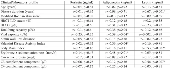
No significant differences were found in resistin, adiponectin and leptin levels when SSc group was di-vided according to ILD extent of < 5% or > 5% and < 20% or >20%. But with regard to pulmonary func-tion tests, we found statistically significant associafunc-tion of serum adiponectin levels with VC (r=0.39, p=0.04). We also demonstrated a significant negative correla-tion between adiponectin level and Valentini disease activity index (r=-0.39, p=0.04); adiponectin levels were found to be significantly lower in active patients than in inactive patients (p=0.04) and negatively correlated with ESR (r=0.48, p=0.01). While resistin le -vel manifes ted a significant increase in patients with digital ulcers (p=0.03), it showed no relationship with other investigated parameters. Serum concentrations of leptin were associated with the duration of SSc symptoms other than Raynaud’s phenomenon (r=0.67, p<0.01), BMI (r=0.53, p<0.01), C3 level (r=0.56, p<0.01) and were significantly higher in smokers (p=0.04). However, we did not demonstrate any signi -ficant associations between median resistin, leptin and adiponectin levels and the type of treatment and other clinical status/disease activity parameters of SSc
patients, including the presence of arthritis, gastroin-testinal symptoms, modified Rodnan score and CRP levels. The correlations between the clinical profile of SSc patients and serum levels of the three adipokines are summarized in Table III.
dIscussIon
The associations between adipokines and the activity and symptoms of SSc suffer from many gaps in the lite -rature. In the present pilot study we tried to supple-ment the existing data in this area of research.
In this study we did not observe statistically signifi -cant differences in serum resistin level, either between SSc patients and the control or between the two SSc subtypes. This is in agreement with the previous study, where serum resistin levels were comparable between dcSSc, lcSSc and control subjects19. It is also in line
with observations of Pehlivan et al., that although pa-tients with SSc had higher resistin levels than the con-trol group, the difference did not reach statistical sig-nificance29. Moreover, other authors report that in
pa-tients with SLE resistin measurements did not differ between patients and controls16. On the other hand,
re-sistin was found to be abundant in another inflamma-tAble I. clInIcAl, rAdIologIcAl And lAborAtory chArActerIstIcs of systemIc sclerosIs PAtIents
Disease duration (months) 54.1 ± 68**
Raynaud’s phenomenon 28 (96.6)
Digital ulcers 11 (37.9)
Gastrointestinal symptoms 15 (51.7)
Arthritis 8 (27.6)
Myositis 1 (3.4)
Pulmonary arterial hypertension 1 (3.4)
Renal involvement 1 (3.4)
Modified Rodnan skin score 13 (16)*
Antinuclear antibodies 25 (86.2)
Valentini disease activity index 2 (2.5)*
n(%) index >3 7 (24.1)
HRCT Extent of disease, % 15 (23.7)*
Extent of reticular pattern, % 10 (15)*
Extent of ground glass, % 5 (12.5)*
Presence of honeycombing 10 (34.5)
DLCO (p%) 58.4 (16.5)*
n(%) < 80% 27 (93.1)
Total lung capacity (p%) 108.2 (18.1)*
n(%) < 80% 1 (3.45)
Vital capacity (p%) 98.3 (19.7)*
n(%) < 80% 4 (13.8)
Six-minute walk test, distance (m) 375 (90)*
Erythrocyte sedimentation rate (mm/h), n(%) > 15 25 (20)*, 21 (72.4)
C-reactive protein (mg/l), n (%) > 5 0 (4.6)*, 7 (24.1)
C3 complement component (mg/l), n (%) < 900 1162 (236)*, 1 (3.4) C4 complement component (mg/l), n (%) < 200 191 (66)*, 16 (55.2)
Treatment with cyclophosphamide 9 (31)
Treatment with methotrexate 5 (17.2)
Treatment with azathioprine 2 (6.9)
Current smokers 6 (20.7)
Data presented as the median (interquartile range)*, mean ± standard deviation** or as n (%)
tory disease, RA17. These observations lead us to a
hy-pothesis, that increased resistin concentration is the result of severe inflammation, and the patient group in this study did not show high elevation in the systemic markers of inflammation, like ESR or CRP. However, we cannot exclude a bias caused by a limited number of subjects in this study. Further, we did not find an as-sociation between resistin level and radiological and functional parameters of the lung involvement. There is convincing literature evidence of the role of resistin in the pathogenesis of PAH associated with SSc19. Also
the results of Angelini et al. suggest that resistin-like molecule (RELM)-beta is involved in the development of PAH in SSc patients30. The reason for the lack of
re-lationship between resistin and pulmonary parame-ters found in our study can be the fact that PAH, sig-nificant lung functional impairment and abnormali-ties in HRCT coexisted in only one patient. However,
we found that serum resistin level correlated with the presence of digital ulcers in SSc patients. Although the results indicate a weak correlation, they are concordant with the data of a recent study, which demons -trated a higher prevalence of digital ulcers in SSc pa-tients with elevated serum resistin levels than in those with normal levels29. Further, previous results showed
that resistin exert direct effect to promote endothelial cell activation by promoting endothelin-1 release31.
Re-cently it was also reported that plasma endothelin-1 level was higher in SSc patients with digital ulcers than in a group without them32. Our results can thus
sup-port a link between resistin and the development of ulcers in SSc patients. They also indirectly implicate a possible role of resistin in the process of aberrant an-giogenesis, one of the main features of SSc.
In this study we observed lower serum leptin levels in SSc patients than in healthy subjects, though not tAble II. AdIPokInes levels In serum of systemIc sclerosIs PAtIents And heAlthy subjects
SSc Healthy controls
Resistin (ng/ml), median (IQR) 7.9 (2.3) 6.8 (2.4)
Leptin (ng/ml), median (IQR) 9.9 (11.6) 14.5 (22.1)
Adiponectin (mg/ml), median (IQR) 23.2 (20.2) 22.1 (10.9)
IQR, interquartile range; SSc, systemic sclerosis
tAble III. correlAtIons between serum levels of AdIPokInes And clInIcAl/lAborAtory PArAmeters In systemIc sclerosIs PAtIents
Clinical/laboratory profile Resistin (ng/ml) Adiponectin (mg/ml) Leptin (ng/ml)
Age (years) r=0.04, p=0.84 r=0.02, p=0.92 r=0.13, p=0.51
Disease duration (years) r=0.01, p=0.95 r=-0.08, p=0.71 r=0.67, p=0.001* Modified Rodnan skin score r=0.04, p=0.85 r=-0.3, p=0.12 r=-0.09, p=0.65 HRCT ILD extent (%) r= -0.1, p=0.65 r=-0.12, p=0.58 r=0.2, p=0.38
DLCO (p%) r= -0.1, p=0.6 r=0.31, p=0.12 r=-0.23, p=0.27
Total lung capacity p(%) r= -0.1, p=0.6 r=0.38, p=0.05 r=-0.12, p=0.56 Vital capacity (p%) r= -0.23, p=0.25 r=0.39, p=0.04* r=-0.002, p=0.99 6-min walk test distance r=0.05, p=0.82 r=-0.14, p=0.56 r=-0.02, p=0.92 Valentini Disease Activity Index r=-0.02, p=0.93 r=-0.39, p=0.04* r=0.16, p=0.41 Body Mass Index r=0.27, p=0.16 r=-0.16, p=0.41 r=0.53, p=0.002* Erythrocyte sedimentation rate (mm/h) r=0.14, p=0.47 r=-0.48, p=0.01* r=-0.05, p=0.81 C-reactive protein (mg/l) r=0.2, p=0.31 r=-0.07, p=0.72 r=0.12, p=0.55 C3 complement component (g/l) r=0.06, p=0.76 r=0.12, p=0.56 r=0.56, p=0.007* C4 complement component (g/l) r=-0.07, p=0.73 r=-0.25, p=0.24 r=-0.05, p=0.81 *p < 0.05; HRCT, high resolution computed tomography of the lungs; ILD, interstitial lung disease; DLCO, diffusing capacity of the lung for carbon monoxide
down to significant levels, which is consistent with the results of a previous study33. However, literature data
focusing on the serum leptin level are controversial. Kotulska et al. reported a significantly lower serum lep-tin level in SSc patients34. On the contrary, the results
of the two recent studies showed significantly higher levels of circulating leptin in SSc subjects with respect to healthy controls29,35. The reason for the conflicting
literature data can be heterogeneity of SSc, a limited number of patients, differences in disease duration and activity, and in the treatment of the disease. A correla-tion of leptin level with C3 and BMI found in this study confirmed previous results obtained in SSc patients33, 34. Similarly to the previous studies, we failed to find
any association of circulating leptin with other clinical and laboratory parameters of the SSc33,35. A
relation-ship between decreased serum leptin and BMI noted in SSc patients can be the result of frequent gastroin-testinal tract involvement in this disease. Vernooy et al. observed that leptin expression is enhanced in bronchial epithelial cells and alveolar macrophages of ex-smokers compared with never-smokers and they also confirmed the presence of leptin signaling path-way in lung epithelial cells36. The observation
indi-rectly supports the correlation between serum leptin levels and smoking found in our study.
Serum adiponectin levels were comparable between SSc and healthy subjects, which is in accordance with the preceding study20. Moreover, the investigation of
serum adiponectin levels and their association with clinical and laboratory parameters in SSc revealed sig-nificantly lower adiponectin levels in patients with ac-tive disease as it was evaluated using Valentini activity score27. Although the association was weak and should
be treated with caution, this finding harmonizes well with the anti-inflammatory and anti-fibrotic functions of this adipocytokine37. However, we did not observe
statistically significant differences in serum adiponectin concentrations between dcSSc and lcSSc subgroups. It is in conflict with earlier studies, where adiponectin levels were decreased in dcSSc subjects20.
This discrepancy can be the result of immunosup-pressive treatment in more than half of the investigat-ed group, whereas in the citinvestigat-ed study patients treatinvestigat-ed with corticosteroids or other immunosuppressants pri-or to their first visits were excluded. Further, we did not observe any relationship between circulating adiponectin and radiological and functional parame-ters of lung involvement except VC. Because the re-duction in serum adiponectin levels is associated with
the initiation of the fibrotic response, but not with the maintenance, the data from two previous studies reported by the same team of researchers first demons -trated that serum adiponectin levels inversely corre-late with the activity of skin sclerosis but not with the severity of ILD (in dcSSc patients), and in the second study serum adiponectin levels were significantly de-creased in dcSSc patients with active ILD compared with healthy controls [38]. We found active disease in only 7 out of 29 subjects, and early SSc was present in less than a half of the whole group, which may explain the lack of statistically significant association of adiponectin level with skin and lung fibrosis in our study. However, we cannot exclude the influence of immunosuppressive therapy or a bias caused by the limited number of patients in our study.
conclusIons
In summary, our results did not establish whether re-sistin, adiponectin or leptin in SSc patients are involved in the process of lung fibrosis. Instead, we showed that: 1) the concentration of resistin is related to the presence of digital vasculopathy, 2) serum adipo nectin reflects disease activity characterized by Valentini disease activi -ty index, and 3) serum leptin is associated with disease duration. To conclude, measurement of serum adipokines can help us to better understand the di sease process and further research in this field in a larger sam-ple of patients is of great importance.
corresPondence to
Olewicz-Gawlik A
Department of Rheumatology and Clinical Immunology, Poznan University of Medical Sciences
Poznan, Poland E-mail: [email protected]
references
1. Chung L, Lin J, Furst DE, Fiorentino D. Systemic and localized scleroderma. Clin Dermatol 2006;24:374-392.
2. Hassoun PM. Lung involvement in systemic sclerosis. Presse Med 2011;40:e3-e17.
3. Kos K, Wilding JP. Adipokines: emerging therapeutic targets. Curr Opin Investig Drugs 2009;10:1061-1068.
4. Patel L, Buckels AC, Kinghorn IJ, et al. Resistin is expressed in human macrophages and directly regulated by PPAR gamma ac-tivators. Biochem Biophys Res Commun 2003;300:472-476. 5. Bokarewa M, Nagaev I, Dahlberg L, Smith U, Tarkowski A.
Re-sistin, an adipokine with potent proinflammatory properties. J Immunol 2005;174:5789-5795.
6. Steppan CM, Bailey ST, Bhat S, et al. The hormone resistin links obesity to diabetes. Nature 2001;409:307-312.
7. Kaser S, Kaser A, Sandhofer A, Ebenbichler CF, Tilg H, Patsch JR. Resistin messenger-RNA expression is increased by proin-flammatory cytokines in vitro. Biochem Biophys Res Commun 2003;309:286-290.
8. Bokarewa M, Nagaev I, Dahlberg L, Smith U, Tarkowski A. Re-sistin, an adipokine with potent proinflammatory properties. J Immunol 2005;174:5789-5795.
9. Son YM, Ahn SM, Kim GR, et al. Resistin enhances the expan-sion of regulatory T cells through modulation of dendritic cells. BMC Immunol 2010;11:33.
10. Carbone F, La Rocca C, Matarese G. Immunological functions of leptin and adiponectin. Biochimie 2012;94:2082-2088. 11. Fasshauer M, Kralisch S, Klier M, et al. Adiponectin gene
ex-pression and secretion is inhibited by interleukin-6 in 3T3-L1 adipocytes. Biochem Biophys Res Commun 2003;301:1045--1050.
12. Zhang F, Basinski MB, Beals JM, et al. Crystal structure of the obese protein leptin-E100. Nature 1997;387:206-209. 13. Procaccini C, Jirillo E, Matarese G. Leptin as an
immunomo-dulator. Mol Aspects Med 2012;33:35-45.
14. Bruno A, Pace E, Chanez P, et al. Leptin and leptin receptor ex-pression in asthma. J Allergy Clin Immunol 2009;124:230-237 15. Miller M, Cho JY, Pham A, Ramsdell J, Broide DH. Adiponec-tin and functional adiponecAdiponec-tin receptor 1 are expressed by air-way epithelial cells in chronic obstructive pulmonary disease. J Immunol 2009;182:684-691.
16. Almehed K, d’Elia HF, Bokarewa M, Carlsten H. Role of resis-tin as a marker of inflammation in systemic lupus erythemato-sus. Arthritis Res Ther 2008;10:R15.
17. Migita K, Maeda Y, Miyashita T, et al. The serum levels of re-sistin in rheumatoid arthritis patients. Clin Exp Rheumatol 2006;24:698-701.
18. Johnston A, Arnadottir S, Gudjonsson JE, et al. Obesity in pso-riasis: leptin and resistin as mediators of cutaneous inflamma-tion. Br J Dermatol 2008;159:342-350.
19. Masui Y, Asano Y, Akamata K, et al. Serum resistin levels: a pos-sible correlation with pulmonary vascular involvement in pa-tients with systemic sclerosis. Rheumatol Int 2013. [Epub ahead of print]
20. Masui Y, Asano Y, Shibata S, et al. Serum adiponectin levels in-versely correlate with the activity of progressive skin sclerosis in patients with diffuse cutaneous systemic sclerosis. J Eur Acad Dermatol Venereol JEADV 2012;26: 354–360.
21. Fang F, Liu L, Yang Y, et al. The adipokine adiponectin has po-tent anti-fibrotic effects mediated via adenosine monophos-phate-activated protein kinase: novel target for fibrosis thera-py. Arthritis Res Ther 2012;14:R229.
22. Jain M, Budinger GR, Lo A, et al. Leptin promotes fibroproli-ferative acute respiratory distress syndrome by inhibiting pe-roxisome proliferator-activated receptor-g. Am J Respir Crit Care Med. 2011;183:1490-1498.
23. Masi, AT, Rodnan, GP, Medsger, TA, et al. Preliminary criteria for the classification of systemic sclerosis (scleroderma). Arth-ritis Rheum 1980;23:581-590.
24. van den Hoogen F, Khanna D, Fransen J, et al. 2013 classifica-tion criteria for systemic sclerosis: an American college of rheu-matology/European league against rheumatism collaborative initiative. Ann Rheum Dis. 2013;72:1747-1755.
25. LeRoy, EC, Black, C, Fleischmajer, R, et al. Scleroderma (sys-temic sclerosis): classification, subsets and pathogenesis. J. Rheumatol 1988;15:202-205.
26. Clements, P, Lachenbruch, P, Siebold, J, et al. Inter and in-traobserver variability of total skin thickness score (modified Rodnan TSS) in systemic sclerosis. J. Rheumatol 1995;22:1281--1285.
27. Valentini G, D’Angelo S, Della Rossa A, Bencivelli W, Bombar-dieri S. European Scleroderma Study Group to define disease activity criteria for systemic sclerosis. IV. Assessment of skin thickening by modified Rodnan skin score. Ann Rheum Dis 2003; 62:904–905
28. Goh NS, Desai SR, Veeraraghavan S, et al. Interstitial lung di-sease in systemic sclerosis: a simple staging system. Am J Res-pir Crit Care Med 2008; 177, 1248-1254.
29. Pehlivan Y, Onat AM, Ceylan N, et al. Serum leptin, resistin and TNF-a levels in patients with systemic sclerosis: the role of adipokines in scleroderma. Int J Rheum Dis 2012;15:374-379. 30. Angelini DJ, Su Q, Yamaji-Kegan K, et al. Resistin-like mole-cule-beta in scleroderma-associated pulmonary hypertension. Am J Respir Cell Mol Biol 2009;41:553-561.
31. Verma S, Li SH, Wang CH, et al. Resistin promotes endothelial cell activation: further evidence of adipokine-endothelial inte-raction. Circulation 2003;108:736-740.
32. Kim HS, Park MK, Kim HY, Park SH. Capillary dimension mea-sured by computer-based digitalized image correlated with plasma endothelin-1 levels in patients with systemic sclerosis. Clin Rheumatol 2010;29:247-254.
33. Budulgan M, Dilek B, Da SB, et al. Relationship between serum leptin level and disease activity in patients with systemic scle-rosis. Clin Rheumatol 2013. [Epub ahead of print]
34. Kotulska A, Kucharz EJ, Brzezi ska-Wcisło L, Wadas U. A de-creased serum leptin level in patients with systemic sclerosis. Clin Rheumatol 2001;20:300-302.
35. Riccieri V, Stefanantoni K, Vasile M, et al. Abnormal plasma le-vels of different angiogenic molecules are associated with dif-ferent clinical manifestations in patients with systemic sclero-sis. Clin Exp Rheumatol 2011;29:S46-S52.
36. Vernooy JH, Drummen NE, van Suylen RJ, et al. Enhanced pul-monary leptin expression in patients with severe COPD and asymptomatic smokers. Thorax 2009;64:26-32.
37. Fantuzzi G. Adiponectin and inflammation: consensus and controversy. J Allergy Clin Immunol 2008;121:326–330. 38. Masui Y, Asano Y, Takahashi T, et al. Clinical significance of
monitoring serum adiponectin levels during intravenous pul-se cyclophosphamide therapy in interstitial lung dipul-seapul-se asso-ciated with systemic sclerosis. Mod Rheumatol. 2013;23:323--329.