Cell cycle kinetics, apoptosis rates
and gene expressions of
MDR-1
,
TP53
,
BCL-2
and
BAX
in transmissible venereal
tumour cells and their association with
therapy response
M. M. Flórez
1,2, H. B. Fêo
1, G. N. da Silva
3, R. S. Yamatogi
4, A. J. Aguiar
1,
J. P. Araújo Jr.
4and N. S. Rocha
11Department of Veterinary Clinics, Faculty of Veterinary Medicine, São Paulo State University – UNESP,
Botucatu, Brazil
2Veterinary Pathology Research Group, Faculty of Agricultural Sciences, Universidad de Caldas, Manizales,
Colombia
3Department of Clinical Analysis. Pharmacy School, Universidade Federal de Ouro Preto, Ouro Preto, Brazil 4Department of Microbiology and immunology, Institute of Biosciences of Botucatu (IBB) and Biotechnology
Institute (IBTEC), São Pablo State University – UNESP, Botucatu, Brazil
Abstract
Transmissible venereal tumour (TVT) generally presents different degrees of aggressiveness, which makes them unresponsive to conventional treatment protocols. This implies a progressive alteration of their biological profile. This study aimed to evaluate the cytotoxicity, cell survival, apoptosis and cell cycle alterations in TVT cell cultures subjected to treatment with vincristine. Similarly, it assessed possible implications of MDR-1, TP53, BCL-2, and BAX gene expressions in eight TVT primary cultures for both resistance to chemotherapy and biological behaviour. When comparing TVT cells receiving vincristine to those untreated, a statistical difference related to increased cytotoxicity and decreased survival rates, and alterations in G1 and S cell cycle phases were found but without detectable differences in apoptosis. Increased MDR-1 gene expression was observed after treatment. The groups did not differ statistically in relation to the TP53, BAX and BCL-2 genes. Although preliminary, the findings suggest that such augmented expression is related to tumour malignancy and
chemotherapy resistance.
Keywords
apoptosis, chemotherapy, cytotoxicity, MDR-1, toxicogenomic
Introduction
Despite its uncertain origin, transmissible vene-real tumour (TVT) is a malignancy classified morphologically as a round cell neoplasm with plasmacytoid or lymphocytic aspect.1 The cells with lymphocytic aspect are small and have a regular shape with a round core, which has coarse chromatin and one or two nucleoli. The cytoplasm is sparse and finely granulated, with vacuoles in the cell periphery. The cells with plasmacytoid aspect
are bulky and present an irregular contour, with eccentric nucleus and abundant cytoplasm.1–3
According to this development, we may observe a nodular and friable tissue with hemorrhagic areas, often slightly marked and presenting ulcerations. This tumour may present in solitary or multiple form, presenting aspect of cauliflower or plates, with the presence of a serosanguineous secretion and possible secondary bacterial infection. Ani-mals can also present itching and show behavioural
Correspondence address: Mauricio M. Flórez Department of Veterinary Clinics
Faculty of Veterinary Medicine
São Paulo State University – UNESP Botucatu SP Brazil e-mail:
changes, often becoming aggressive or apathetic, lethargic and anorexic. In more advanced cases, with perineal tumour progression, urinary reten-tion may be observed.3,4
The transplantation occurs when intact host tumour cells lose the ability to express molecules of major histocompatibility complex (MHC) class I and II, enabling tissue transposition to a healthy animal by contact between skin and/or damaged mucous membranes. The characteristics of canine coitus allows prolonged contact abrasions on gen-ital mucous membranes, making the coitus an efficient transmission mode. Once a tumour is established, its proliferation may occur by reaching other locations, which eventually develops into metastases.2–4
Spontaneous regression is documented in cases of experimental TVT, but the same is not rou-tinely reported in clinical care.3,5The tumour
spon-taneously regresses in healthy animals, and such regression is associated with the infiltration of lym-phocytes and plasma cells as well as necrosis and apoptosis. Despite Higgins reporting spontaneous tumour regression, numerous clinical studies have not registered this event. In addition, the chronic presence of tumours for periods of 4 years opposes to this theory.3,4
The therapeutic protocol established for TVT, namely a weekly application of vincristine sulphate as a single agent (four to eight intravenous shots), is the most widely used for tumour regression.2–4 Despite a description that 90% of dogs responded positively to this treatment, the presence of signs and symptoms suggesting serious side effects leads to a treatment interruption in a high number of cases.5,6
One suitable drug against vincristine-resistant tumours is doxorubicin.7 Vinblastine, prednisone or their combination, is less often applied because of greater side effects.8,9 Therefore, TVT therapy
is currently restricted to a small number of drugs, which are sometimes insufficient because of specific tumour resistances.5,6,10
The progressive increase of TVTs with high per-centages of aggressiveness and variable response to chemotherapy, including resistance, is partially due to the high expression of p-glycoprotein (Pgp) by tumour cells, which leads to the expulsion
of chemotherapeutic.11–13 Thus, treatment cost increases, as do side effects such as anorexia, nephrotoxicity and myelosuppression.
Similarly, other mechanisms related to ther-apy resistance and variable biological behaviour of tumours include changes in pro- and anti-apoptotic genes from the familyBCL-2, and DNA repair sys-tems, which are associated with the family TP53. However, this aspect has been scarcely studied in TVT.
Pgp, also known as ABCB1, is produced by the gene MDR-1. This protein has a molecular mass of 170 kDa, 12 transmembrane domains and 2 ATP bond sites, and belongs to the group of ABC proteins (ATP-binding cassette). They are energy-dependent channels involved in the transfer of biological molecules through the mem-brane against concentration gradient, with high expression in tumour tissues previously exposed to drugs.14,15
Pgp expresses in different tissues,16,17 and
defends cells from cytotoxic agents under normal conditions.17,18 However, an overexpression of
Pgp not only induces to a multidrug resistance19 by reducing intracellular drug concentration to non-lethal levels,15,20,21but also seems to play a role
in preventing early apoptosis in tumour cells.22The
TP53gene is one that often mutates, including in human cancer. Several reports indicate alteration of TP53 in about the half of studied tumours.23 From a clinical point of view,TP53inactivation or mutation is a usual and severe molecular event for most tumours. There are mutant TP53forms dis-playing longer half-life times, oncogenic potential and negative effects on the unique types, leading to chemoresistance.24
Substantial evidence supports the hypothesis that the expression of MDR-1 (Pgp) is regulated by some mutants of p53 protein, and suggests that the response to chemotherapy or radiation may depend in part onTP53status before treatment.25
ATP53mutation has been described in dogs.28 In TVT, Choi and Kim described the first reports about its mutation.29 Sánchez-Servín et al. sup-ported further findings.30Nonetheless, in spite of the gene mutation evidence in the tumour, the abil-ity of this feature to generate some changes in the protein function remains unknown.31
According to Sánchez-Servínet al.determining the role ofTP53polymorphisms in the pathogenesis and response to chemotherapy is still necessary in relation to TVT.30In addition, Stockmannet al.and others emphasise the need to assess the effects of expression of the protein and its family members on TVT cells, by relating the findings to prognosis and possible treatment options.32
The genesBAXand BCL-2belong to the same family that comprises about 25 genes. They encode proteins regulating permeability of the outer membrane of mitochondria, and are subdi-vided according to their domain and function in apoptotic (Bax, Bad, Bak and Bok, etc.), and anti-apoptotic (BCL-2itself,BCL-XLandBCL-W) processes.14,33,34 Because BCL-2 overexpression
was identified in B cell malignancies, it has been observed in different types of tumours and cell lines in both humans and other animals.35–37 Its involvement in cancer has also been determined.14 However, reports on BCL-2 participation in car-cinogenesis are heterogeneous. On the one hand, overexpression confers resistance of many cell types to drugs and radiation.34,37 On the other
hand, overexpression also relates to low and favourable malignant phenotype prognoses.38
In TVT, Bcl-2 family protein expression has been identified. According to Stockmann et al.
Bcl-2 overexpression is independent of the TVT development stage.32Previously, Frenzelet al. sug-gested that overexpression of Bcl-2 may promote the acquisition of functions associated with tumour progression and survival.39 Similarly, Amaral
et al. emphasise that less aggressive TVT have a high rate of apoptosis, which may lead to better prognosis.40
Thus, culture studies have formidably increased the understanding of the pathogenesis of certain cancers, and provided a basis for developing new methods of tumour diagnosis and treatment.41As to TVT, the success of culturing has been described
by other researchers,42–47 although few analyses were made in their studies.
It is known that some TVTs have varying degrees of aggressiveness and resistance to chemotherapy. This demonstrates the need for a specific treatment for each type of tumour, which would minimize cost and side effects by avoiding excessive use of chemotherapy.6,8,11
Finally, considering the importance of achiev-ing further compression of TVT evolution and pathogenicity, this study aimed to evaluate the cytotoxicity, cell survival, apoptosis and cell cycle alterations in TVT cell cultures subjected to treat-ment with vincristine. Similarly, it assessed possible implications ofMDR-1,TP53, BCL-2andBAXgene expressions for both resistance to chemotherapy and biological behaviour, in TVT primary cultures.
Results
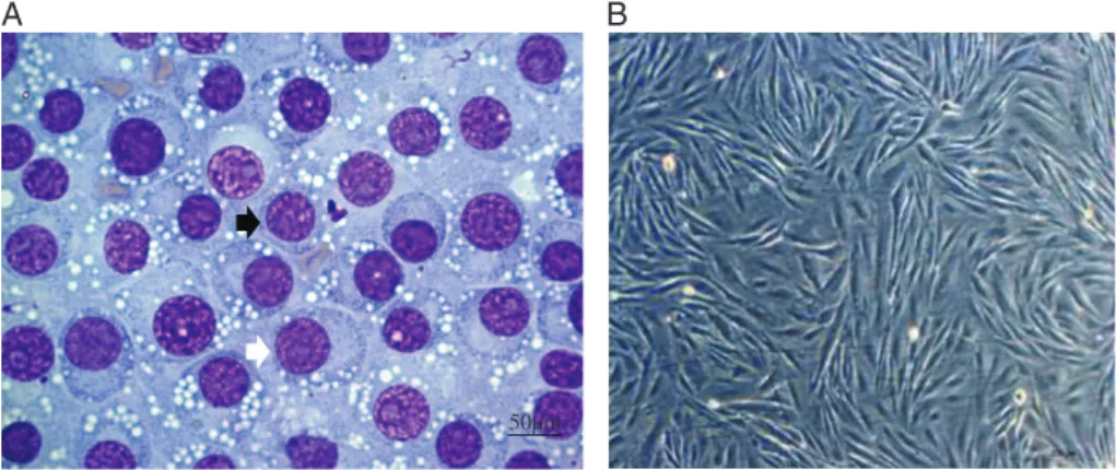
Eight cell tumour cultures were isolated (Fig. 1). After establishing subcultures, cells were character-ized by immunocytochemistry, presenting positive values for vimentin, lysozyme, alpha-antitrypsin and negative for CD3 and CD79𝛼. Chromosome
analysis revealed numbers ranging from 56 to 70. None of them exhibited the same number as dog somatic cells (data being prepared for publication).
Cytotoxicity and survival in TVT cells
The cytotoxicity test showed greater cytotoxicity and high significance in treated cells at 0.25, 0.5 and 1μM (P<0.01), when compared with control
samples. On the other hand, when comparing treat-ments, there was greater cytotoxicity at 1μM than 0.25μM (P<0.05) (Table 1, Fig. 2).
In relation to survival analysis, treated cells displayed lower levels and higher significance (P<0.01). In addition, no difference among
differ-ent concdiffer-entrations was found (P>0.05) (Table 1,
Fig. 3).
Apoptosis
A B
50µm
Figure 1. Isolation of TVT cells. (A) Cytology in TVT (×40 obj). Black arrow: lymphocytic standard (round cells, little cytoplasm and high nucleus:cytoplasm ratio). White arrow: plasmacytoid standard (ovoid cells, broad cytoplasm and eccentric nuclei). (B) TVT cell culture, thirdpassage, form ranging from spindle to oval, bar 200μm.
Table 1. Percent viability (cytotoxicity) and TVT cell survival after vincristine treatment
Test Negative control 0.25𝛍M 0.5𝛍M 1𝛍M
Percentage of viable cells (cytotoxicity) 100a 55.3
±14b 48.1
±18b,c 39.8 ±17c
Percentage of survival 100a 40.7
±16b 35.8
±18b 33.5 ±17b
Data expressed as average±of the standard deviation. Different letters indicate statistical differences a,b (P<0.01); b,c (P<0.05).
Control 0.25 0.5 1
0 50 100 150
Vincristine concentrations ***
**
Cell Proliferation %
Figure 2. Percentage of TVT cell viability after 24-h vincristine treatment. A statistical difference between controls and treatments can be noted *** (P<0.01), between 0.25 and 0.5μM ** (P<0.05).
Cell cycle
In this analysis, the cell cycle phases G1, G2, S and sub G1 were taken into account. Treated cell analysis showed a significant decrease in G1, for all treatments and in S phases, only in T3 (1μM), at respective significance levels of P<0.01 and
P<0.05. In the phase sub G1, a significant increase
(P<0.01) was also found in treated cells. In
addi-tion, a difference between T3 compared with T1 (P<0.05) was evident. Similarly, differences in
Control 0.25 0.5 1
0 50 100 150
***
Vincristine concentrations
Survival %
Figure 3. Percentage of TVT cell survival 5 days after vincristine treatment. A statistical difference between control and treatments can be observed ***(P<0.01).
G2 phase remained undetectable in any treatment (P>0.05) (Table 3).
Gene expression
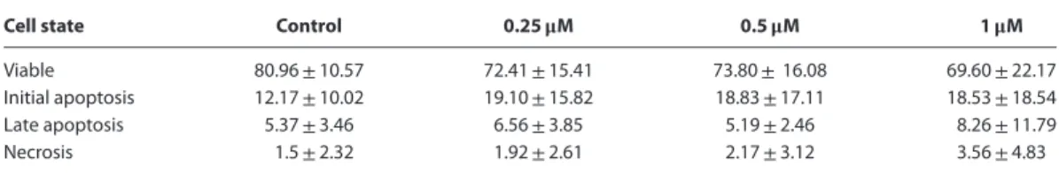
Table 2. Percentage of viable TVT cells in apoptosis and necrosis after vincristine treatment
Cell state Control 0.25𝛍M 0.5𝛍M 1𝛍M
Viable 80.96±10.57 72.41±15.41 73.80±16.08 69.60±22.17 Initial apoptosis 12.17±10.02 19.10±15.82 18.83±17.11 18.53±18.54 Late apoptosis 5.37±3.46 6.56±3.85 5.19±2.46 8.26±11.79
Necrosis 1.5±2.32 1.92±2.61 2.17±3.12 3.56±4.83
Data expressed as average±standard deviation. Statistical comparisons data showed no significant differences (P>0.05). Table 3. Kinetics of the cell cycle in TVT cells 48 h after vincristine treatment
Cell state Control 0.25𝛍M 0.5𝛍M 1𝛍M
G1 (%) 63.7±15.17a 43.39±15.8b 41.94±16.74b 39.50±14.69b
S(%) 2.9±1.51a 2.15±0.69ab 2.4±1.07ab 1.67±0.6b
G2 (%) 19.98±12.08a 23.28
±11.94a 19.07
±7.74a 16.34 ±7.59a
Sub G1 (%) 13.42±7.91a 31.17±13.52b 36.59±15.17bc 42.49±17.29c
Data expressed as average±standard deviation. Different letters indicate statistical differences a,b (P<0.01); b,c (P<0.05).
Controle 0.25 0.5 1 0
1 2 3 4
**
Vincristine concentrations μM
mRNA Relative expression
Figure 4. mRNA relative expressionof MDR-1in TVT cells after vincristine treatment. Endogenous control by RPS5N and RPS19. Evidence of a statistical difference between treated and control cells **P<0.05.
treated and control cells (Fig. 4). As toTP53,BAX
and BCL-2 gene expressions, statistical analysis revealed no significant differences in groups with and without treatment (P>0.05). Is important to
note that the QR value for TP53 expression was
> 2.00 in samples with and without treatment
(Fig. 5).
Discussion
A high-level of Pgp (MDR-1) expression in tumours is associated with a reduced susceptibility to ther-apy in dogs,48–53 and humans.15,20,21 Therefore,
patients with this type of alteration require a greater
Controle 0.25 0.5 1
Vincristine concentrations μM
mRNA Relative expression
0 1 2 3 4 5
Figure 5. mRNA relative expression ofTP53in TVT cells after vincristine treatment. Endogenous control by RPS5 and RPS19. No evidence of a statistical difference between treated and control cells was found atP>0.05.
number of doses or higher drug concentrations dur-ing treatment.
In this investigation, cells subjected to vincristine displayed a variable expression of theMDR-1gene. These results are important for two reasons. First, this allows demonstration of variability in TVT cells as a response to therapy. Second, results are an indi-cation of an existing modulator effect of vincristine onMDR-1gene expression.
authors of this work indicates that TVTs with a sig-nificantly greater immunoreactivity to the staining with anti-glycoprotein-p antibody was also directly related to a partial response to chemotherapy.11,12
Given these findings, in some TVT cultures,
MDR-1 expression may be modulated by vin-cristine, besides being an important mechanism for the regulation of drug resistance. Such a position is based on the action mechanisms of MDR-1. As stated, Pgp (MDR-1) carries cytotoxic agents such as vincristine to the outside of cell,21 which reduces its levels to non-lethal concentrations.17,18
Consequently, Pgp (MDR-1) overexpression is associated with drug resistance.20 Furthermore, under this condition, a significant influence on the apoptosis mechanisms in the tumour cells can be observed, namely preventing early apoptosis in tumour cells.22
On the other hand, a lowerMDR-1expression has been reported. However, this condition is still controversial given its description as a leading fac-tor for an increased exposure to toxic agents, which eventually raises the risk of developing alterations in the cell genome. This would cause different grades of malignancy,22a condition that was also observed in cells through this study, specifically in those untreated. For this reason, further studies are still needed to identify the function of TVT cell condi-tions.
TVT cells with low levels of Pgp expression may require chemotherapy to increase its expres-sion as in other tumours.55Moreover, multiclonal-ity in TVT cells is possible due to its primary origin, including clonal selection when creating cultures. However, further studies are necessary to clarify these aspects because this is the first veterinary oncological study to identifyMDR-1gene expres-sion in TVT by means of real-time reverse tran-scription (RT-qPCR) at thein vitrolevel.
TheTP53expression found in cells was generally high, a condition associated with poor prognosis in lymphomas,57and breast cancer.58Nevertheless, in other tumours a variable behaviour is described because of their functions.TP53induces p53 acti-vation that contributes to apoptosis or autophagy.59 Furthermore, this protein participates in different
phases of the cell cycle such as the G1 phase of trans-activation by p21, and the G2/M by blocking cell entry to mitosis.27
P53 produced byTP53may present a dual mech-anism in cells, that is, protector and inducer of apoptosis.27 Given the results, the occurrence of these mechanisms may also occur in TVT cells. In the first case, this process can cause a longer cell survival, and increase the possibility of malignant transformation.
TP53 overexpression has been considered a marker for the presence of mutations25; in TVT, researchers have already described gene mutations.29–31 Thus, the results of this research regardingTP53may be related to what has already been disclosed. Therefore, further investigations should be pursued.
Another apoptotic pathway stimulated by
TP53isBAX gene expression.34,60A high
expres-sion of this gene in all cells also exhibiting high
TP53expression was expected during the present investigation, because the latter promotes apop-tosis through BAX upregulation and BCL-2
downregulation.61 However, this condition was not observed. In this case, the presence of TP53
mutations in TVT cells is possible, because the loss of TP53activity in cancer cells has been associ-ated with limited BAX production and apoptosis activation by the mitochondrial pathway.62,63
Finally, in reference to gene expression of the
BCL-2 family, our results are unprecedented in TVT; mRNA expression of BCL-2 and BAX did not differ between groups. In this tumour, only the expression of Bcl-2 protein has been shown so far. With respect to Stockmann et al., the high expression of Bcl-2 protein was revealed by immunohistochemistry.38 Nonetheless, identify-ing the implications of this condition within the tumour is necessary.
In other studies, BCL-2 overexpression was found by analysing expression of the same genes in other types of tumours in humans and other animals.14,35–37,63,64 This condition confers to
that present poor malignancy and favourable prognosis.34 On the basis of this results, such implications are improbable in TVT.
At the end of this research,in vitroresults in TVT cells also differed from descriptions in the literature of vincristine as an inducer ofBAXexpression in the process of activating cell death mechanisms.66 Such a divergence should still be investigated to determine whether these interactions have some association with tumour biological behaviour.
The Cell Proliferation Kit II (XTT) test, per-formed in the analysis, is one of the most com-monly used colorimetric indicators of cell viability, being able to assess the mitochondrial cell function in accordance with the enzymatic reduction of the tetrazolium salt by mitochondrial dehydrogenases in viable cells.67However, this test does not eas-ily differentiate between cytocidal activity and the cytostatic compound. In relation to the cited work, vincristine used in the tests is a drug of proven cyto-static effect68,69that induces apoptosis or cell death
after a prolonged arrest of the cell cycle.69–71 Despite this evidence about vincristine during the experimental apoptosis analysis, no differences were observed between treated and control cells after 48 h of treatment. However, a detailed cell cycle analysis in samples receiving treatment showed a decrease in the percentage of cells in G1 phase and a significant increase in sub G1 phase, with the latter being dependent on concentration. In other words, an anti-proliferative effect was present. In any case, differences in the number of cells with proliferative capacity (G2 phase) were not found. All results demonstrated the existence of a cell sensitivity to the test compound.
By design, vincristine concentrations in this work were close to the therapeutic dose in vitro.72–75 Similarly, time was spent just for allowing vincristine to develop its cytostatic potential.66,74,76Thus, results agreed in relation to
cell behaviour when facing antitumour agent. Taking into account the above data, the descrip-tion of vincristine antitumour potential varies according to concentration, exposure time,71and the number of cells in the mitosis phase during the exposure period.71,77 Researchers using the drug
for solid tumour treatment, and other cell lines of different TVT, also described high percentages
of apoptosis.71,77,78 In addition, Shi et al., when
comparing various cancer cell lines observed that death, as a response to antimitotic treatment, varies widely.79
In TVT, variable responses to treatment are also described. Hsiaoet al.reported on TVT apoptosis, both in neoplasias healing spontaneously and in those receiving treatment.80 However, the two cases associated this condition with the presence of cytotoxic T cells. On the other hand, Stettner
et al. described the contrary, because apoptosis was observed in tumours with scarce presence of such cells.81This raises questions about whether a residual activity of cytotoxic T cells is also sufficient to generate apoptosis.
Accordingly, apoptosis has not been confirmed as the main mechanism of TVT cellular death induced by vincristine. Similarly, new research studies are needed, since the evidence in the present work ties this drug to other death mech-anisms. Researchers found that cells undergoing prolonged arrest of mitosis by drugs can follow generally two pathways: going into a typical process of apoptosis from the beginning; or passing to a dropout process of ‘slippage’, in which cells depart from mitosis and return to G1 phase, maintaining a tetraploid state.65,82
Nevertheless, researchers describing such a mechanism differ as to the possible development of these cells. For example, Jordanet al.reported that cells undergoing this process die at the next interphase, or after one or more cycles.74 Other scientists such as Panvichianet al.indicate possible death during mitosis.83However, Chen and Hor-witz disclose a continued and abnormal division of these cells.84
Furthermore, Orthet al.in treating MCF7 cells resistant to apoptosis, found that after the ‘slip-page’ process cells showed DNA damage, which induced p53 and p21, as well as other types of cycle inhibitors, to deprive cells of a new mitosis.85Based on these results, it may be proposed that some types of cells showing partial apoptosis depend on a com-bination with DNA damage to induce their death.
of these mechanisms, which characterizes unprece-dented outcomes. Thus, investigating the influence of types of death other than apoptosis in TVT cells may give rise to new therapeutic options.
As the results of G2 phase, investigators, such as Mujagicet al.treated sarcomas with vincristine, and described a transient accumulation of cells related to this phase, peaking 4 and 8 h after treatment.73 After this period, levels decreased, including drug presence. It is reported that even cells are more susceptible to the lethal effects of vincristine as they progress from S phase to G2.68,73,86 Taking into
account the findings of this work, even in the case of another type of tumour, similar events may occur in TVT cells.
Moreover, evaluations of cytotoxicity and sur-vival are essential for providing information about cellular damage.87 In this study, vincristine was employed to demonstrate how TVT cells behave in an environment devoid of immune system and extracellular matrix. In this case, cells decreased metabolism, and exhibited high cytotoxicity and low survival. To the best of our knowledge, the literature has no reference toin vitroresults sim-ilar to TVT culture. Nonetheless, findings for other cancers concluded that such a situation is related to the action mechanism of vinca alkaloids, which inhibits cell proliferation and causes mitosis blockage and cell metabolism.71,88 Therefore, it
is believed that similar mechanisms must occur in TVT cells, because vincristine is a universal chemotherapy drug.
Authors describe response rates ranging from 8 to 100% when vincristine is used for the treatment of tumoursin vitro, corroborating this work.89,90
Divergences found in this study include a vari-able cytotoxicity level at concentrations of 0.25 and 1μM, in which treatment with the highest drug concentration produced the highest cyto-toxicity levels, thus establishing an association with antitumour potential of vincristine. In this case, vincristine acts according to its concentration and exposure time.71,73 Nevertheless, vincristine
at a high concentration completely abolishes microtube depolymerization.66,71,73 Moreover, a
high-vincristine concentration provokes lethal cytotoxicity.71 Thus, in this work, higher doses
resulted in lower cell metabolism, a finding that can account for the observed outcomes.
Conclusions
Given the conditions under which this research was carried out, the following conclusion can be estab-lished:MDR-1gene expression (Pgp) was higher in TVT cells with lower cytotoxicity and higher sur-vival levels after chemotherapy with vincristine.
However, these cells present high expression of theTP53gene. In this case, the presence ofTP53
mutations in TVT cells is possible. TheBAXgenes andBCL-2showed no significant changes in expres-sion after the treatment. This fact must be clarified in order to identify the association with chemother-apy resistance and tumour malignancy. Cell death in TVT cultures, after treatment with vincristine, may be linked to other mechanisms of cell death such as mitotic catastrophe, beyond the possibility that these cells may possess a mechanism favouring the escape from mitosis.
Finally, the identification of higher expression ofMDR-1in TVT cells could help to improve the therapy in animals presenting this type of tumours, which would minimize cost and side effects by avoiding excessive use of vincristine and/or other treatments.
Methods
Tumour collection
This study was submitted to the Ethics Com-mittee on Animal Use (CEUA), Faculty of Veterinary Medicine and Animal Science, Botu-catu – UNESP, obtaining a favourable opinion (Protocol 223/2011). The experimental protocol included epidemiological study based on informa-tion from owners on the animals’ medical history, behavioural habits, reproductive history, contact with other dogs, population density in the living area, and previous treatments.
the predominant cell type, namely plasmacytoid, lymphocytoid or mixed, as described by Amaral
et al.1
Once TVT diagnosis had been confirmed, the animals were anaesthetized for total cleansing of the tumour site, where sample collection was car-ried out by incisional biopsy, obtaining fragments of approximately 1 cm3. All samples were taken from patients before undergoing chemotherapy. The samples were stored in saline and phosphate solution (PBS) pH 7.4 in RNA Later (Qiagen®, Venlo, Limburg, the Netherlands), until processing of the material.
TVT primary culture and compound test concentration
Insulations of TVT cultures were made according to the protocol described by Bassani-Silvaet al.and Hsiao et al.46,91 Thus, aseptic fragments of TVT,
placed in saline PBS pH 7.4 (Invitrogen, Life Tech-nology, Waltham, MA, USA), were transported to the Laboratory forin vitroFertilization and Cellular Cultures in the Department of Animal Reproduc-tion and Veterinary Radiology FMVZ – UNESP, Botucatu Campus.
There, samples were ground using a stainless steel scalpel. Subsequently the material was transferred to a trypsin solution (TrypLE Select; Invitrogen, Life Technology) at 37.5∘C, and kept for 40 min with a magnetic homogenizer, after which the solu-tion was passed through a filter of 70μm (70μm Falcon®cell strainers, Corning, NY, USA). Cells resulting from this process were placed in a falcon tube over a Percoll gradient to 42% (Amersham Pharmacia Biotech, Piscataway, NJ, USA), and cen-trifuged (820×g, 4∘C, 25 min). TVT cells located in the air–liquid interphase were collected, and then the pellet was resuspended and conditioned in 25 cm two flasks (Sarstedt, Germany) with 5 mL of DMEM high glucose culture (Dulbecco’s modified essential medium – Gibco). This material was supplemented with 10% foetal calf serum (FCS) (Gibco, Life Technologies), and with the combi-nation of 100 U mL−1penicillin and 100 mg mL−1
streptomycin (Life Technologies, Gibco) and 3μg mL−1 amphotericin B (Life Technologies,
Gibco). The initial isolation purity was confirmed
using Hemacolor (Merck, Whitehouse Station, NJ, USA).
Following, samples were maintained in a CO2 incubator at 5%, moisture 95% and a temperature of 37.5∘C. Cell viability and concentration were assessed by an exclusion test using trypan blue, and cells resuspended in DMEM high glucose culture (DMEM – Gibco, Life Technologies).
It is important to highlight that, in order to ver-ify that cells coming from cultures as pertaining to TVT, samples were subjected to immunocyto-chemistry as well as analyses of chromosome num-bers in the Animal Genetics Laboratory of the Insti-tute of Biosciences UNESP – Botucatu (data not yet published).
Vincristine (Sigma-Aldrich, St. Louis, MO, USA), at different concentrations (0.25, 0.5 and 1.0 μM L−1), was utilized for crop treatment. Taking
into account the molecular weight of the drug (923.04μmol L−1), compound test concentrations
were 0.023, 0.046 and 0.092μmol L−1.
Cytotoxicity and cell survival tests
For evaluation of cytotoxicity and cell survival, XTT (Roche Diagnostics, Mannheim, Germany) was used. First, cells were seeded in 96-well plates at respective cell concentrations of 5×10³to 1.5×10³ for the cytotoxicity and survival tests. After 24 h, cells were treated with 0.25, 0.5 and 1.0μM L−1
vin-cristine (Sigma-Aldrich) for 24 h. Untreated cells were used as control.
For the cytotoxicity test, immediately after treat-ment with test compound, cells were washed with 1×Hanks solution (0.4 g KCl, 0.06 g KH2PO4, 0.04 g Na2HPO4, 0.35 g NaHCO3, 1 g glucose and 8 g NaCl in 1 L·H2O). Next, 180 μL of DEM with-out phenol red (Invitrogen, Life Technologies), and 20μL of XTT (XTT labelling solution 1 mL/20μL of electron-coupling reagent) were added to the cells, being further incubated for 2 h at 370∘C in 5% CO
2. Subsequently, the medium was removed with XTT, and taken for reading.
performed using the XTT kit in the same manner already described for the cytotoxicity assay.
Dye absorption was read by enzyme-linked immunosorbent assay (ELISA) system, Spectra Count, at 450 and 690 nm wavelength, with the result being proportional to the number of viable cells in the test sample. All tests were performed in triplicate.
Cell cycle kinetics and apoptosis detection
For both tests, 2×10⁵cells were seeded in 6-well plates. After 24 h cells were treated with 0.25, 0.5 and 1.0μM L−1of vincristine (Sigma-Aldrich).
After 48 h, they were washed with Hanks solution (0.4 g KCl, 0.06 g KH2PO4, 0.04 g Na2HPO4, 0.35 g NaHCO3, 1 g glucose and 8 g NaCl in 1 L·H2O), detached with trypsin (TrypLE Select – Invitrogen, Life Technologies) and then resuspended in fresh medium.
To assess the cell cycle, samples were centrifuged at 600-g for 10 min. The pellet was resuspended in 200 of HSF (50 mcg propidium iodide, 0.1% sodium citrate, 0.1% Triton X-100), stored on ice, and pro-tected from light for 30 min. The percentages of cells in G0/G1, S and G2/M were measured using the software GUAVA Cytosoft, version 4.2.1.
For apoptosis, cells were resuspended in 100μL of Guava nexin reagent kit (Millipore Merck®, Darmstadt, Germany), protected from light for 20 min, and immediately analysed in easyCyte Guava flow cytometer (Millipore). Annexin V was utilized to identify the externalization of phos-phatidylserine on the cell membrane, as well as 7-ADD as an indicator of cell membrane integrity. The analysis was performed using the software Guava System. Five thousands cells per sample were analysed.
Cell status was defined as follows: unstained cells – viable; cells stained only with Annexin – initial apoptosis; cells stained with 7-AAD and annexin – late apoptotic; and cells stained with only 7-AAD – necrosis. All analyses were per-formed in triplicate.
MDR-1,TP53,BCL-2andBAXexpression
by qPCR
Total RNA from cultures was extracted using the kit RNeasy Mini (Qiagen) according to the man-ufacturer’s instructions. After being purified, RNA was treated with RQ1 RNase-free DNase (Promega, Madison, WI, USA) for 30 min at 37∘C to avoid false positive results arising from the genomic DNA amplification. The quality of the extracted RNA was evaluated on 2% agarose gel stained with ethid-ium bromide, and analysed using the equipment NanoVue (GE Healthcare). The samples were stored in a freezer at−80∘C.
The complementary DNA (cDNA) synthesis was performed to 1μg of RNA using the kit High Capac-ity (Applied Biosystems®). The reaction was per-formed with 6μL of Random Primer (10×), 6μL of RT buffer (10×), 2.5μL dNTPs (25×), 3μL of Multi-Scribe (50μm mL−1) and RNase-free H
2O, accord-ing to the manufacturer’s protocol. The reaction was incubated at 25∘C for 10 min, then at 37∘C for 120 min, and finally stored at 4∘C. Samples were kept at a temperature of−20∘C.
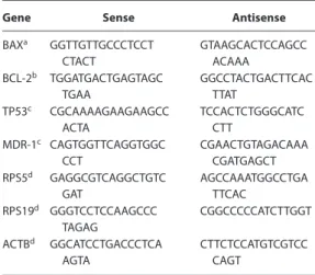
The qPCR steps were performed in an automatic thermocycler (ABI Prism 7500 Sequence Detec-tion System FAST, Applied Biosystems). A sequence amplification of primers is detailed in Table 4. The qPCR reaction consisted of 4μL of sample cDNA, 200 nM of each primer, 10μL GoTaq qPCR Mas-ter Mix (Promega), and nuclease-free waMas-ter, giving a final volume of 20μL.
Table 4. Sense and antisense genes used in RT-qPCR
Gene Sense Antisense
BAXa GGTTGTTGCCCTCCT
CTACT
GTAAGCACTCCAGCC ACAAA
BCL-2b TGGATGACTGAGTAGC
TGAA
GGCCTACTGACTTCAC TTAT
TP53c CGCAAAAGAAGAAGCC
ACTA
TCCACTCTGGGCATC CTT
MDR-1c CAGTGGTTCAGGTGGC
CCT
CGAACTGTAGACAAA CGATGAGCT RPS5d GAGGCGTCAGGCTGTC
GAT
AGCCAAATGGCCTGA TTCAC
RPS19d GGGTCCTCCAAGCCC
TAGAG
CGGCCCCCATCTTGGT
ACTBd GGCATCCTGACCCTCA
AGTA
CTTCTCCATGTCGTCC CAGT
aSanoet al.2005.93 bKlopfleischet al.2009.94 cCulmseeet al.2004.95 dBrinkhofet al.2006.96
stable endogenous control from the three endoge-nous genes tested (RPS5, RPS19 and ACTB).92 All reactions were performed in duplicate. A QR value <0.5 was defined as decreased expression,
whereas QR > 2.00 was considered increased
expression.
Statistical analysis
Descriptive statistics were performed to analyse the results. Data fromin vitroassays were expressed as an average percentage±of the standard deviation, and compared by Kruskal–Wallis test, followed by multiple comparisons with Dunn’s test. ANOVA test was conducted for the gene expression analysis, followed by the Tukey multiple comparisons test,97 using the programme GraphPad Prism 5.0. Values ofP<0.05 were considered significant.
Acknowledgements
The authors would like to thank the Fundação de Amparo à Pesquisa do Estado de São Paulo (FAPESP), São Paulo, SP, Brazil for its financial sup-port to develop this project. This sponsor did not have any influence on the study design, on the col-lection, analysis and interpretation of data, or on the writing of the manuscript and decision to sub-mit for publication. Proc 2012/19285-2. M. M. F. designed the study, analysed and interpreted the
data, made a critical review of the manuscript and wrote the manuscript; H. B. F. critically revised and formatted the manuscript; G. N. S. made a critical review of and wrote the manuscript; J. A. A. sub-stantially contributed to conceiving the study; R. Y. helped to draft the study; J. P. A. substantially contributed to conceiving the study, acquired the data and helped produce the draft; N. S. R. super-vised and coordinated the study, provided clini-cal and pathology advice, and criticlini-cally revised the manuscript. All authors read and approved the final manuscript.
Conflict of interests
The authors declare that they have no competing interests.
References
1. Amaral AS, Bassanil-Silva S, Ferreira I, Fonseca LS, Andrade FH, Gaspar LFJ,et al.Cytomorphological characterization of transmissible canine venereal tumor.RPCV2007;103: 563–564.
2. Drumond KO, Quessada AM, Silva SMMS, Costa FAL, Silva LS, de Pinho FA,et al.Transmissible venereal tumor treated with autohemotherapy.Acta Sci Vet2013;41: 1107.
3. Ganguly B, Das U and Das AK. Canine
transmissible venereal tumour: a review.Vet Comp Oncol2013;11: 1–12.
4. Erünal-Maral N, Findik M and Asalan S. Use of exfoliative cytology for diagnosis of transmissible venereal tumour and controlling the recovery period in the bitch.Dtsch Tierarztl Wochenschr
2000;107: 175–180.
5. Sousa J, Saito V, Nardi AB, Rodaski S, Guérios S and Bacila M. Características e incidência do tumor venéreo transmissível (TVT) em cães e eficiência da quimioterapia e outros tratamentos.Arch Vet Sci
2000;5: 41–48.
6. Athanasius EC, Kene RO and Anyanwu HC. Comparative efficacy of surgery, vincristine sulphate and combined therapy of levamisole and Bacille Calmette Guerin vaccine in the treatment of transmissible venereal tumour-infected dogs.Comp Clin Pathol2014;23: 1263–1267.
7. Rogers KS. Transmissible venereal tumour.
Compend Contin Educ Pract Vet1997;19: 1036–1045.
vagino plasty in female and subtotal penile amputation and scrotal ablation in male dogs. In:
Proceeding of 48th Kasetsart University Annual Conference: Veterinary Medicine, Bangkok, Kasetsart, 2010: 191–200.
9. Sudjaidee P, Theewasutrakul P, Techarungchaikul S, Ponglowhapan S and Chatdarong K. Treatment of canine transmissible venereal tumor using Vincristine Sulfate combined with L-Asparaginase in clinical vincristine-resistant cases: a case report.
Thai J Vet Med2012;42: 117–122.
10. Andrade SMF.Manual de terapêutica veterinária. 3rdedn., São Paulo, ROCA, 2008.
11. Gaspar LFJ, Amaral AS, Bassani-Silva S and Rocha NS. Imunorreatividade à glicoproteína-p nos diferentes tipos citomorfológicos de tumor venéreo transmissível canino.Vet em Foco2009;6: 1–6. 12. Gaspar LFJ, Ferreira I, Colodel MM, Brandão CVS
and Rocha NS. Spontaneus canine transmissible venereal tumor: cell morphology and influence on P-glycoprotein expression.Turk J Vet Anim Sci
2010;34: 447–454.
13. Gerardi DG, Tinucci-Costa M, Silveira ACT and Moro JV. Expression of P-glycoprotein, multidrug resistance associated protein,
glutathione-S-transferase pi and p53 in canine transmissible venereal tumor.Pesq Vet Bras2014;
34: 71–78.
14. Mendelsohn J, Howley I and Thompson G.The Molecular Basis of Cancer. 3rdedn., Saunders,
Elsevier, 2008.
15. Hodges LM, Markova SM, Chinn LW, Gow JM, Kroetz DL, Klein TE,et al.Very important pharmacogene summary: ABCB1 (MDR1, P-glycoprotein).Pharmacogenet Genomics2011;21: 152–161.
16. Alexandrova R. Multidrug resistance and p-glycoprotein.Exp Path Parasitol1998;1: 62–66. 17. Thomas H and Coley H. Overcoming multidrug
resistance in cancer: an update on the clinical strategy of inhibiting p-glycoprotein.Cancer Control2003;10: 159–165.
18. Maia RC and Rumjanek VM. Mecanismos moleculares de resistência a múltiplas drogas. In:
Oncologia Molecular. CG Ferreira and JC Rocha Eds., São Paulo, Atheneu, 2004: 113–122.
19. Szakács G, Paterson JK, Ludwig JA, Booth-Genthe C and Gottesman MM. Targeting multidrug resistance in cancer.Nat Rev Drug Discov2006;5: 219–234. 20. Tsujimura S and Tanaka Y. Treatment strategy
based on targeting P-glycoprotein on peripheral lymphocytes in patients with systemic autoimmune disease.Clin Exp Nephrol2012;16: 102–108.
21. Binkhathlan Z and Lavasanifar A. P-glycoprotein inhibition as a therapeutic approach for overcoming multidrug resistance in cancer: current status and future perspectives.Curr Cancer Drug Targets2013;
13: 326–346.
22. Wang LH, Song YB, Zheng WL, Jiang L and Ma WL. The association between polymorphisms in the MDR1 gene and risk of cancer: a systematic review and pooled analysis of 52 case–control studies.
Cancer Cell Int2013;13: 46.
23. Soussi T and Béroud C. Assessing TP53 status in human tumours to evaluate clinical outcome.Nat Rev Cancer2001;1: 233–240.
24. Ozaki T and Nakagawara A. p53: the attractive tumor suppressor in the cancer research field.J Biomed Biotechnol2011;2011: 13.
25. Moro JV, Tinucci-Costa M, Silveira AC, Gerardi DG and Alessi AC. Reactivity of p53 protein in canine transmissible venereal tumor.Arq Bras Med Vet Zootec2010;62: 318–323.
26. Harris CC and Hollstein M. Clinical implications of the p53 tumor-suppressor gene.N Engl J Med1993;
329: 1318–1327.
27. Suzuki K and Matsubara H. Recent advances in p53 research and cancer treatment.J Biomed Biotechnol
2011;2011: 7.
28. Kanayan N, Okuda M, Toyama N, Oikawa T, Inokuma H, Morimoto M,et al.Detection of the anti-P53 antibodies in dogs with tumors.J Vet Med Sci2002;64: 973–979.
29. Choi YK and Kim CJ. Sequence analysis of canine LINE-1 elements and p53 in canine transmissible venereal tumor.J Vet Sci2002;3: 285–292. 30. Sánchez-Servín A, Martínez S, Córdova-Alarcon E
and Fajardo R. TP53 polymorphisms allow for genetic sub-grouping of the canine transmissible venereal tumor.J Vet Sci2009;10: 353–355. 31. Vázquez-Mota N, Simón-Martínez J,
Córdova-Alarcon E, Lagunes L and Fajardo R. The T963C mutation of TP53 gene does not participate in the clonal origin of canine TVT.Vet Res Commun
2008;32: 187–191.
32. Stockmann D, Ferrari HF, Andrade AL, Cardoso TC and Luvizotto MC. Detection of the tumour suppressor gene TP53 and expression of p53, Bcl-2 and p63 proteins in canine transmissible venereal tumor.Vet Comp Oncol2011a;9: 251–259. 33. Chao DT and Korsmeyer SJ. BCL-2 family: regulators of cell death – review.Annu Rev Immunol1998;16: 395–419.
35. Delfino AB, Barreto EC, Jr ETS, Mendonça RG and Ornellas MH. O envolvimento de genes e proteínas na regulação da apoptose – carcinogênese.Rev Bras Cancerol1997;43: 173–186.
36. Fett-Conte AC and Salles ABCF. A importância do gene p53 na carcinogênes humana.Rev Bras Hematol Hemoter2002;24: 85–89.
37. Zhou H, Chen J, Meagher JL, Yang CY, Aguilar A, Liu L,et al.Design of Bcl-2 and Bcl-xL inhibitors with subnanomolar binding affinities based upon a new scaffold.J Med Chem2012;55: 4664–4682. 38. Stockmann D, Ferrari HF, Andrade AL, Lopes RA,
Cardoso TC and Luvizotto MC. Canine transmissible venereal tumors: aspects related to programmed cell death.Braz J Vet Pathol2011b;4: 67–75.
39. Frenzel A, Grespi F, Chmelewskijwand W and Villunger A. Bcl2 family proteins in carcinogenesis and the treatment of cancer.Apoptosis2009;14: 584–596.
40. Amaral AS, Ferreira I, Colodel MM, Fávero DM and Rocha NS. DNA damage in canine transmissible venereal tumor.Rev Lus Ciên Med Vet2011;4: 1–5. 41. Athanasou NA. Cell and organ culture in the
understanding of bone and its neoplams. In:
Pathology of Bone and Joint Neoplams. TR Helliwell Ed., Philadelphia, W.B. Saunders, 1999: 34–58. 42. Adams EW, Carter LP and Sapp WJ. Growth and
maintenance of the canine venereal tumor in continuous culture.Cancer Res1968;28: 753–757. 43. Mohanty GC and Rajya BS. Growth and
morphological characteristics of canine venereal tumor cell in vitro.Vet Pathol1977;14: 420–425. 44. Okamoto Y, Fujinaga T, Tajima M, Hoshi N, Otomo
K and Koike T. Isolation and cultivation of canine transmisible sarcoma cells.Nihon Juigaku Zasshi
1988;50: 9–13.
45. Beschorner WE, Hess AD, Nerenberg ST and Epstein RB. Isolation and characterization of canine venereal tumor-associated inhibitory and blocking factors.Cancer Res1979;39: 3920–3927. 46. Bassani-Silva S, Sforcin JM, Amaral AS and Sousa
NR. Propolis effect in vitro on canine transmissible venereal tumour cells.RPCV2007;102: 261–265. 47. Montoya FL, Bersano OP, Ferreira LJ, Pedraza FJ
and Rocha NS. Caracterización inmunofenotípica de cultivos primarios de tumor venéreo transmisible canino.Rev Col Cienc Pec2013;3 Suppl: 380. 48. Bergman PJ, Ogilvie GK and Powers BE.
Monoclonal antibody C219 immunochemistry against P-glycoprotein: sequential analysis and predictive ability in dogs with lymphoma.J Vet Inter Med1996;10: 354–359.
49. Lee JJ, Hughes CS, Fine RL and Page RL. P-glycoprotein expression in canine lymphoma: a relevant, intermediate model of multidrug resistance.Cancer1996;77: 1892–1898.
50. Steingold SF, Sharp NJ, McGahan MC, Hughes CS, Dunn SE and Page RL. Characterization of canine MDR1 mRNA: its abundance in drug resistant cell lines and in vivo.Anticancer Res1998;18: 393–400. 51. Honscha KU, Schirmer A, Reischauer A, Schoon
HA, Einspanier A and Gäbel G. Expression of ABC-transport proteins in canine mammary cancer: consequences for chemotherapy.Reprod Domest Anim2009;44(Suppl. 2): 218–223. 52. Zandvliet M, Teske E and Schrickx JA. Multi-drug
resistance in a canine lymphoid cell line due to increased P-glycoprotein expression, a potential model for drug-resistant canine lymphoma.Toxicol In Vitro2014;28: 1498–1506.
53. Breier A, Štefanková S, Barancík M and Tribulová N. Time dependence of [3h]-vincristine
accumulation by L1210 mouse leukemic cells. Effect of P-glycoprotein overexpression.Gen Physiol Biophys1994;13: 287–298.
54. Burkhart CA, Kavallaris M and Band Horwitz S. The role of beta-tubulin isotypes in resistance to antimitotic drugs.Biochim Biophys Acta2001;1471: O1–O9.
55. Ambudkar SV, Kimchi-Sarfaty C, Sauna ZE and Gottesman MM. P-glycoprotein: from genomics to mechanism.Oncogene2003;22: 7468–7485. 56. Sulová Z, Mislovicová D, Gibalová L, Vajcnerová Z,
Poláková E, Uhrík B,et al.Vincristine-induced overexpression of P-glycoprotein in L1210 cells is associated with remodeling of cell surface saccharides.J Proteome Res2009;8: 513–520. 57. Veldhoen N, Stewart J, Brown R and Milner J.
Mutations of the p53 gene in canine lymphoma and evidence for germ line p53 mutations in the dog.
Oncogene1998;16: 249–255.
58. Lee CH, Kim WH, Lim JH, Kang MS, Kim DY and Kweon OK. Mutation and overexpression of p53 as a prognostic factor in canine mammary tumors.J Vet Sci2004;5: 63–69.
59. Sui X, Jin L, Huang X, Geng S, He C and Hu X. p53 signaling and autophagy in cancer: a revolutionary strategy could be developed for cancer treatment.
Autophagy2011;7: 565–571.
60. Lacroix M, Toillon RA and Leclercq G. p53 and breast cancer, an update.Endocr Relat Cancer2006;
13: 293–325.
62. Liu FS, Jan YJ, Lai CR, Twu NF, Lu CH, Hung MJ,
et al.Expression analysis of apoptosis-related markers TP53, BCL-2, BAX and c-MYC in female genital tract sarcomas.J Chin Med Assoc2008;71: 628–634.
63. Fernald K and Kurokawa M. Evading apoptosis in cancer.Trends Cell Biol2013;23: 620–633. 64. Llambi F and Green DR. Apoptosis and
oncogenesis: give and take in the BCL-2 family.
Curr Opin Genet Dev2011;21: 12–20.
65. Topham CH and Taylor SS. Mitosis and apoptosis: how is the balance set?Curr Opin Cell Biol2013;25: 780–785.
66. Groth-Pedersen L, Ostenfeld MS, Høyer-Hansen M, Nylandsted J and Jäättelä M. Vincristine induces dramatic lysosomal changes and sensitizes cancer cells to lysosome-destabilizing siramesine.Cancer Res2007;67: 2217–2225.
67. Mosmann T. Rapid colormetric assay for cellular growth and survival: application to proliferation and cytotoxicity assays.J Immunol Methods1983;
65: 55–63.
68. Jordan MA, Thrower D and Wilson L. Mechanism of inhibition of cell proliferation by Vinca alkaloids.
Cancer Res1991;51: 2212–2222.
69. Rixe O and Fojo T. Is cell death a critical end point for anticancer therapies or is cytostasis sufficient?
Clin Cancer Res2007;13: 7280–7287.
70. Galán-Malo P, Vela L, Gonzalo O, Calvo-Sanjuán R, Gracia-Fleta L, Naval J,et al.Cell fate after mitotic arrest in different tumor cells is determined by the balance between slippage and apoptotic threshold.
Toxicol Appl Pharmacol2012;258: 384–393. 71. Silverman JA and Deitcher SR. MARQIBO®
(vincristine sulfate liposome injection) improves the pharmacokinetics and pharmacodynamics of vincristine.Cancer Chemother Pharmacol2013;71: 555–564.
72. Dixon GJ, Dulmadge EA, Mulligan LT and Mellett LB. Cell culture bioassay for vincristine sulfate in sera from mice, rats, dogs, and monkeys.Cancer Res
1969;29: 1810–1813.
73. Mujagic H, Chen SS and Geist R. Effects of vincristine on cell survival, cell cycle progression and mitotic accumulation in Asynchronously growing sarcoma 180 cells.Cancer Res1983;43: 3591–3597.
74. Jordan MA, Wendell K, Gardiner S, Derry WB, Copp H and Wilson L. Mitotic block induced in HeLa cells by low concentrations of paclitaxel (Taxol) results in abnormal mitotic exit and apoptotic cell death.Cancer Res1996;56: 816–825. 75. Tanaka NM, Maeda MSCF, Pereira LI, Araújo RC,
Kemper B, Gonçalves RC,et al.Atividade do sulfato
de vincristina em cultivos primários de tumores mamários caninos.Rev Acad Ciên Anim2013;11: 87–96.
76. Jackson DV and Bender RA. Cytotoxic thresholds of vincristine in a murine and a human leukemia cell line in vitro.Cancer Res1979;39: 4346–4349. 77. Ehrhardt H, Pannert L, Pfeiffer S, Wachter F,
Amtmann E and Jeremias I. Enhanced anti-tumour effects of Vinca alkaloids given separately from cytostatic therapies.Br J Pharmacol2013;168: 1558–1569.
78. He D, Wu H, Ding L and Li Y. Combination of BCL11A siRNA with vincristine increases the apoptosis of SUDHL6 cells.Eur J Med Res2014;19: 34.
79. Shi J, Orth JD and Mitchison T. Cell type variation in responses to antimitotic drugs that target microtubules and kinesin-5.Cancer Res2008;68: 3269–3276.
80. Hsiao YW, Liao KW, Hung SW and Chu RM. Effect tumor infiltrating lymphocytes on the expression of MHC molecules in canine transmissible venereal tumor cells.Vet Immunol Immunopathol2002;87: 19–27.
81. Stettner N, Brenner O, Eilam R and Harmelin A. Pegylated liposomal doxorubicin as a
chemotherapeutic agent for treatment of canine transmissible venereal tumor in murine models.J Vet Med Sci2005;6: 1133–1139.
82. Gascoigne KE and Taylor SS. How do anti-mitotic drugs kill cancer cells?J Cell Sci2009;122: 2579–2585.
83. Panvichian R, Orth K, Day ML, Day KC, Pilat MJ and Pienta KJ. Paclitaxel-associated
multimininucleation is permitted by the inhibition of caspase activation: a potential early step in drug resistance.Cancer Res1998;58: 4667–4672. 84. Chen JG and Horwitz SB. Differential mitotic
responses to microtubule -stabilizing and -destabilizing drugs.Cancer Res2002;62: 1935–1938.
85. Orth JD, Loewer A, Lahav G and Mitchison TJ. Prolonged mitotic arrest triggers partial activation of apoptosis, resulting in DNA damage and p53 induction.Mol Biol Cell2012;23: 567–576. 86. Huang Y, Fang Y and Wu J. Regulation of Vinca
alkaloid-induced apoptosis by NF-kappaB/IkappaB pathway in human tumor cells.Mol Cancer Ther
2004;3: 271–277.
89. Gidding CE, Kellie SJ, Kamps WA and Graaf SS. Vincristine revisited.Crit Rev Oncol Hematol1999;
29: 267–287.
90. Morales-Ramírez P, Vallarino-Kelly T and Cruz-Vallejo V. Kinetics of micronucleus induction and cytotoxicity caused by distinct antineoplastics and alkylating agents in vivo.Toxicol Lett2014;224: 319–325.
91. Hsiao YW, Liao KW, Chung TF, Liu CH, Hsu CD and Chu RM. Interactions of host IL-6 and IFN-gamma and cancer-derived TGF-beta1 on MHC molecule expression during tumor spontaneous regression.Cancer Immunol Immunother2008;57: 1091–1104.
92. Larionov A, Krause A and Miller W. A standart curve based method for relative real time PCR data processing.BMC Bioinformat2005;6: 62. 93. Sano J, Oguma K, Kano R, Yazawa M, Tsujimoto H
and Hasegawa A. High expression of Bcl-xL in
delayed apoptosis of canine neutrophils induced by lipopolysaccharide.Res Vet Sci2005;78: 183–187. 94. Klopfleisch R and Gruber AD. Differential
expression of cell cycle regulators p21, p27 and p53 in metastasizing canine mammary
adenocarcinomas versus normal mammary glands.
Res Vet Sci2009;87: 91–96.
95. Culmsee K, Gruber A, Himmelstjerna G and Nolte I. Quantification of MDR-1 gene expression in canine tissues by real-time reverse transcription quantitative polymerase chain reaction.Res Vet Sci
2004;77: 223–229.
96. Brinkhof B, Spee B, Rothuizen J and Penning L. Development and evaluation of canine reference genes for accurate quantification of gene expression.
Anal Biochem2006;356: 36–43. 97. Pagano M and Gauvreau K.Princípios de