Braz. J. of Develop.,Curitiba, v. 6, n. 11, p.85867-85883, nov. 2020. ISSN 2525-8761
Lentinus villosus Klotzsch (1833) AM 169: a natural and renewable source of
alkaline protease
Lentinus villosus Klotzsch (1833) AM 169: uma fonte natural e renovável de
protease alcalina
DOI:10.34117/bjdv6n11-127
Recebimento dos originais: 06/10/2020 Aceitação para publicação: 06/11/2020
Rodrigo da Silva Braga
Graduado em Farmácia
Instituição: Faculdade Estácio do Amazonas
Endereço: Av. Constantino Nery, 3693 (Bairro Chapada) 69025-315 Manaus – AM E-mail: bragarodrigo355@gmail.com
Érica Caroline de Matos Brito
Graduada em Nutrição
Instituição: Faculdade Estácio do Amazonas
Endereço: Av. Constantino Nery, 3693 (Bairro Chapada) 69025-315 Manaus – AM E-mail: ericacarolinemb@gmail.com
Raiane Áila Teixeira Souza
Doutora em Biotecnologia
Instituição: Faculdade Estácio do Amazonas
Endereço: Av. Constantino Nery, 3693 (Bairro Chapada) 69025-315 Manaus – AM E-mail: aila.raiane@gmail.com
Maria Francisca Simas Teixeira
Doutora em Ciências Biológicas
Instituição: Universidade Federal do Amazonas
Endereço: Av. Gen. Rodrigo Octávio 6200 (Bairro Coroado I) 69080-900 Manaus - AM E-mail: mteixeira@ufam.edu.br
Salomão Rocha Martim
Doutor em Biodiversidade e Biotecnologia Instituição: Universidade Nilton Lins
Endereço: Av. Prof. Nilton Lins, 3259 (Bairro Flores) 69058-030, Manaus – AM E-mail: salomao.martim@gmail.com
Braz. J. of Develop.,Curitiba, v. 6, n. 11, p.85867-85883, nov. 2020. ISSN 2525-8761
ABSTRACT
Mushrooms have stood out as sources of proteases with industrial applicability. The objective of this work was to evaluate the parameters that influence the synthesis of proteases by Lentinus villosus Klotzsch (1833) AM 169 and to characterize these enzymes. The matrix culture, prepared on potato dextrose agar supplemented with yeast extract, was maintained for 8 days at 25 ° C. Submerged fermentation was carried out in GYPG (glucose, yeast extract, peptone and gelatin). Azocasein solution (1%, w/v) was used as an enzyme substrate. The best conditions for protease production were: initial pH of the culture medium (6), fermentation time (12 days), age of the inoculum (6 days) and size of the inoculum (10%). The proteases were active at pH 8, at 60 ° C, stable at pH (5 to 9) and temperature (30 °C to 60 °C). These enzymes were classified as cysteine proteases, stimulated by Fe2+ and Mn2+.
Lentinus villosus synthesizes proteases with potential for application in the detergent, textile, leather
processing and bioremediation processes.
Keywords: basidiomycetes, peptidases, submerged fermentation. RESUMO
Os cogumelos têm se destacado como fontes de proteases com aplicabilidade industrial. O objetivo desse trabalho foi avaliar os parâmetros que influenciam na síntese de proteases por Lentinus villosus Klotzsch (1833) AM 169 e caracterizar essas enzimas. A cultura matriz, preparada em ágar batata dextrose suplementado com extrato de levedura, foi mantida por 8 dias a 25 °C. A fermentação submersa foi realizada em GYPG (glicose, extrato de levedura, peptona e gelatina). Solução de azocaseína (1%, p/v) foi utilizada como substrato enzimático. As melhores condições para a produção de protease foram: pH inicial do meio de cultura (6), tempo de fermentação (12 dias), idade do inóculo (6 dias) e tamanho do inóculo (10%). As proteases foram ativas em pH 8, a 60 °C, estáveis ao pH (5 a 9) e à temperatura (30 °C a 60 °C). Estas enzimas foram classificadas como cisteíno proteases, estimuladas por Fe2+ e Mn2+. Lentinus villosus sintetiza proteases com potencial para aplicação nas indústrias de detergente, têxtil, no beneficiamento do couro e em processos de biorremediação.
Braz. J. of Develop.,Curitiba, v. 6, n. 11, p.85867-85883, nov. 2020. ISSN 2525-8761
1 INTRODUCTION
Proteases, peptidases, proteinases or proteolytic enzymes are hydrolases that catalyze the breakdown of peptide bonds in proteins (Gurumallesh et al., 2019). These biocatalysts play physiological functions essential for the maintenance of the life of living beings, such as cell signaling, inflammation, apoptosis, blood coagulation and hormone processing (Bond, 2019; Boon et al., 2020). Due to broad biotechnological applications, proteases correspond to 65% of the enzyme market (Kummari & Prasad, 2015; Lanka et al., 2017). In the industrial sector, the peptidases are used in the manufacture of cheeses, beverages, softening of meats and bakery products. They are also used in wastewater treatment, bioremediation processes and medical diagnostics (Ahmed et al., 2017).
The alkaline proteases are a group of proteolytic enzymes with significant catalytic activity at pH > 8,0 (Srilakshmi et al., 2014). These biocatalysts have outstanding economic importance in the production of detergents, but can also be used in the textile and chemical industries, leather processing and bioremediation (Sharma et al., 2017; Zhou et al., 2020).
In nature, vegetables, animals and microorganisms, including bacteria and filamentous fungi are sources of proteolytic enzymes (Aljammas et al., 2018; Souza et al., 2019). The production of enzymes synthesized by microorganisms has acquired space in the market and industrial relevance, since proteases extracted from plants and animals are not supplying the commercial demand (Razzaq
et al., 2019). In addition, proteases of microbial origin are Generally Recognized as Safe (GRAS)
according to the US Food and Drug Administration (Tavano et al., 2018).
Species of filamentous fungi are considered excellent sources of proteases because they are easy to culture and secrete extracellular enzymes, conditions that facilitate the production of proteolytic biocatalysts on a large scale (Inácio et al., 2015). Among the filamentous fungi, mushrooms have been highlighted as sources of proteases with potential for industrial application (Martim et al., 2017a).
The submerged fermentation technology has been used for the production of proteases by different species of mushrooms: Lentinus citrinus Walleyn & Rammeloo (1994) (Kirsch et al., 2013),
L. crinitus (L.) Fr. 1825 (Magalhães et al., 2019), Pleurotus albidus (Berk.) Pegler 1983 (Martim et al., 2017b) and Termitomyces clypeatus R. Heim 1951 (Majumder et al., 2014). This work aimed to
evaluate the parameters that influence the synthesis of proteases by Lentinus villosus Klotzsch (1833) and to characterize these biocatalysts of industrial importance.
Braz. J. of Develop.,Curitiba, v. 6, n. 11, p.85867-85883, nov. 2020. ISSN 2525-8761
2 MATERIAL AND METHODS
2.1 MUSHROOM
Lentinus villosus AM 169, from the Cultures Collection of the Parasitology Department of
Federal University of Amazonas, was cultivated in Petri dishes containing PDA culture medium [potato dextrose agar supplemented with 0,5% (w/v) yeast extract]. The cultures were kept at 25 ºC for eight days.
2.2 PROTEASES PRODUCTION BY SUBMERGED FERMENTATION
For the proteases production in liquid medium, three 80mm (Ø) mycelial fragments were inoculated in 125 mL Erlenmeyer flasks containing 50 mL of GYPG liquid medium (20 g of glucose, 5 g of yeast extract, 5 g of peptone and 5 g of gelatin per 1000 ml of water). The liquid medium, pH 5.6, was sterilized at 121 °C for 15 minutes. The fermentation was conducted at 30 °C, 150 rpm. After five days, the biomass was filtered off under vacuum on Whatman number 1 filter paper, followed by centrifugation at 4 °C (8,000 x g / 5 minutes). The proteolytic activity was determined in the crude extract.
2.3 DETERMINATION OF PROTEOLYTIC ACTIVITY
Proteolytic activity was determined according to Leighton et al. (1973). As a substrate, 250 μl
of 1% (w/v) azocasein in 150 μL Tris-HCl buffer of the crude enzyme extract was used. The reaction mixture was maintained for 60 minutes at 25 °C and the reaction was interrupted by addition of 1.2 mL of 10% (w/v) trichloroacetic acid followed by centrifugation at 4 °C (8,000 xg/ 5 minutes). From the recovered supernatant was removed 0.8 mL for homogenization with 1.4 mL of 1M NaOH. The blank was prepared maintaining the same conditions as the sample. One unit of proteolytic activity was defined as the amount of enzyme required to produce an absorbance change of 0.01 in one hour.
2.4 STANDARDIZATION OF FERMENTATION PARAMETERS FOR PROTEASE PRODUCTION
In order to evaluate the optimal conditions of proteases production by L. villosus, the initial pH of the culture medium (4, 5, 6, 7 and 8), influence of inoculum age (6, 8, 10, 12 and 14 days), fermentation time (2, 4, 6, 8, 10, 12 and 14 days) and inoculum size (2%, 4%, 8%, 10% and 20%) were analyzed.
Braz. J. of Develop.,Curitiba, v. 6, n. 11, p.85867-85883, nov. 2020. ISSN 2525-8761 2.5 PARTIAL CHARACTERIZATION OF PROTEASES
The proteases obtained under the optimum conditions of fermentation were characterized for optimum pH and temperature, pH and temperature stability and effects of ions and inhibitors as described by Martim et al. (2017b).
For optimal pH testing, proteolytic activity was determined at 25 °C at different pH values using the following buffer solutions (0.1 M): sodium acetate (pH 5 and 6), Tris-HCl (pH 7 and 8) and Glycine-NaOH (pH 9 and 10). The optimum temperature was determined by incubating the crude extract at different temperatures (30, 40, 50, 60, 70 and 80 °C).
For pH stability, the crude extract was diluted (1:1) in the following 0.1 M buffer solutions: sodium acetate (pH 5 and 6), Tris-HCl (pH 7 and 8) and Glycine-NaOH (pH 9 and 10) and maintained at 25 °C for 24 hours. At thermal stability, the extract was incubated at different temperatures (30, 40, 50, 60, 70 and 80 °C) for 1 hour. Azocasein 1% (w/v) dissolved in Tris-HCl buffer was used as substrate. Relative activities were determined under optimum pH and temperature conditions.
The effect of inhibitors on enzyme activity was investigated using 10 mM phenylmethylsulfonyl fluoride (PMSF), 10 mM ethylenediaminetetraacetic acid (EDTA), 10 mM iodoacetic acid and 0.1 mM pepstatin in the reaction mixture. The metal ions used in the 10 mM concentration in the reaction mixture were CaCl2, CuSO4, FeSO4, MgSO4, MnSO4, ZnSO4, NaCl and
KCl. The samples were incubated under optimum pH and temperature conditions. The residual enzyme activity was compared to the control (incubated without inhibitors and metal ions) and corresponded to 100% activity.
3 STATISTICAL ANALYSIS
In all experiments the data were submitted to analysis of variance and the means were compared by the Tukey method (p ˂ 0.05) using Minitab program, version 18.0 (Minitab, 2017). All analyzes were performed in triplicate.
4 RESULTS AND DISCUSSION
4.1 STANDARDIZATION OF FERMENTATION PARAMETERS FOR PROTEASE PRODUCTION
The synthesis of proteases by microorganisms using the submerged fermentation technology is influenced by environmental factors and nutritional characteristics of the culture medium (Ibrahim et
Braz. J. of Develop.,Curitiba, v. 6, n. 11, p.85867-85883, nov. 2020. ISSN 2525-8761
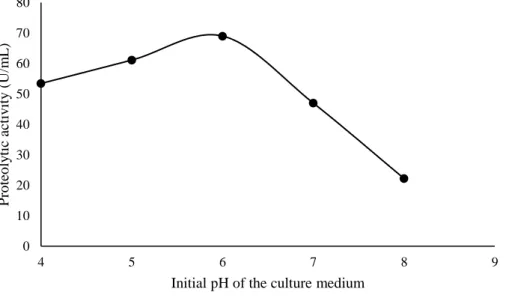
villosus is shown in figure 1. Lentinus villosus synthesized significant quantitative of proteases at pH
6 (69.0 U/mL). There was a decrease in proteolytic activity at pH 7 and 8, as shown in figure 2. This same pH value was considered the optimal pH for protease production by Rhizopus stolonifer (Ehrenb.) Vuill. (1902) (Gais et al., 2009). Daudi et al. (2015) and Bano et al. (2016) reported that at pH 5.5 and 6.5 there was maximum protease production by Mucor pusillus Lindt (1886) and P. eryngii (DC.) Quél. 1872, respectively. The pH of the culture medium interferes with enzyme production, enzyme activity and nutrient transport across the cell membrane (Sharma et al., 2017).
Figure 1: Influence of the pH of the culture medium on the protease production by Lentinus villosus.
Under the conditions evaluated, the maximum protease production (130.0 U/mL) by L. villosus was verified with six-day inoculum cultured in PDA (Figure 2). Boukhalfa-Lezzar et al. (2014) and Martim et al. (2017b) observed that the five-day inoculum favored the production of proteases by
Aspergillus oryzae (Ahlb.) (1884) 2220 and P. albidus, respectively. Irfan et al. (2011) reported that R. oligosporus Saito (1905) M-30 produced maximum quantitative of proteolytic enzymes when using
7-day old inoculant. The optimal age of the inoculum to the production of enzymes varies according to the physiological characteristics of the microorganism and the environmental conditions of the bioprocess (Maldonado et al., 2014).
0 10 20 30 40 50 60 70 80 4 5 6 7 8 9 P ro teo ly tic ac tiv ity ( U/m L )
Braz. J. of Develop.,Curitiba, v. 6, n. 11, p.85867-85883, nov. 2020. ISSN 2525-8761 Figure 2. Influence of inoculum age on protease production by Lentinus villosus.
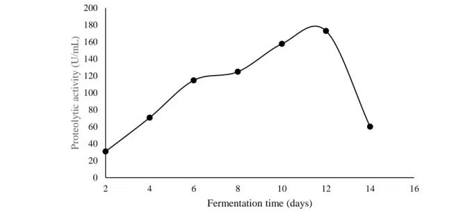
Fermentation time is a parameter that significantly influences the protease production by filamentous fungi. In the present study, it was observed that L. villosus synthesized and excreted proteases in all conditions evaluated. However, maximum peptidase production was verified at 12 days of fermentation (172.88 U/mL), as shown in figure 3. Brito et al. (2019) when evaluating the production of proteases by L. crinitus DPUA 1693 also reported a similar result. The significant protease production by P. ostreatus (Jacq.) P. Kumm. (1871) and P. eryngii in liquid medium was verified at 14 and 4 days, as reported by Sales-Campos et al. (2010) and Bano et al. (2016), respectively. Protease production increases with incubation time to some extent. Then, there is a reduction in the synthesis of proteolytic enzymes due to the insufficient availability of some nutrients in the growth medium or decomposition of the protease (Niyonzima & More, 2013).
0 20 40 60 80 100 120 140 6 7 8 9 10 11 12 13 14 15 P ro teo ly tic ac tiv ity ( U/m L )
Braz. J. of Develop.,Curitiba, v. 6, n. 11, p.85867-85883, nov. 2020. ISSN 2525-8761 Figure 3. Influence of fermentation time on the protease production by Lentinus villosus.
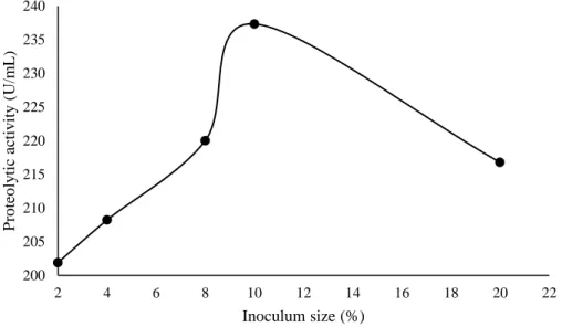
The influence of inoculum size on the protease production by L. villosus is shown in figure 4. In the conditions evaluated, it was observed that 10% inoculum (w/v) stimulated the maximum protease production (237.33 U/mL) by L. villosus. However, the use of 20% inoculum decreased the protease production by 8.65%. Martim et al. (2017b) and Brito et al. (2019) also observed that the synthesis of proteases by P. albidus and L. crinitus was stimulated with the use of 10% inoculum. On the other hand, Benluvankar et al. (2015) reported that the 5% inoculum stimulated the protease production of by Penicillium sp. Link (1809) LCJ228. The use of high concentrations of inoculum favors the rapid consumption of nutrients, resulting in a reduction in the synthesis of alkaline proteases by filamentous fungi (Niyonzima & More, 2013).
0 20 40 60 80 100 120 140 160 180 200 2 4 6 8 10 12 14 16 P ro teo ly tic ac tiv ity ( U/m L )
Braz. J. of Develop.,Curitiba, v. 6, n. 11, p.85867-85883, nov. 2020. ISSN 2525-8761 Figure 4: Influence of inoculum size on the protease production by Lentinus villosus
4.2 PARTIAL CHARACTERIZATION OF PROTEASES
In this study, the proteases of L. villosus had the optimal activity at pH 8.0, therefore classified as alkaline. There was an activity reduction of 23.64% and 18.9% at pH 9 and 10, respectively, as shown in Figure 5. Sun et al. (2011) and Ravikumar et al. (2012) reported that the proteases of Amanita
farinosa Schwein. (1822) and P. sajor-caju (Fr.) Singer (1951) also demonstrate maximum catalytic
activity at pH 8.0, respectively. Zhang et al. (2011) reported that the peptidases of T. albuminosus (Berk.) R. Heim (1941) have significant protease activity at pH 10.6. A change in the hydrogen potential value, in addition to the ideal pH, can lead to protonation or deprotonation of enzyme side groups. Under these conditions, there may be structural changes in the enzymes, resulting in decreased enzyme activity (Chittoor et al., 2016).
200 205 210 215 220 225 230 235 240 2 4 6 8 10 12 14 16 18 20 22 P ro teo ly tic ac tiv ity ( U/m L ) Inoculum size (%)
Braz. J. of Develop.,Curitiba, v. 6, n. 11, p.85867-85883, nov. 2020. ISSN 2525-8761 Figure 5. Effect of pH on the proteolytic activity of Lentinus villosus.
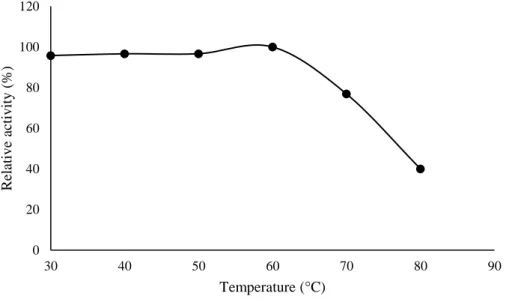
Temperature influences the growth and production of biocomposites by different species of microorganisms. In this study it was observed that the proteases of L. villosus had maximum catalytic activity at 60 °C. Relative activities of 95.98%, 96.43% and 96.61% were observed at temperatures of 30 °C, 40 °C and 50 °C, respectively. A lower relative activity (12.19%) was observed at 70 °C, as shown in Figure 6. Similar results were verified by Ravikumar et al. (2012) and Genier et al. (2015), which reported the maximum catalytic activity of Pleurotus sajor-caju and P. ostreatus proteases at 60 °C, respectively. Brito et al. (2019) reported that L. crinitus proteases demonstrated significant activity at 50 °C. At high temperatures, weak hydrogen and hydrophobic bonds break, which maintain the structure protease. Under these temperature conditions, the enzyme is denatured, resulting in loss of enzyme activity (Oueslati & Mounirhaouala, 2014; Martim et al., 2017b).
0 20 40 60 80 100 120 5 6 7 8 9 10 11 R elativ e ac tiv ity ( %) pH
Braz. J. of Develop.,Curitiba, v. 6, n. 11, p.85867-85883, nov. 2020. ISSN 2525-8761 Figure 6. Effect of temperature on the proteolytic activity of Lentinus villosus.
The proteases of L. villosus demonstrated stability in the range of pH 5 to 8, with values of proteolytic activity higher than 90%. The enzyme presented 86.78% of activity at pH 10 (Figure 7). Brito et al. (2019) found that L. crinitus proteases maintained stability in the range of 30 °C to 60 °C, with relative activities greater than 80%. El-Baky et al. (2011) reported that proteases of Piptoporus
soloniensis (Dubois) Pilát (1937) retained 70% activity in the range of pH 3 to 5. Silva et al. (2017)
report that the proteases of Phanerochaete chrysosporium Burds. (1974) were stable in the range of pH 3 to 8.
Figure 7. Effect of pH on the enzymatic stability of Lentinus villosus.
0 20 40 60 80 100 120 30 40 50 60 70 80 90 R elativ e ac tiv ity ( %) Temperature (°C) 0 20 40 60 80 100 120 5 6 7 8 9 10 11 R elativ e ac tiv ity ( %) pH
Braz. J. of Develop.,Curitiba, v. 6, n. 11, p.85867-85883, nov. 2020. ISSN 2525-8761 The effect of temperature on the enzymatic stability of L. villosus is shown in figure 8. Under the conditions evaluated, the proteases of L. villosus maintained stability greater than 90% in the range of 30 °C to 60 °C. However, at the temperature of 70 °C there was a decrease in activity, with retention of only 23.14% of catalytic activity (Figure 8). Martim et al. (2017b) and Brito et al. (2019) found similar results for P. albidus and L. crinitus proteases, respectively. Leonhardt et al. (2016) observed that proteases of P. pulmonarius (Fr.) Quél. (1872) maintained stability greater than 80% in the temperature range of 20 °C to 40 °C.
Figure 8. Effect of temperature on the enzymatic stability of Lentinus villosus.
The effect of inhibitors and metal ions on the proteolytic activity of L. villosus is shown in Table 1. Iodoacetic acid caused a reduction of 20.29% in the proteolytic activity of L. villosus, suggesting the presence of a greater amount of cysteine protease in the enzymatic extract of this mushroom. In the presence of EDTA, PMSF and Pepstatin the proteases maintained catalytic activity of 100%, 99.58% and 99.47%, respectively. Machado et al. (2017), Martim et al. (2017a) and Brito et al. (2019) reported inhibition of proteolytic activity of P. ostreatoroseus Singer (1961), P. albidus and L. crinitus in 95%, 62% and 11%, respectively, in the presence of iodoacetic acid.
In the present study, ions Fe2+ and Mn2+ stimulated L. villosus protease activity in 77.25% and 45.75% respectively. On the other hand, the ions Zn2+, Ca2+, K+ and Na+ promoted reduction of the enzymatic activity in 9.23%, 8.77%, 6.05% and 5.21%, respectively. The ion Cu2+ caused greater inhibition in the enzymatic activity, reducing the catalytic activity in 53.68%. Zhang et al. (2010) also reported that protease catalytic activity of the Helvella lacunosa Afzel. (1783) was stimulated in the
0 20 40 60 80 100 120 30 40 50 60 70 80 90 R elativ e ac tiv ity ( %) Temperature (°C)
Braz. J. of Develop.,Curitiba, v. 6, n. 11, p.85867-85883, nov. 2020. ISSN 2525-8761 presence of Fe2+ and Mn2+, but in the presence of Cu2+, Hg2+ and Fe3+ there was a reduction of protease activity. Magalhães et al. (2019) observed a 29.43% increase in the proteolytic activity of L. crinitus in the presence of Mn2+. On the other hand Shivashankar and Premkumari (2014) observed that Fe2+ ions did not influence the proteolytic activity of Hypsizygus ulmarius (Bull.) (1984), whereas Zn2+, Ca2+ and Cu2+ completely inhibited the proteolytic activity.
5 CONCLUSION
Lentinus villosus produces predominantly cysteine proteases in liquid medium. The synthesis
of these proteolytic enzymes is influenced by the initial pH of the culture medium, fermentation time, age and size of the inoculum. Proteases have maximum catalytic activity at pH 8 to 60 ° C, stability at pH and temperature. These biocatalysts have the potential for application in detergents, textiles, leather production and bioremediation processes.
ACKNOWLEDGMENTS
The authors would like to thank the DPUA Culture Collection and the Laboratory of Industrial and Medical Microbiology, at the University Federal University of Amazonas (UFAM).
Table 1. Effect of inhibitors and metal ions on the protease activity of Lentinus villosus.
Chemical products Concentration (mM) Relative activity (%)
Control 10 100 EDTA 10 94,79 ± 2,55d PMSF 10 93,63 ± 0,38d Pepstatin 1 93,44 ± 1,45d Iodoacetic acid 10 79.71 ± 0,38f FeSO4 10 177,25 ± 1,92a MnSO4 10 145,75 ± 1,38b MgSO4 10 100,00 ± 2,14c KCl 10 94,79 ± 2,55d NaCl 10 93,95 ± 1,072d CaCl2 10 91,23 ± 2,14e ZnSO4 10 90,77 ± 1,33e CuSO4 10 46,32 ± 0,66g
Braz. J. of Develop.,Curitiba, v. 6, n. 11, p.85867-85883, nov. 2020. ISSN 2525-8761
REFERENCES
AHMED, N. S. A., S. G. MOHAMMED, A. A. E HUSSEIN & M. A. E. SIDDIG, 2017. Optimization of conditions for protease production from Aspergillus niger under solid state fermentation. Global
Advanced Research Journal of Microbiology 6(4): 21-29.
ALJAMMAS, H. A., H. A. FATHI & W. ALKHALAF, 2018. Study the influence of culture conditions on rennin production by Rhizomucor miehei using solid-state fermentations. Journal of Genetic
Engineering and Biotechnology 16: 213-216. DOI: https://doi.org/10.1016/j.jgeb.2017.10.004
BANO, S., M. U. DAHOT & S. H. A. NAQVI, 2016. Optimization of culture conditions for the production of protease by Pleurotus eryngii. Pakistan Journal of Biotechnology 13(3): 193-198.
BENLUVANKAR, V., G. R. JEBAPRIYA & J. JOEL GNANADOSS, 2015. Protease production by
Penicillium sp. LCJ228 under solid state fermentation using groundnut oilcake as substrate. International of Journal of Life Science & Pharma Research 5(1): 12-19.
BOND, J. S., 2019. Proteases: History, discovery, and roles in health and disease. Journal of
Biological Chemistry 294(5): 1643-1651. DOI: https://doi.org/10.1074/jbc. TM118.004156
BOON, L., E. UGARTE-BERZAL, J. VANDOOREN & G. OPDENAKKER, 2020. Protease propeptide structures, mechanisms of activation, and functions. Critical Reviews in Biochemistry and
Molecular Biology 55: 1-55. DOI: https://doi.org/10.1080/10409238.2020.1742090
BOUKHALFA-LEZZAR, H., H. LEGHLIMI, E. COPINET, F. DUCHIRON & A. MECHAKRA-MAZA, 2014. Utilization of tomato pomace as a substrate for neutral protease production by
Aspergillus oryzae 2220 on solid-state fermentation. International Journal of Advanced Research
2(11): 338-346.
BRITO, E. C. M., R. S. BRAGA, M. F. S. TEIXEIRA & S. R. MARTIM, 2019. Produção e caracterização parcial de proteases aspárticas sintetizadas por Lentinus crinitus (L.) Fr. 1825 DPUA 1693 (Polyporaceae). Boletim do Museu Paraense Emílio Goeldi. Ciências Naturais 14(3): 463-472.
CHITTOOR, J. THAZ, L. BALAJI & G. JAYARAMAN, 2016. Optimization of Parameters that Affect the Activity of the Alkaline Protease from Halotolerant Bacterium, Bacillus acquimaris VITP4, by the Application of Response Surface Methodology and Evaluation of the Storage Stability of the Enzyme.
Iranian Journal of Biotechnology 14(1): 23-32. DOI: https://doi:10.15171/ijb.1269
DAUDI, S., H. MUKTHAR, A. U. REHMAN & I. U. HAQ, 2015. Production of rennin-like acid protease by Mucor pusillus through submerged fermentation. Pakistan Journal of Botany 47(3): 1121-1127.
EL-BAKY, H. A., D. LINKE, M. NIMTZ & R. G. BERGER, 2011. PsoP1, a Milk-Clotting Aspartic Peptidase From the Basidiomycete Fungus Piptoporus Soloniensis. Journal of Agricultural and Food
Braz. J. of Develop.,Curitiba, v. 6, n. 11, p.85867-85883, nov. 2020. ISSN 2525-8761 GAIS, S., F. FAZOUANE & A. MECHAKRA, 2009. Production of Milk Clotting Protease by
Rhizopus stolonifer through Optimization of Culture Conditions. International Journal of Biological, Biomolecular, Agricultural, Food and Biotechnological Engineering 3(6): 340-344.
GENIER, H. L. A., F. E. F. SOARES, J. H. QUEIROZ, A. S. GOUVEIA, J. V. ARAÚJO, F. R. BRAGA, I. R. PINHEIRO & M. C. M. KASUYA, 2015. Activity of the fungus Pleurotus ostreatus and of its proteases on Panagrellus sp. Larvae. African Journal of Biotechnology 14(17): 1496-1503. DOI: https://doi.org/10.5897/AJB2015.14447
GURUMALLESH, P., K. ALAGU, B. RAMAKRISHNAN & S. MUTHUSAMY, 2019.
International Journal of Biological Macromolecules 128: 254-267. DOI:
https://doi.org/10.1016/j.ijbiomac.2019.01.081
IBRAHIM, A. S. S., A. A. AL-SALAMAH, Y. B. ELBADAWI, M. A. EL-TAYEB & S. S. S. IBRAHIM, 2015. Production of extracellular alkaline protease by new halotolerant alkaliphilic
Bacillus sp. NPST-AK15 isolated from hyper saline soda lakes. Electronic Journal of Biotechnology
18: 236-243. DOI: http://dx.doi.org/10.1016/j.ejbt.2015.04.001
INÁCIO, F. D., R. O. FERREIRA, C. A. V. ARAÚJO, T. BRUGNARI, R. CASTOLDI, R. M. PERALTA & C. G. M. SOUZA, 2015. Production of enzymes and biotransformation of orange waste by oyster mushroom, Pleurotus pulmonarius (Fr.) Quél. Advances in Microbiology 5: 1-8. DOI: http://dx.doi.org/10.4236/aim.2015.51001
IRFAN, M., A. RAUF, Q. SYED, M. NADEEM & S. BAIG, 2011. Exploitation of Different Agro-residues for Acid protease Production by Rhizopus sp. in Koji Fermentation. Food & Biotechnology
Research Center (FBRC) Pakistan Council of Scientific & Industrial 5(1): 43-52. DOI:
http://doi:10.5455/ijavms.20110215080443
KIRSCH, L. S., V. C. S. EBINUMA & M. F. S. TEIXEIRA, 2013. Mycelial biomass and biochemical properties of proteases produced by Lentinus citrinus DPUA 1535 (Higher Basidiomycetes) in Submerged Cultivation. International Journal of Medicinal Mushrooms 15(5): 505-515. DOI: http://dx.doi.org/10.1615/IntJMedMushr.v15.i5.80
KUMMARI, S. & S. PRASAD, 2015. Isolation and Screening of Protease Producing Bacteria from Soil. International Journal of Emerging Trends in Science and Technology 2 (5): 2516-2519.
LANKA, S., C. ANJALI & M. PYDIPALI, 2017. Enhanced production of alkaline protease by
Aspergillus niger DEF 1 isolated from dairy form effluent and determination of its fibrinolytic ability.
African Journal of Microbiology Research 11(11): 440-449, 2017. DOI:
https://doi.org/10.5897/AJMR2016-8379
LEIGHTON, T. J., R. H. DOI, R. A. J. WARREN & R. A. KELLN, 1973. The relationship of serine protease activity to RNA polymerase modification and sporulation in Bacillus subtilis. Journal of
Molecular Biology 76(1): 103-122. DOI: https://doi.org/10.1016/0022-2836(73)90083-1
LEONHARDT, R. H., U. KRINGS, R. BERGER & D. LINKE, 2016. Heterologous production of the stain solving peptidase PPP1 from Pleurotus pulmonarius. Bioprocess and Biosystems Engineering 39(5): 845-853. DOI: https://doi.org/10.1007/s00449-016-1564-2.
Braz. J. of Develop.,Curitiba, v. 6, n. 11, p.85867-85883, nov. 2020. ISSN 2525-8761 MACHADO, A. R. G., S. R. MARTIM, M. M. ALECRIM & M. F. S. TEIXEIRA, 2017. Production and characterization of proteases from edible mushrooms cultivated on amazonic tubers. African
Journal of Biotechnology 16(46): 2160-2166. DOI: https://doi.org/10.5897/AJB2017.16154
MAGALHÃES, A. A. S., T. A. SILVA, M. F. S. TEIXEIRA, R. F. CRUZ FILHO, S. D. SILVA, D. M. D. GOMES & J. O. PEREIRA, 2019. Produção e caracterização de enzimas proteolíticas de
Lentinus crinitus (L.) Fr. 1825 DPUA 1693 do bioma amazônico (Polyporaceae). Boletim do Museu Paraense Emílio Goeldi. Ciências Naturais 14(3): 453-461.
MAJUMDER, R., S. P. BANIK, L. RAMRAKHUIANI & S. KHOWALA, 2014. Bioremediation by alkaline protease (AkP) from edible mushroom Termitomyces clypeatus: optimization approach based on statistical design and characterization for diverse applications. Journal of Chemical Technology
and Biotechnology 90(10): 1886-1896. DOI: https://doi.org/10.1002/jctb.4500
MALDONADO, R. R., J. F. M. BURKERT, E. AGUIAR-OLIVEIRA, L. DURRANT, M. A. MAZUTTI, F.MAUGERI FILHO & M. I. RODRIGUES, 2014. Elucidation of the effects of inoculum size and age on lipase production by Geotrichum candidum. Biotecnología Aplicada 31(3): 216-221.
MARTIM, S. R., L. S. C. SILVA, L. B. SOUZA, E. J. CARMO, M. M. ALECRIM, M. C. VASCONCELLOS, I. M. A. OLIVEIRA & M. F. S. TEIXEIRA, 2017a. Pleurotus albidus: A new source of milk-clotting proteases. African Journal of Microbiology Research 11(17): 660-667. DOI: https://doi.org/10.5897/AJMR2017.8520
MARTIM, S. R., L. S. C. SILVA, M. M. ALECRIM, B. C. SOUZA, I. M. A. OLIVEIRA & M. F. S. TEIXEIRA, 2017b. Proteases ácidas de cogumelo comestível da Amazônia para aplicabilidade industrial. Boletim do Museu Paraense Emílio Goeldi. Ciências Naturais 12(3): 353-362.
MINITAB (2018). Minitab statistical software. LEAD Technologies, Inc. Version 18.0, 2017. NIYONZIMA, F. N. & S. S. MORE, 2013. Screening and optimization of cultural parameters for an alkaline protease production by Aspergillus terreus GR. under submerged fermentation. International
Journal of Pharma and Bio Sciences 4(1): 1016-1028.
OUESLATI, A. & MOUNIRHAOUALA, 2014. Operating conditions effects onenzyme activity: case enzyme protease. Journal of Engineering Research and Applications 4(9): 33-37.
RAVIKUMAR, G., D. GOMATHI, M. KALAISELVI & C. A. UMA, 2012. Protease from the medicinal mushroom Pleurotus sajor-caju; production, purification and partial characterization. Asian
Pacific Journal of Tropical Biomedicine 2(1): 411-417. DOI:
https://doi.org/10.1016/S2221-1691(12)60198-1
RAZZAQ, A., S. SHAMSI, A. ALI, Q. ALI, M. SAJJAD, A. MALIK, & M. ASHRAF, 2019. Microbial Proteases Applications. Frontiers in Bioengineering and Biotechnology 7(110): 1-20. DOI: https://doi: 10.3389/fbioe.2019.00110
SALES-CAMPOS, C., L. M. ARAUJO, M. T. A. MINHONI & M. C. N. ANDRADE, 2010. Análise Físico-química e Composição Nutricional da Matéria Prima e de Substratos pré e pós Cultivo de
Braz. J. of Develop.,Curitiba, v. 6, n. 11, p.85867-85883, nov. 2020. ISSN 2525-8761 SHARMA, K. M., R. KUMAR, S. PANWAR & A. KUMAR, 2017. Microbial alkaline proteases: Optimization of production parameters and their properties. Journal of Genetic Engineering and
Biotechnology 15(1): 115-126. DOI: https://doi.org/10.1016/j.jgeb.2017.02.001
SHIVASHANKAR, M. & B. PREMKUMARI, 2014. Enzyme kinetics of protease from hypsizygus
ulmarius. International Journal of Pharma and Bio Sciences 5(2): 746-754.
SILVA, R. R., L. C. G. OLIVEIRA, M. A. JULIANO, L. JULIANO, A. H. C. OLIVEIRA, J. C. R. & H. CABRAL, 2017. Biochemical and milk-clotting properties and mapping of catalytic subsites of an extracellular aspartic peptidase from basidiomycete fungus Phanerochaete chrysosporium. Food
Chemistry 225: 45-54. DOI: https://doi.org/10.1016/j.foodchem.2017.01.009
SOUZA, I. H. S., NOGUEIRA, J. P., DUTRA, R. P. & A. C. FREITAS, 2019. Stability study of pre-purified protease obtained from Aspergillus oryzae NRRL 1911 by solid state fermentation with canola cake as substrate at different pH and temperature. Brazilian Journal of Development 5(10): 18538-18552. DOI:10.34117/bjdv5n10-106
SRILAKSHMI, J., J. MADHAVI, S. LAVANYA & K. AMMANI, 2014. Commercial Potential of Fungal Protease: Past, Present and Future Prospects. Journal of Pharmaceutical, Chemical and
Biological Sciences 2(4): 218-234.
SUN, J., Y. ZHAO, H. CHAI, H. WANG & T. B. NG, 2011. A novel alkaline protease with antiproliferative activity from fresh fruiting bodies of the toxic wild mushroom Amanita farinose. Acta
Biochimica Polonica 58(4): 567-572.
TAVANO, O. L., A. BERENGUER-MURCIA, F. SECUNDO & R. FERNANDEZ-LAFUENTE, 2018. Biotechnological Applications of Proteases in Food Technology. Comprehensive Reviews in
Food Science and Food Safety 17: 412-436. DOI: https://doi.org/10.1111/1541-4337.12326
ZHANG, G., H. WANG, X. ZHANG & T. NG, 2010. Helvellisin, a novel alkaline protease from the wild ascomycete mushroom Helvella lacunosa. Journal of Bioscience and Bioengineering, 109(1): 20-24. DOI: https://doi.org/10.1016/j.jbiosc.2009.06.022
ZHANG, G., H. WANG, X. ZHANG & T. NG, 2011. A novel alkaline protease from wild edible mushroom Termitomyces albuminosus. Acta Biochimica Polonica 52(2): 269-263.
ZHOU, C., H. ZHOU, D. LI, H. ZHANG, H. WANG & F. LU, 2020. Optimized expression and enhanced production of alkaline protease by genetically modified Bacillus licheniformis 2709.