Effect of accelerated corneal crosslinking on ocular
response analyzer waveform-derived parameters in
progressive keratoconus
Efeito do crosslimking corneano acelerado nos parâmetros
derivados da forma de onda do analisador de resposta ocular
no ceratocone progressivo
Mehmet Murat Uzel1, Mustafa Koc1, Cigdem Can1, Sibel Polat1, Pelin Yılmazbaş1, Dilek Ileri1
1. Ophthalmology Department, Ulucanlar Eye Training and Research Hospital, Ankara, Turkey.
Submitted for publication: October 5, 2017 Accepted for publication: May 19, 2018
Funding: No specific financial support was available for this study.
Disclosure of potential conflicts of interest: None of the authors have any potential conflicts of interest to disclose.
Corresponding author: Mehmet Murat Uzel.
Ulucanlar Eye Training and Research Hospital. 06780, Altindag-Ankara, TURKEY E-mail: [email protected]
Approved by the following research ethics committee: Numune Research and Training Hospital (#E15-681).
ABSTRACT | Purpose: To evaluate the effect of accelerated corneal crosslinking on corneal biomechanics with an ocular response analyzer in patients with progressive keratoconus.
Methods: In this retrospective study, 50 eyes of 45 patients with progressive keratoconus who underwent accelerated corneal crosslinking were evaluated with ocular response analyzer wave-form parameters before and one year after corneal crosslinking. Paired two-tailed Student’s t-test was performed to compare the parameters before vs. after corneal crosslinking. Results: Mean patient age was 17.6 ± 3.6 (range 9-25) years. A significant increase was observed in p1 area, p2 area, h2, and dive2 values. No significant difference in corneal hysteresis, corneal resistance factor, or other waveform-derived parameters was observed at one year postoperatively. Conclusion: For estimating the effect of accelerated corneal crosslinking on corneal biomechanics, parameters such as p1 area, p2 area, h2, and dive2 are more sensitive than corneal hysteresis and corneal resistance factor. These results may help us to find out which corneal crosslinking method is most effective for stiffening the cornea.
Keywords: Keratoconus; Cornea/physiopathology; Corneal cross-linking; Corneal hysteresis; Diagnostic techniques, ophthalmo-logical
RESUMO | Objetivo: Avaliar o efeito do cross-linking corneano acelerado na biomecânica corneana com analisador de resposta ocular em pacientes com ceratocone progressivo. Métodos: Neste
estudo retrospectivo, 50 olhos de 45 pacientes com ceratocone progressivo submetidos à cross-linking corneano acelerado foram avaliados com os parâmetros da forma de onda do analisador de resposta ocular antes e um ano após o tratamento com cross-linking corneano. O teste t de Student pareado bicaudal foi realizado para comparar os parâmetros antes e depois do cross-linking corneano. Resultados: A média de idade dos pacientes foi de 17,6 ± 3,6 (variação de 9 a 25) anos. Um aumento significativo foi observado nos valores de p1area, p2area, h2 e dive2. Nenhuma diferença significativa foi encontrada na histerese da córnea, fator de resistência da córnea ou outros parâmetros derivados da forma de onda foi observada em um ano de pós-operatório.
Conclusão: Para estimar o efeito do cross-linking corneano acelerado na biomecânica corneana, parâmentros como p1area, p2area, h2 e dive2 são mais sensíveis que histerese da córnea e fator de resistência corneana. Esses resultados podem nos ajudar a descobrir qual método cross-linking corneano é mais eficaz no enrijecimento da córnea.
Descritores: Ceratocone; Córnea/fisiopatologia; Cross-linking corneano; Histerese da córnea; Técnicas de diagnóstico oftal-mológico
INTRODUCTION
Keratoconus is a degenerative disease that causes the steepening of the cornea and progressive corneal thinning, leading to the deterioration of visual quality and irregular astigmatism(1). Histopathological changes to the stroma
result in the loss of biomechanical strength of the cor-nea, which leads to ectasia(2). Corneal crosslinking (CXL)
is the only treatment that specifically targets biomecha-nical weakness and may halt the progression of kerato-conus. During the conventional corneal CXL protocol, 3 mW/cm2 ultraviolet-A (UVA) light is applied for 30 min(3).
of the procedure. According to the Bunsen-Roscoe reci-procity law, the effect of treatment is similar when the duration and intensity of illumination are changed, while total energy is conserved (accelerated corneal CXL)(4).
Experimental and clinical evidence show that, in terms of ensuring a safety profile with biomechanical stability, the accelerated corneal CXL protocol is equivalent to the standard protocol(4-6).
The Ocular Response Analyzer (ORA, Reichert Oph-thalmic Instruments, Depew, NY) may be used to measure the corneal hysteresis (CH) and the corneal resistance factor (CRF) to evaluate the biomechanical properties of the cornea in vivo(7). Studies have shown that the CRF
and the CH are significantly reduced in patients with keratoconus(8,9). Although CXL has been shown to
stabi-lize corneal biomechanics ex vivo(10-12), this improvement
in CRF and CH levels has not been demonstrated in vivo(13,14). Numerous studies show that these
second-ge-neration parameters are more sensitive than the CRF and the CH in differentiating early-stage keratoconus from the normal cornea(15-17). The new ORA software
pro-vides waveform-derived parameters and a more detailed analysis of the corneal deformation signal. Similarly, biomechanical changes undetectable with CRF and CH after conventional corneal CXL have been shown to be detectable with the new ORA software(14,18,19). To our
knowledge, no study in the literature evaluates biome-chanical changes after accelerated corneal CXL with waveform-derived parameters. However, the ability to detect the extent of the biomechanical changes in the cornea after corneal CXL might allow for the optimiza-tion of the protocol. The purpose of our study is to assess the effect of accelerated CXL on corneal biomechanics using ORA waveform-derived parameters.
METHODS
This study is a non-randomized, retrospective clinical study. It was performed at the Ulucanlar Eye Training and Research Hospital during the period from December 2013 through June 2015. The study protocol was appro-ved by the ethics committee of Ankara Numune Research and Training Hospital according to the Declaration of Helsinki. Written consent was obtained from adults and parents of children, after a necessary briefing and before performing CXL.
Patients with progressive keratoconus were included in this study. Progression was defined as an increase of 1.0 diopter (D) in the maximum keratometry value over
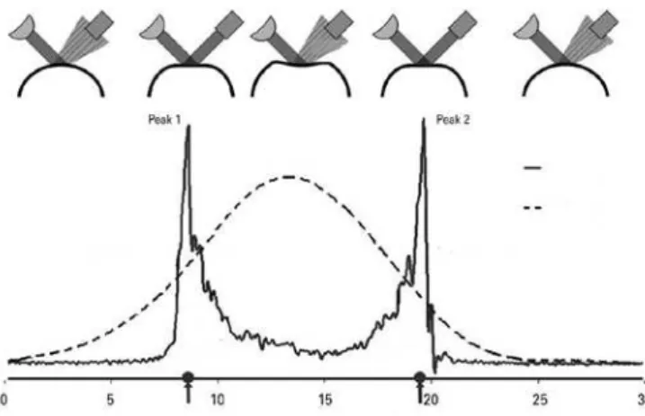
the past three months in children and over the past six months in adults. The exclusion criteria included pre-vious anterior segment surgery, ocular surface problems, corneal scars, and repeated corneal CXL. ORA measure-ments, topographic findings (Pentacam HR, Oculus Optik-geräteGmbH), biomicroscopic findings, uncorrected and corrected distance visual acuity (UDVA and CDVA), and refraction measurements were evaluated preoperatively and 12 months postoperatively. All the measurements were obtained by the same (blinded) technician. The average of three consecutive measurements was used for each parameter. Both UDVA and CDVA were recorded using a Snellen chart. Values were later converted to lo-garithm of minimum angle of resolution (logMAR) values. The ORA is a modified non-contact pneumotonometer that alters corneal curvature via an air pulse and mea-sures aspects of the corneal biomechanical response du-ring an air-puff perturbation. Changes in corneal curvature are evaluated by analyzing the intensity of light reflected by the cornea. As air pressure is gradually increased during each measurement, the cornea begins to flatten. The first applanation occurs when the level of light rea-ching the detector is at its maximum (peak 1= p1). The cornea continues to collapse with increased air pressure and becomes concave at maximum pressure. When this flow of air symmetrically reduces air pressure, the cor-nea moves outward again, and the second applanation occurs (peak 2= p2). This process may be depicted using two plots: an applanation curve and an air pressure curve (Figure 1). The difference b etween the two ap-planation pressures yields the CH value, which indicates the cornea’s degree of viscoelasticity. The CRF is another parameter indicative of corneal viscoelasticity and is obtained via regression analysis of applanation
res and correlates maximally with corneal thickness(7)
(Figure 2). The manufacturer has recently developed new ORA software (version 2.04), which provides 37 new pa-rameters calculated based on the ORA signal waveform. Table 1 describes these second-generation parameters.
Surgical technique
Corneal CXL was performed in the operating room with the patient under topical anesthesia with 0.5%
proparacaine hydrochloride eye drops. Using a smooth spatula, the epithelium was removed from the 8.0-mm treatment zone. Iso-osmolar riboflavin solution (Merribo;
Meran Tıp, Turkey, with dextran, 2 ml 1%) was instilled
in the cornea every two minutes for 30 min. Ultrasound pachymetry (UP-1000, Nidek Co. Ltd.) was performed next. If the corneal thickness was less than 400 µm, one drop of hypoosmolar riboflavin solution (Merribo; Meran
Tıp, Turkey, without dextran, 300 mOsmol/L) was instilled
in the cornea (every 20 s for two minutes). Pachymetry was repeated until the cornea had swollen to thickness >400 µm. The CXL system exposes the cornea to 370-nm
UVA light (Apollon Cross-linking System, Meran Tıp,
Turkey) for 10 min at an irradiance level of 9 mW/cm2.
During the UVA irradiation, riboflavin solution is conti-nually instilled to maintain the corneal saturation balance. At the end of the procedure, the cornea is irrigated with cold water, and a bandage contact lens was applied to minimize the pain.
Statistical analysis
As a result of a priori power analysis via PASS 11 (Power and Sample Size Calculation Software, Version 11), we decided to enroll at least 45 eyes in the study. We enrol-Figure 2. Comparison of corneal hysteresis (CH), corneal resistance factor
(CRF), central corneal thickness corrected CRF and CH after CXL treatment.
Table 1. Description of waveform-derived biomechanical parameters of ocular response analyzer
Parameters Upper 75% peak parameters Upper 50% peak parameters
Area p1 area, p2 area p1 area1, p2 area1
(Areas under peak 1 and peak 2)
Height h1, h2 h11, h21
(Heights of peak1 and peak 2)
Width w1, w2 w11, w21
(Base widths of peak1 and peak 2)
Aspect Ratio aspect1, aspect2 aspect11, aspect21
(Height / width ratios of peak1 and peak 2)
Slope uslope1, dslope1, uslope11, dslope11,
(Base to peak value of peak1 and peak 2) uslope2, dslope2 uslope21, dslope21
Slew Rate slew1, slew2, mslew1, mslew2
-(Aspect ratio of dive 1 and dive 2-maximum single step increase rise of peak 1 and peak 2)
Path path1, path2 path11, path21
(Absolute value of path length around peak 1 and peak 2)
Irregularity aindex, bindex
-(Degree of non-monotonicity of rising and falling edges of peak 1 and peak 2)
Dive dive1, dive2
-(Absolute value of monotonic decrease on downslope part of peak 1 and peak 2)
High Frequency aplhf
led 50 eyes, resulting in study power of 89.2%. Central corneal thickness corrected CH (ccCH) and CRF (ccCRF) calculation methodology have been reported elsewhere(20).
SPSS software, version 20 (IBM, Armonk, NY) was used to perform the statistical analysis. To control the poten-tially confounding effect of CCT, linear regression was applied to other ORA waveform parameters. Transfor-med CRF (DifCRF) and CH (DifCH) were computed as the difference between measured and CCT-predicted CRF and CH, respectively, for each observation in both of the groups. Pearson correlation coefficient was used to eva-luate the correlations among Kmax, elevation anterior, elevation posterior, corneal thickness, and ORA parame-ters. Paired two-tailed Student’s t-test was performed to compare the parameters before vs. after CXL. In multiple comparisons, a correction of the significance level was performed according to the Bonferroni method. For 37 tests comparing the measurements obtained before vs. after CXL, an adjusted p-value of 0.05/37= 0.0013 was considered significant.
RESULTS
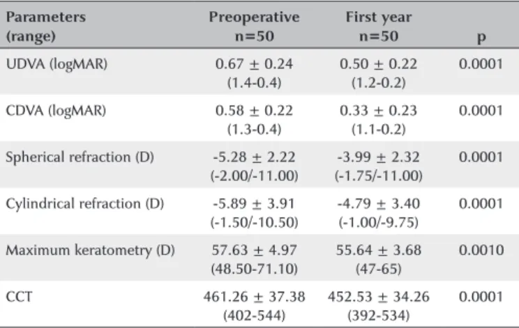
The study included 50 eyes from 45 patients (19 males, 26 females) with mean age of 17.6 ± 3.6 (range 9-25) years. Table 2 presents changes in visual acuity, refraction, and maximum keratometry at one year postoperatively after corneal CXL. Significant improvements in UDVA (0.17 logMAR), CDVA (0.25 logMAR), spherical refraction (1.29 D), cylindrical refraction (1.1 D), and maximum keratometry (1.99 D) were evident (p<0.001 for all).
Table 3 presents the changes in corneal biomechanic parameters after accelerated corneal CXL. Significant
in-Table 2. Preoperative and 1-yr postoperative results for visual acuity, re fraction, corneal thickness, and maximum keratometry
Parameters Preoperative First year
p (range) n=50 n=50
UDVA (logMAR) 0.67 ± 0.24 (1.4-0.4)
0.50 ± 0.22 (1.2-0.2)
0.0001
CDVA (logMAR) 0.58 ± 0.22 (1.3-0.4)
0.33 ± 0.23 (1.1-0.2)
0.0001
Spherical refraction (D) -5.28 ± 2.22 (-2.00/-11.00)
-3.99 ± 2.32 (-1.75/-11.00)
0.0001
Cylindrical refraction (D) -5.89 ± 3.91 (-1.50/-10.50)
-4.79 ± 3.40 (-1.00/-9.75)
0.0001
Maximum keratometry (D) 57.63 ± 4.97 (48.50-71.10)
55.64 ± 3.68 (47-65)
0.0010
CCT 461.26 ± 37.38 (402-544)
452.53 ± 34.26 (392-534)
0.0001
UDVA= uncorrected distance visual acuity; CDVA= corrected distance visual acuity; logMAR= logarithm of the minimal angle of resolution; D= diopter; CCT= central corneal thickness. *P<0.05.
Table 3. Corneal hysteresis, corneal resistance factor, and waveform-derived parameters of baseline and first year after accelerated corneal crosslinking
Parameters Preoperative n=50 First year n=50 p
CH 8.28 ±001.45 8.19 ± 001.70 0.7780
CRF 6.96 ±001.26 6.42 ± 001.64 0.1050
CH-CRF 1.32 ±000.84 1.31 ± 000.74 0.9600
DifCH -0.19 ± 001.31 -0.04 ± 001.33 0.2670
DifCRF -0.17 ± 001.10 -0.05 ± 001.19 0.1210
p1 area 1662.50 ± 686.80 2097.50 ± 588.60 0.0001
p2 area 1004.40 ± 437.50 1434.40 ± 347.80 0.0001
p1 area1 687.10 ± 305.20 716.60 ± 273.60 0.4340
p2 area1 410.90 ± 193.20 444.90 ± 154.00 0.2230
h1 177.60 ± 077.00 186.70 ± 056.90 0.4460
h2 131.90 ± 054.90 181.30 ± 051.00 0.0001
h11 118.40 ± 051.30 123.10 ± 037.90 0.6040
h21 87.90 ± 036.60 94.20 ± 034.00 0.3450
w1 24.13 ±005.92 25.28 ± 007.90 0.3930
w2 21.04 ± 009.67 22.15 ± 007.01 0.5320
w21 9.91 ± 004.53 12.46 ± 005.14 0.0060
w11 12.17 ± 003.96 14.71 ± 004.32 0.0060
aspect1 8.10 ± 004.36 8.14 ± 002.91 0.9060
aspect2 7.75 ± 005.01 7.96 ± 004.34 0.8900
aspect11 11.03 ± 006.12 12.71 ± 004.23 0.0700
aspect21 11.37 ± 008.68 12.77 ± 007.05 0.4320
uslope1 33.47 ± 019.88 38.96 ± 013.28 0.0490
uslope2 38.64 ± 026.05 39.71 ± 021.66 0.7990
dslope1 11.38 ± 006.62 13.68 ± 004.32 0.0180
dslope2 9.84 ± 006.66 10.53 ± 006.00 0.7000
uslope11 32.93 ± 020.90 34.06 ± 013.31 0.6620
uslope21 32.46 ± 025.79 34.63 ± 016.29 0.5820
dslope11 17.52 ± 010.78 19.68 ± 007.91 0.1470
dslope21 16.92 ± 013.97 19.47 ± 012.25 0.2960
slew1 34.26 ± 019.42 35.43 ± 013.42 0.6390
slew2 39.88 ± 025.11 41.36 ± 020.44 0.7230
mslew1 55.12 ± 027.00 56.38 ± 017.87 0.7880
mslew2 54.75 ± 027.68 57.16 ± 027.69 0.7450
path1 22.62 ± 005.19 23.34 ± 005.86 0.587
path2 25.42 ± 007.29 27.17 ± 008.61 0.3030
path11 33.01 ± 008.07 44.08 ± 008.70 0.0050
path21 36.87 ± 011.39 37.49 ± 012.32 0.8190
aindex 8.46 ± 001.98 8.87 ± 002.24 0.4770
bindex 8.40 ± 001.99 8.91 ± 002.44 0.2540
dive1 156.20 ± 079.10 163.20 ± 060.50 0.5810
dive2 95.50 ±051.10 135.50 ± 042.00 0.0001
Aplhf 1.15 ±000.21 1.21 ± 000.15 0.2680
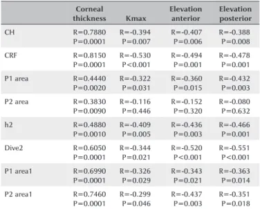
creases in p1 area (26.2%), p2 area (42.8%), h2 (37.7%), and dive2 (41.9%) values were observed (p<0.001 for all); however, no significant differences in CH, CRF, difCH, or difCRF or other waveform-derived parameters were detected at one year postoperatively (p=0.778, p=0.105, p=0.267, p=0.121, respectively). We found positive cor-relations between CH, CRF, p1 area, p2 area, dive2, h2, p1 area1, p2 area1, and corneal thickness. On the other hand, there was a negative correlation between CH, CRF, p1 area, dive2, h2, p1 area1, p2 area1, on one hand, and Kmax, elevation anterior, and elevation posterior, on the other. Table 4 presents statistically significant data.
DISCUSSION
This study evaluated changes in corneal biomechanical properties after accelerated corneal CXL using second-ge-neration waveform-derived parameters. Although no sta-tistically significant difference in CRF or CH was observed, significant increases in p1 area, p2 area, h2, and dive2 parameters were observed. To the best of our knowledge, this is the first study to use these parameters in evalua-ting the effects of accelerated corneal CXL on corneal biomechanics.
Topographic measurements are used to indirectly assess the effects of corneal CXL. It is essential to evaluate the biomechanical changes of the cornea directly in order to evaluate the efficacy of corneal CXL. Currently,
ORA is the device used most widely for evaluating in vivo corneal biomechanical properties. Although the biome-chanical resistance of the cornea was found to increase after corneal CXL in ex vivo studies(10-12), many studies
that used ORA showed that the CRF and the CH remained unchanged after standard or accelerated CXL(13,14,21,22).
There may be several reasons why CRF and CH may not be used to detect biomechanical changes after corneal CXL. First, the biomechanical change may be too small to be detected using the CRF and CH measurements. Second, corneal CXL may change the eye’s elasticity and viscosity(23). The low CRF and CH values observed in
keratoconus are caused by changes in proteoglycan and glycosaminoglycan structure(24). Thus, after corneal CXL,
new covalent bonds form between collagen fibers(25). In
a review, Gatinel suggested that differences between these mechanisms may explain why the CRF and the CH are insufficient to detect biomechanical changes(26).
Accordingly, CXL treatment does not affect corneal bio-mechanics. Any changes to the corneal detected after CXL have been caused by epithelial wound healing. However, we found a strong positive correlation between corneal thickness and CRF and CH. After CXL, there was a decrease in CRF and CH values and an increase in DifCH and Dif CRF values. This may indicate that corneal stiffness tends to increase after CXL. However, this trend was not statistically significant. In addition, we found negative correlations between Kmax, elevation anterior, elevation posterior, and ORA parameters. Although li-near regression did not reveal any significant difference, the results presented above show clearly that corneal shape and thickness affect ORA parameters.
Therefore, new in vivo methods to detect such bio-mechanical changes are being studied. One such method is applanation resonance tonometry. Using applanation resonance tonometry, Rehnman et al.(27) showed that CH
increases after conventional corneal CXL. In another study using inverse finite element modeling, corneal stiffness increased after corneal CXL(28).
The new ORA software provides 37 parameters, allowing for more detailed analysis of the corneal de-formation signal waveform. Each parameter describes a morphologic feature of the waveform. Many studies have shown that these parameters are useful in distinguishing between keratoconus and normal patients, as well in identifying early and severe keratoconus(15-17). Spoerl et
al.(18) showed a significant increase in p2 area, h2, and
dive2 parameters at the one year follow-up after stan-dard CXL treatment. They found that the most prominent Table 4. Correlation between cornea-related parameters and ORA
pa-ra meters in patients with kepa-ratoconus
Corneal thickness Kmax Elevation anterior Elevation posterior CH R=0.7880 P=0.0001 R=-0.394 P=0.007 R=-0.407 P=0.006 R=-0.388 P=0.008 CRF R=0.8150 P=0.0001 R=-0.530 P<0.001 R=-0.494 P=0.001 R=-0.478 P=0.001
P1 area R=0.4440 P=0.0020 R=-0.322 P=0.031 R=-0.360 P=0.015 R=-0.432 P=0.003
P2 area R=0.3830 P=0.0090 R=-0.116 P=0.446 R=-0.152 P=0.320 R=-0.080 P=0.632 h2 R=0.4880 P=0.0010 R=-0.409 P=0.005 R=-0.436 P=0.003 R=-0.466 P=0.001 Dive2 R=0.6050 P=0.0001 R=-0.344 P=0.021 R=-0.520 P<0.001 R=-0.551 P<0.001
P1 area1 R=0.6990 P=0.0001 R=-0.326 P=0.029 R=-0.343 P=0.021 R=-0.363 P=0.014
P2 area1 R=0.7460 P=0.0001 R=-0.299 P=0.046 R=-0.437 P=0.003 R=-0.351 P=0.018
increase was detected in the p2 area (35%), with values approaching those observed for healthy corneas. Spoerl et al.(18) suggested that an increase in p2 area could
indi-cate an improvement in the corneal shear stiffness. The authors argued that this parameter is more successful than CRF and CH for visualizing changes in corneal bio-mechanics. In a study by Vinciguerra et al.(14), a signi
fi-cant increase in peak 1 and peak 2 was observed in the first year after conventional corneal CXL. Similarly, in our study, increases in p1 area, p2 area, h2, and dive2 were observed after accelerated corneal CXL. The rate of increase in peak 2-related parameters is about 40%, which is more pronounced than the rate of increase in peak 1-related parameters. This ratio is close to the rate of increase reported by authors who performed an ex vivo study (33%). Increases in stiffness after CXL in hu-man donor cornea were assessed with optical coherence elastography(12).
An increase in the intensity of light reaching the de-tector manifests as elevation at peaks. After corneal CXL, decreased aberrations, increased homogeneity, and corneal flattening may increase the amount of light reflected from the cornea. Increases in peak height (h2) and the area under the peak (p1 area, p2 area) may be related to the increase in reflected light. Hallahan et al.(19)
followed 24 eyes for three months after conventional cor-neal CXL, and no significant difference was found in any of the 37 parameters investigated. In this study, the small sample size and short follow-up (three months) preven-ted the measurements of any increase in biomechanical resistance after corneal CXL. Notably, epithelial remo-deling during the first few months postoperatively may alter corneal biomechanics and cause light scattering throughout the cornea, which may lead to inaccurate measurements(14,29). In a study by Vinciguerra et al.(14),
in-creases in peak 1 and peak 2 were not significant during the early postoperative period but became significant at six months.
Although the new second-generation waveform pa-rameters are not real measured biomechanical parame-ters, the signal provided by biomechanical waveform analysis provides a morphologically unique fingerprint for each eye and may contain valuable clinical informa-tion(30). Therefore, these parameters could be a sensitive
indicator of corneal morphology. Waveform-derived parameters may be considered to be more sensitive than CRF and CH in visualizing changes in corneal biomecha-nics after CXL.
Clinical studies have shown that corneal CXL is effective in improving visual acuity, reducing corneal steepness, and stabilizing keratoconus. Changes in visual acuity and corneal topography caused by accelerated CXL treatment have similarly been reported by other studies in the literature(31,32). Thus, the accelerated corneal CXL
proto-col may have succeeded in discontinuing and partially reversing the progression of keratoconus.
In conclusion, parameters p1 area, p2 area, h2, and dive2 appear to be more sensitive than CRF and CH for detecting corneal biomechanical changes after accelerated corneal CXL. Therefore, these parameters may help to determine which CXL method is more effective for increa-sing biomechanical resistance of the cornea.
REFERENCES
1. Rabinowitz YS. Keratoconus. Surv Ophthalmol. 1998;42(4):297-319. 2. Touboul D, Bénard A, Mahmoud AM, Gallois A, Colin J, Roberts
CJ. Early biomechanical keratoconus pattern measured with an ocular response analyzer: curve analysis. J Cataract Refract Surg. 2011;37(12):2144-50.
3. Wollensak G, Spoerl E, Seiler T. Riboflavin/ultraviolet-a-induced colla-gen crosslinking for the treatment of keratoconus. Am J Ophthalmol. 2003;135(5):620-7.
4. Kymionis GD, Grentzelos MA, Kankariya VP, Liakopoulos DA, Portaliou DM, Tsoulnaras KI, et al. Safety of high-intensity corneal collagen crosslinking. J Cataract Refract Surg. 2014;40(8):1337-40. 5. Schumacher S, Oeftiger L, Mrochen M. Equivalence of biomecha-nical changes induced by rapid and standard corneal cross-linking, using riboflavin and ultraviolet radiation. Invest Ophthalmol Vis Sci. 2011;52(12):9048-52.
6. Gatzioufas Z, Richoz O, Brugnoli E, Hafezi F. Safety profile of high-fluence corneal collagen cross-linking for progressive kerato-conus: preliminary results from a prospective cohort study. J Refract Surg. 2013;29(12):846-8.
7. Luce DA. Determining in vivo biomechanical properties of the cor-nea with an ocular response analyzer. J Cataract Refract Surg. 2005; 31(1):156-62.
8. Fontes BM, Ambrósio R Jr, Velarde GC, Nosé W. Ocular response analyzer measurements in keratoconus with normal central corneal thickness compared with matched normal control eyes. J Refract Surg. 2011;27(3):209-15.
9. Touboul D, Roberts C, Kérautret J, Garra C, Maurice-Tison S, Sau-busse E, et al. Correlations between corneal hysteresis, intraocular pressure, and corneal central pachymetry. J Cataract Refract Surg. 2008;34(4):616-22.
10. Wollensak G, Spoerl E, Seiler T. Stress-strain measurements of human and porcine corneas after riboflavin-ultraviolet-A-induced cross-linking. J Cataract Refract Surg. 2003;29(9):1780-5.
11. Wollensak G, Iomdina E. Long-term biomechanical properties of rabbit cornea after photodynamic collagen crosslinking. Acta Ophthalmol. 2009;87(1):48-51.
13. Goldich Y, Barkana Y, Morad Y, Hartstein M, Avni I, Zadok D. Can we measure corneal biomechanical changes after collagen cross-lin king in eyes with keratoconus?-a pilot study. Cornea. 2009; 28(5):498-502.
14. Vinciguerra P, Albè E, Mahmoud AM, Trazza S, Hafezi F, Roberts CJ. Intra- and postoperative variation in ocular response analyzer parameters in keratoconic eyes after corneal cross-linking. J Refract Surg. 2010;26(9):669-76.
15. Luz A, Lopes B, Hallahan KM, Valbon B, Ramos I, Faria-Correia F, et al. Enhanced Combined Tomography and Biomechanics Data for Distinguishing Forme Fruste Keratoconus. J Refract Surg. 2016; 32(7):479-94.
16. Luz A, Lopes B, Hallahan KM, Valbon B, Fontes B, Schor P, et al. Discriminant value of custom ocular response analyzer waveform derivatives in forme fruste keratoconus. Am J Ophthalmol. 2016; 164:14-21.
17. Ventura BV, Machado AP, Ambrósio R Jr, Ribeiro G, Araújo LN, Luz A, et al. Analysis of waveform-derived ORA parameters in early forms of keratoconus and normal corneas. J Refract Surg. 2013; 29(9):637-43.
18. Spoerl E, Terai N, Scholz F, Raiskup F, Pillunat LE. Detection of bio-mechanical changes after corneal cross-linking using Ocular Response Analyzer software. J Refract Surg. 2011;27(6):452-7.
19. Hallahan KM, Rocha K, Roy AS, Randleman JB, Stulting RD, Dupps WJ Jr. Effects of corneal cross-linking on ocular response analyzer waveform-derived variables in keratoconus and postrefractive surgery ectasia. Eye Contact Lens. 2014;40(6):339-44.
20. Galletti JG, Pförtner T, Bonthoux FF. Improved keratoconus de-tection by ocular response analyzer testing after consideration of corneal thickness as a confounding factor. J Refract Surg. 2012; 28(3):202-8.
21. Hashemi H, Fotouhi A, Miraftab M, Bahrmandy H, Seyedian MA, Amanzadeh K, et al. Short-term comparison of accelerated and standard methods of corneal collagen crosslinking. J Cataract Re-fract Surg. 2015;41(3):533-40.
22. Tomita M, Mita M, Huseynova T. Accelerated versus conventional corneal collagen crosslinking. J Cataract Refract Surg. 2014;40(6): 1013-20.
23. Glass DH, Roberts CJ, Litsky AS, Weber PA. A viscoelastic biome-chanical model of the cornea describing the effect of viscosity and elasticity on hysteresis. Invest Ophthalmol Vis Sci. 2008;49(9): 3919-26.
24. Akhtar S, Bron AJ, Salvi SM, Hawksworth NR, Tuft SJ, Meek KM. Ultrastructural analysis of collagen fibrils and proteoglycans in keratoconus. Acta Ophthalmol. 2008;86(7):764-72.
25. Spoerl E, Wollensak G, Seiler T. Increased resistance of crosslinked cornea against enzymatic digestion. Curr Eye Res. 2004;29(1):35-40. 26. Gatinel D. Reevaluating the effectiveness of corneal collagen
cross-linking and its true biomechanical effect in human eyes. Int J Keratoconus Ectatic Corneal Dis. 2017;6(1):34-41.
27. Beckman Rehnman J, Behndig A, Hallberg P, Lindén C. Increased corneal hysteresis after corneal collagen crosslinking: a study based on applanation resonance technology. JAMA Ophthalmol. 2014;132(12):1426-32.
28. Sinha Roy A, Rocha KM, Randleman JB, Stulting RD, Dupps WJ Jr. Inverse computational analysis of in vivo corneal elastic modulus change after collagen crosslinking for keratoconus. Exp Eye Res. 2013;113:92-104. Erratum in: Exp Eye Res. 2016;145:472. 29. Koç M, Uzel MM, Koban Y, Durukan I, Tekin K, Ylmazbaş P.
Comparison of results of accelerated corneal cross-linking with hypo-osmolar riboflavin solution performed on corneas thicker and thinner than 400 µm. Cornea. 2016;35(2):151-6.
30. Franco S, Lira M. Biomechanical properties of the cornea measu-red by the Ocular Response Analyzer and their association with intraocular pressure and the central corneal curvature. Clin Exp Optom. 2009;92(6):469-75.
31. Elbaz U, Shen C, Lichtinger A, Zauberman NA, Goldich Y, Chan CC, et al. Accelerated (9-mW/cm2) corneal collagen crosslinking
for keratoconus-A 1-year follow-up. Cornea. 2014;33(8):769-73. 32. Koc M, Uzel MM, Tekin K, Kosekahya P, Ozulken K, Yilmazbas P.