Abstract
Submitted: May 9, 2017 Modification: October 31, 2017 Accepted: November 22, 2017
Immediate effects of temporary
bite-raising with light-cured
orthodontic band cement on the
electromyographic response of
masticatory muscles
Objective: To assess the immediate effects of temporary bite-raising using light-cured orthodontic band cement on the superficial masseter and anterior temporalis electromyography (EMG) activity in healthy adults. Materials and Methods: Surface EMG signals were recorded bilaterally from the superficial masseter and anterior temporalis muscles of 30 volunteers with a normal occlusion, before and after having temporary bite-raising. The bite-raising was done by adding light-cured orthodontic band cement (3x5x2 mm WxLxH) on the lingual cusps of both upper first molars. The measurements were recorded (i) at rest, (ii) while clenching in centric occluding position and (iii) while chewing on an artificial test food. The EMG activity at rest and during clenching, the maximum voltage, and the duration of the identified EMG signal burst while chewing the artificial test food before and after temporary bite-raising were statistically compared using the paired t-test or the Wilcoxon signed-rank test based on the normality of the variables. The significance level was set at 5%. Results: After temporary bite-raising, we found no significant change in integral EMG activity at rest position for the superficial masseter (mean difference (MD)=7.5 μVs) and for the anterior temporalis muscle (MD=36.8 μVs); however, the integral EMG activity during clenching was significantly reduced for the superficial masseter (MD=201.2 μVs) and for the anterior temporalis muscle (MD=151.8 μVs). During mastication, the maximum voltage of the identified burst was significantly reduced on the preferred chewing side of the superficial masseter and anterior temporalis muscles (MD=127.9 and 47.7 μV, respectively), while no significant change was found for the duration of the identified burst (MD=-34.1 and 3.4 ms, respectively) after temporary bite-raising. Conclusion: The results point to an altered neuromuscular behavior during clenching and chewing immediately after temporary bite-raising with light-cured orthodontic band cement. This information is relevant for orthodontists to inform their patients what will happen to their masticatory muscle activity when this bite-raising method is used.
Keywords: Electromyography. Masticatory muscles. Vertical dimension.
Orthodontics. Darin PATIVETPINYO1
Weera SUPRONSINCHAI2 Chidsanu CHANGSIRIPUN1
1 Faculty of Dentistry, Department of Orthodontics, Chulalongkorn University, Bangkok, Thailand. 2 Faculty of Dentistry, Department of Physiology, Chulalongkorn University, Bangkok, Thailand.
Corresponding address: Chidsanu Changsiripun Department of Orthodontics, Faculty of Dentistry,
Chulalongkorn University, Henri-Dunant Road, Wangmai, Patumwan, Bangkok, Thailand 10330. Phone: +66 2 218 8932 - Fax: +66 2 218 8953
Introduction
Temporary bite-raising or bite-opening is often
needed in patients undergoing orthodontic treatment,
such as in deep overbite and/or crossbite cases.
Bite-raising is used to prevent the brackets from being
sheared off, and to eliminate occlusal interferences,
allowing unobstructed tooth movement18. Moreover,
bite-raising allows the proper positioning of the
brackets during the early treatment phase by keeping
specific groups of teeth out of occlusion and preventing
full jaw closure14. One method of performing
bite-raising is adding a light-cured orthodontic band cement
to the occlusal surfaces of the posterior molars or
lingual surfaces of the anterior teeth1,16. This procedure
is hygienic, minimizes bulkiness, reduces interference
with speech, and is less intrusive on the tongue space
compared to a conventional removable bite plate. In
addition, when bite-raising is used in patients with
multi-brackets on every tooth, the orthodontic tooth
movement is done without interference from the
acrylic plate, and is easy to place on the tooth surfaces
in one visit. Build-ups for posterior teeth using this
method were recently reported to be an effective
alternative treatment for anterior open bite in adults23.
Several studies indicate that increased occlusal jaw
opening may lead to changes on the electromyographic
(EMG) activity of the jaw muscles and there are
several studies on the consequence of bite-raising
by other means on the EMG activity4,11. However, we
do not know of any published studies on the effects
of opening the bite by adding light-cured orthodontic
band cement on the occlusal surfaces of the posterior
teeth, on jaw muscle activity. Despite the increase on
the vertical dimension of this method being similar to
other methods, it results in only two occlusal contact
areas. This might affect the EMG activity of the jaw
muscles differently.
The objective of this study was to examine the
immediate masseter and temporalis muscle EMG
activity before and after opening the bite with
light-cured orthodontic band cement on the lingual cusps of
both upper first molars during physiologic rest position, maximum voluntary clenching (MVC), and chewing on artificial test food in normal occlusion participants.
The null hypothesis was that bite-raising using this
method does not immediately affect the masseter or
temporalis muscle EMG activity at rest, during MVC,
and chewing.
Materials and methods
Participants
Forty-two volunteers, randomly recruited from
Dentistry students of the Chulalongkorn University,
received a clinical examination. Twelve volunteers
were excluded due to having an edentulous area
(n=2), overjet >3 mm (n=4), overbite >3 mm (n=5), or open bite (n=1). Thus, 30 volunteers (11 men, 19 women; age range from 18–25 years) participated in
this study. The participants were dentate with a Class I
occlusion, an overjet less than 3 mm, and an overbite
less than 3 mm (mean overbite 2.35±0.65 mm, mean
± S.D.). The participants had no edentulous area, no
significant facial deformity, and no temporomandibular
symptoms based on the assessment protocol of
Schiffman, et al.19 (2016). The participants had no
previous orthodontic treatment in the past 3 years.
No participant missed an appointment or had dental
treatment during the experimental period. The study
protocol was approved by the Human Research Ethics
Committee of the Faculty of Dentistry, Chulalongkorn
University (HREC-DCU 2015-090). All participants
signed informed consent forms.
Protocol for measuring muscle activity
The EMG measurements were performed on two
appointments, with a week interval between each.
The objective of the study and the measurement
procedure were explained to the participants on the
first appointment, we also measured the EMG activity
without any intervention.
During the second appointment, we performed a
baseline recording at rest and during MVC. The bite
of the participant was then temporarily raised by
placing light-cured orthodontic band cement on the
lingual cusp of the upper first molars. After polishing,
rinsing, drying, and isolating the teeth, Ultra
Band-Lok BLUE (Reliance Orthodontic Products, Inc., Itasca,
IL) was applied in a thermoplastic sheet template
(3x5x2 mm WxLxH trapezoid-shaped slot) and placed
on the tooth. We used a light-curing unit (600 mW
continuous output) to cure the material. The occlusion
was balanced by identifying an occlusal contact on
each side with shimstock foil and locating the specific contact with the eight-micron thick articulating film
according to the technique described by Anderson,
Schulte and Aeppli3 (1993). Representative intraoral
are shown in Figure 1. The mean overbite after
temporary bite-raising was -0.57±0.70 mm (mean±
S.D.). Consequently, the mean interincisal distance
change was 2.90±0.20 mm (mean ± S.D.). After the
EMG recordings, the bite-raising material was removed
using adhesive removing pliers.
Surface EMG recordings
Surface EMGs were simultaneously recorded from
the left and right superficial masseter and anterior
temporalis muscles by a researcher using the ML866
PowerLab 4/35 (ADInstruments Pty Ltd., Bella Vista,
Australia), following the protocol indicated by the
manufacturer. The participants were seated upright
on a chair with their trunk perpendicular to the floor, both feet on the floor, hands resting on their lap, and
looking forward with their head unsupported in a quiet
place listening to relaxing music20. We cleaned the skin
with isopropyl alcohol (70%), and gently applied a
small amount of abrasive gel (ADInstruments Pty Ltd.)
on it, which lowered the electrode-skin impedance
at the electrode site with minimal discomfort or
irritation. Self-adhesive disposable ECG electrodes
(ADInstruments Pty Ltd.) were placed bilaterally on
the skin of the subject overlying the muscles and fixed
with Nexcare Sensitive Skin Tape (3M, St. Paul, MN).
For the anterior temporalis muscles, the electrodes
were placed 1.5–2 cm posterior to the lateral canthus
of the eye, slightly above the zygomatic arch. For
the superficial masseter muscles, the electrodes
were placed on the midpoint of the bisection of the
anteroposterior and inferosuperior dimensions of the
muscle belly, which was determined by asking the
subject to clench the teeth according to the method
described by Teenier, Throckmorton and Ellis21 (1991).
The inter-electrode distances were recorded for each
subject for the left and right sides and used to place the
electrode in the latter phase. A ground electrode was
placed on the sternal end of the clavicle. External noise
was controlled to avoid artifacts caused by smiling or
other facial expressions.
After placing the electrodes and practicing for the
measurement, the subjects were given a five minutes
rest period to relax and for the skin to absorb the
conductive gel. The EMG recordings were then made
as follows:
Test A - physiological rest position
The purpose of this test was to monitor and
quantify the amount of electrical activity generated
by the superficial masseter and anterior temporalis
muscles when at rest.
Test B - maximum voluntary clench (MVC)
The subjects were instructed to clench their teeth
in centric occluding position using as much force as
possible without causing pain and to hold this force
intensity until instructed to relax after 2 seconds.
Test C - mastication
The subjects were instructed to chew an artificial
test food for 20 strokes on their preferred chewing
side. When the subjects were not sure of their
preferred chewing side, they were instructed to use
the right side. The artificial test food was made with
a dental impression material, OptoSil(Heraeus Kulzer
GmbH, Hanau, Germany), using the standardized
production of an artificial test food protocol2 and cut into four quarters.
We performed two experimental sessions as
recommended in a previous study8, the first session
was for the participants to become used to the EMG
measurement procedure and the second session was
for data collection. The data obtained in the second
session were used for analysis. For test A and test
B, the measurements were repeated three times
and each recording took 6–8 seconds for test A and
2 seconds for test B followed by a one minute rest
period21. For test C, the measurement was done while
chewing the artificial test food.
The digital signal was analyzed using LabChart
software (ADInstruments Pty Ltd.). For test A and B,
the area of the integral EMG curve (duration x mean
voltage, μVs), was computed for each muscle. The left
and right sides values were pooled for each muscle.
The mean integral EMG activity of the three trials for
each muscle was expressed as mean and standard
deviation. To normalize the EMG signals, the
root-mean-square (RMS) value of the EMG signals during
the 2 seconds window was calculated and expressed
as the percentage of the EMG activity during MVC in
centric occlusion.
For test C, the signal was full-wave rectified and smoothed by taking the RMS for each 100 ms interval. The start and the end of each burst were defined as the EMG signal reaching a level 10% above or below
the mean area relative to the baseline signal9. The
maximum voltage (μV) and duration (ms) of the identified burst were computed for each muscle. The first chewing cycle was excluded from the statistical
analysis since research suggests that subjects use
the first chewing cycle to gather information about an
item15. The mean of cycles 2–5 were used for statistical
comparisons.
Intra-examiner reliability
The baseline recordings for both muscles at rest
were used to evaluate intra-examiner reliability, they
were conducted in two visits, with a week interval
between them. Both recordings were performed by
the same examiner and in the same period of the day,
according to the procedure mentioned above.
Statistical analysis
Statistical analysis was performed using the
software SPSS version 17.00 (SPSS Inc., Chicago, IL, USA). The normality of the variables was verified
by the Kolmogorov-Smirnov test. For comparisons
between before and after bite-raising, the paired t-test
or the Wilcoxon signed-rank test was used based on
the normality of the data. We tested 16 pairs of
before-after data. The paired t-test was performed for the
integral superficial masseter and anterior temporalis muscles EMG activity during MVC, the normalized
superficial masseter muscle EMG activity during
MVC, the maximum voltage of the identified burst on the superficial masseter preferred chewing side, the duration of the identified burst on the superficial
masseter and anterior temporalis preferred chewing
side, and the duration of the identified burst on the
anterior temporalis non-chewing side. The Wilcoxon
signed-rank test was performed on the integral
superficial masseter and anterior temporalis muscles EMG activity during rest, the normalized superficial
masseter and anterior temporalis muscles EMG activity
during rest, the normalized anterior temporalis muscle
EMG activity during MVC, the maximum voltage of the identified burst on the superficial masseter non-chewing side, the maximum voltage of the identified
burst on the anterior temporalis preferred chewing
side and non-chewing side, and the duration of the
identified burst on the superficial masseter non-chewing side. Significant differences were defined
as p<0.05. The effect size was calculated for the
significant comparisons [(effect size=SD(Δ)*δ/√N; α=0.05, β=0.20, N=Sample size, SD(Δ)=Standard
Deviation of the change in the outcome, the standard
T value (with degrees of freedom) corresponding to
α=2.05 and parameter (δ)=2.90)]17. Intra-examiner reliability between the two visits was evaluated by
intraclass correlation. The intraclass correlation
coefficient (ICC) was calculated based on a single
rating, absolute-agreement, 2-way mixed-effects
model. ICC values <0.5 indicate poor reliability, values
between 0.5–0.75 indicate moderate reliability, values between 0.75–0.9 indicate good reliability, and values >0.9 indicate excellent reliability12.
Results
The intraclass correlation coefficient for the
superficial masseter and anterior temporalis muscles measurements were 0.93 and 0.91, respectively,
indicating an excellent reproducibility of the EMG
measurements. No significant differences were
found between the superficial masseter integral EMG
activity or normalized EMG activity before and after
also found for the anterior temporalis muscle at rest.
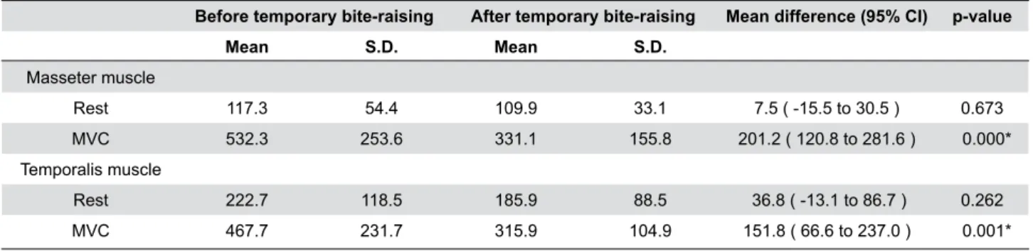
However, integral EMG activity and normalized EMG
activity for both muscles was significantly reduced during clenching after temporary bite-raising (Table 1
and 2). The effect size of the integral and normalized
EMG activity for the superficial masseter muscle was 114.0 μVs and 16.7%, respectively. The effect size
of the integral and normalized EMG activity for the
anterior temporalis muscle was 120.8 μVs and 21.4%,
respectively.
Representative EMG recordings for the superficial
masseter and anterior temporalis muscles during
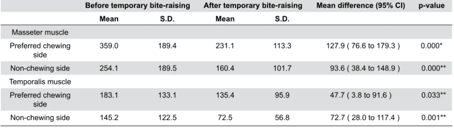
mastication are shown in Figure 2. The maximum
voltage in both muscles was significantly reduced on
both the preferred chewing side and non-chewing side
after temporary bite-raising (Table 3). The effect size
of the maximum voltage for the superficial masseter
muscle on the preferred chewing side and the
non-Before temporary bite-raising After temporary bite-raising Mean difference (95% CI) p-value
Mean S.D. Mean S.D.
Masseter muscle
Rest 117.3 54.4 109.9 33.1 7.5 ( -15.5 to 30.5 ) 0.673 MVC 532.3 253.6 331.1 155.8 201.2 ( 120.8 to 281.6 ) 0.000* Temporalis muscle
Rest 222.7 118.5 185.9 88.5 36.8 ( -13.1 to 86.7 ) 0.262 MVC 467.7 231.7 315.9 104.9 151.8 ( 66.6 to 237.0 ) 0.001*
CI: confidence interval; MVC: maximum voluntary clench *Significant difference (p<0.05, paired t-test)
Table 1- Integral electromiography (EMG) activity (μVs) during rest and MVC before and after temporary bite-raising
Before temporary bite-raising After temporary bite-raising Mean difference (95% CI) p-value
Mean S.D. Mean S.D.
Masseter muscle
Rest 24.4 16.8 23.2 13.8 1.2 ( -2.8 to 5.1 ) 0.572 MVC 104.0 22.4 73.8 30.7 30.2 ( 18.4 to 42.0 ) 0.000* Temporalis muscle
Rest 38.8 25.5 35.8 17.2 3.0 ( -5.4 to 11.4 ) 0.704 MVC 109.0 28.6 76.5 36.6 32.4 ( 17.3 to 47.5 ) 0.000**
CI: confidence interval; MVC: maximum voluntary clench *Significant difference (p<0.05, paired t-test)
**Significant difference (p<0.05, Wilcoxon Signed Ranks Test)
Table 2- Normalized electromiography (EMG) activity (%) during rest and MVC before and after temporary bite-raising
Figure 2- Representative masseter and temporalis muscle electromiography (EMGs)during mastication (test C) before and after temporary
chewing side was 72.8 μV and 78.3 μV, respectively.
The effect size of the maximum voltage for the anterior
temporalis muscle on the preferred chewing side
and the non-chewing side was 62.2 μV and 63.3 μV, respectively. However, no significant difference was found for its duration (Table 4).
Discussion
This study investigated the immediate effect of
temporary bite-raising using light-cured orthodontic
band cement on the occlusal surface of the upper first molars on the EMG activity of the superficial masseter
and anterior temporalis muscles. We found that there
was no change in EMG activity at rest, however, EMG
activity was reduced during MCV and chewing. Based
on these results, the null hypothesis was not rejected
at rest, but it was rejected during MVC and chewing.
Except for sex, the factors affecting masticatory
muscle EMG activity, such as age, facial type,
malocclusion, and the EMG recording period, were
controlled in this study5,25. Men and women were not
on even numbers in our study. However, the paired
t-test or Wilcoxon Signed-Ranks Test was used for
within-subject and within-muscle comparisons, thus,
the variable EMG responses between participants were
eliminated.
The difference was not significant on EMG activity
for both muscles at rest, despite being reduced
immediately after temporary bite-raising. The cause
for this could be the opening distance chosen for
this study, set at 2.5–3 mm, which is close to the
physiological rest position and is usually adequate for
treating orthodontic patients. However, several studies
found that increasing the occlusal vertical dimension
by ≥3 mm by other means affects the EMG activity of the superficial masseter and anterior temporalis
muscles4,11. Therefore, the EMG response to
bite-raising by this method at greater opening distances
should be further investigated.
The EMG activity of both muscles during MVC decreased significantly after temporary bite-raising.
As reported by Jimenez10 (1987), if the occlusion does
not result in mandible stability, the jaw-closing muscles
will contribute to the stabilization by reducing its EMG
activity to avoid damage to other structures. There
were only two occlusal contact areas after temporary Before temporary bite-raising After temporary bite-raising Mean difference (95% CI) p-value
Mean S.D. Mean S.D.
Masseter muscle Preferred chewing
side 359.0 189.4 231.1 113.3 127.9 ( 76.6 to 179.3 ) 0.000* Non-chewing side 254.1 189.5 160.4 101.7 93.6 ( 38.4 to 148.9 ) 0.000** Temporalis muscle
Preferred chewing
side 183.1 133.1 135.4 95.9 47.7 ( 3.8 to 91.6 ) 0.033** Non-chewing side 145.2 122.5 72.5 56.8 72.7 ( 28.0 to 117.4 ) 0.001**
CI: confidence interval
*Significant difference (p<0.05, paired t-test)
**Significant difference (p<0.05, Wilcoxon Signed Ranks Test)
Table 3- Maximum voltage (μV) of the identified burst during mastication before and after temporary bite-raising
Before temporary bite-raising After temporary bite-raising Mean difference (95% CI) p-value
Mean S.D. Mean S.D.
Masseter muscle
Preferred chewing side 410.3 111.5 444.4 101.9 -34.1 ( -80.1 to 11.9 ) 0.141 Non-chewing side 393.0 122.3 443.6 129.9 -50.6 ( -110.3 to 9.1 ) 0.094 Temporalis muscle
Preferred chewing side 427.7 129.8 424.3 137.4 3.4 ( -49.5 to 56.3 ) 0.897 Non-chewing side 392.2 105.2 365.4 120.5 26.8 ( -19.8 to 73.4 ) 0.249
CI: confidence interval
bite-raising on this study. Despite the adjustments on
bite-raising to have even and simultaneous contact
on both sides, it might not be sufficient to produce
mandible stability. This may have resulted in reduced
EMG activity10. Ferrario, et al.7 (2002) found that the
number of occlusal contacts and masseter and anterior
temporalis muscle activity during MVC was significantly
related in young adults. This may explain why the
participants of this study had lower EMG activity during
MVC after temporary bite-raising, when compared with
the values before temporary bite-raising.
Our results are similar those of Dahlstrom and
Haraldson6 (1989), who investigated the superficial
masseter and anterior temporalis muscles EMG activity
using bite plates and suggested that the reduced EMG
activity they observed was probably due to the smaller
occlusal contacts on the bite plates. Chandu, et al.4
(2004) reported similar results with interocclusal
appliances that were constructed by pressure forming
a laminate base and adding posterior acrylic bite blocks
to increase the vertical dimension. However, Wang,
et al.24 (2013) reported an association between jaw
muscle EMG activity and bite force during occluding
movement where greater EMG activity was associated
with a greater bite force. The limited occlusal contact
area results in an uneven distribution of the occlusal
force. Thus, the decreased EMG activity in our study
might result from a reduced bite force by participants
afraid of damaging their teeth. However, we did not
evaluate bite force, thus we cannot conclude that
the decreased EMG activity was due to reduced
bite force. Further investigation including bite force
measurements may assist in defining the cause of the reduced EMG activity during MVC after bite-raising
with this method. The decreased EMG activity after
bite-opening was greater for the superficial masseter
compared to the anterior temporalis, which is consistent
with the study of Koc, et al.11 (2012). This may result
from the different muscle locations and orientations,
resulting in these muscles being physically affected
by changes in jaw position in a different manner.
We do not know of any other studies comparing the
effects on masticatory muscle EMG activity during
mastication after having temporary bite-raising by
adding light-cured orthodontic band cement on the
occlusal surfaces of upper posterior molars. Thus,
comparing our results to previous studies is difficult.
Since bite-raising by this method caused a reduced
occlusal contact area, our results showed that the
superficial masseter and anterior temporalis muscles
maximum voltage on the preferred chewing side was
significantly reduced after temporary bite-raising. This
is consistent with the study of Tomonari, et al.22 (2014)
who found lower EMG activity for both muscles during
chewing on the preferred chewing side of subjects
with reduced occlusal surface contacts. The maximum
voltage for both muscles was significantly reduced on
the non-chewing side after temporary bite-raising. This
could be the result from a protective mechanism to
control jaw balance, since chewing on one side could
cause the mandible to deform and/or tilt around the
sagittal axis13. The increase on interocclusal distance
would cause similar muscle activity on both sides,
which might lift the mandible on the non-chewing side,
causing excessive temporomandibular joint loading.
The short period of investigation is a limitation
of this study. Habituation should be considered
when evaluating the effect of bite-raising, since
muscle physiology and function may adapt if
longer observation periods are allowed. In animal
experiments, bite-raising for 2 weeks significantly reduced masseter muscle spindle sensitivity; however, no significant differences were found between control
and after more than 6 weeks of bite-raising26. However,
a long-term investigation with healthy subjects was not
possible in our study model due to ethical standards.
Therefore, to investigate the precise effect of
bite-raising by this method on masticatory muscle activity,
a future clinical study should evaluate subjects over
an extended period.
Conclusion
Our results revealed that temporary bite-raising
by placing orthodontic band cement on the occlusal
surface of the upper first molars had no immediate effect on EMG activity at rest, however, superficial
masseter and anterior temporalis muscles EMG
activity reduced during MVC and mastication. This
information is useful for orthodontists to inform their
patients about what will happen to their masticatory
muscle activity when this bite-raising method is used.
Furthermore, the effects of this type of bite-raising
on more clinical-related aspects, such as masticatory
performance and masticatory ability should be
Acknowledgements
This study was supported by Faculty Research Grant
(DRF 59007), Faculty of Dentistry, Chulalongkorn University. We are grateful to Professor Vincent Everts
and Dr. Ruben Pauwels for their valuable suggestions.
We also thank Dr. Kevin Tompkins for language
revision. No conflict of interests declared.
References
1- Aguilar GC, Oropeza SG. Bilateral posterior telescopic crossbite correction through the use of Goshgarian palatal bar and bite turbos. Rev Mex Ortod. 2016;4(2):109-16.
2- Albert TE, Buschang PH, Throckmorton GS. Masticatory performance: a protocol for standardized production of an artificial test food. J Oral Rehabil. 2003;30(7):720-2.
3- Anderson GC, Schulte JK, Aeppli DM. Reliability of the evaluation
of occlusal contacts in the intercuspal position. J Prosthet Dent. 1993;70(4):320-3.
4- Chandu A, Suvinen TI, Reade PC, Borromeo GL. The effect of an interocclusal appliance on bite force and masseter electromyography
in asymptomatic subjects and patients with temporomandibular pain and dysfunction. J Oral Rehabil. 2004;31(6):530-7.
5- Custodio W, Gomes SG, Faot F, Garcia RC, Del Bel Cury AA.
Occlusal force, electromyographic activity of masticatory muscles and mandibular flexure of subjects with different facial types. J Appl Oral Sci. 2011;19(4):343-9.
6- Dahlstrom L, Haraldson T. Immediate electromyographic response in
masseter and temporal muscles to bite plates and stabilization splints. Scand J Dent Res. 1989;97(6):533-8.
7- Ferrario VF, Serrao G, Dellavia C, Caruso E, Sforza C. Relationship between the number of occlusal contacts and masticatory muscle activity in healthy young adults. Cranio. 2002;20(2):91-8.
8- Fueki K, Sugiura T, Yoshida E, Igarashi Y. Association between food
mixing ability and electromyographic activity of jaw-closing muscles during chewing of a wax cube. J Oral Rehabil. 2008;35(5):345-52. 9- Fueki K, Yoshida E, Sugiura T, Igarashi Y. Comparison of electromyographic activity of jaw-closing muscles between mixing
ability test and masticatory performance test. J Prosthodont Res. 2009;53(2):72-7.
10- Jimenez ID. Dental stability and maximal masticatory muscle activity. J Oral Rehabil. 1987;14(6):591-8.
11- Koc D, Dogan A, Bek B, Yucel M. Effects of increasing the jaw opening on the maximum bite force and electromyographic activities of jaw muscles. J Dent Sci. 2012;7(1):14-9.
12- Koo TK, Li MY. A guideline of selecting and reporting intraclass correlation coefficients for reliability research. J Chiropr Med. 2016;15(2):155-63.
13- Kuboki T, Azuma Y, Orsini MG, Takenami Y, Yamashita A. Effects of sustained unilateral molar clenching on the temporomandibular
joint space. Oral Surg Oral Med Oral Pathol Oral Radiol Endod. 1996;82(6):616-24.
14- Littlewood SJ, Tait AG, Mandall NA, Lewis DH. The role of removable appliances in contemporary orthodontics. Br Dent J. 2001;191(6):304-10.
15- Peyron MA, Lassauzay C, Woda A. Effects of increased hardness on jaw movement and muscle activity during chewing of visco-elastic model foods. Exp Brain Res. 2002;142(1):41-51.
16- Proffit WR, Fields HW. Treatment in preadolescent children: what is different? In: Proffit WR, Fields HW, Sarver DM, editors. Contemporary orthodontics. 5th ed. St. Louis: Mosby Elsevier; 2013. p. 410.
17- Rosner B. Hypothesis testing: one-sample inference. In: Rosner B. Fundamentals of Biostatistics. 7th ed. Boston: Brooks/Cole, Cengage
Learning; 2010. p. 269-326.
18- Roy AS, Singh GK, Tandon P, De N. An interim bite raiser. Int J Orthod Milwaukee. 2013;24(2):63-4.
19- Schiffman E, Ohrbach R, Truelove E, Look J, Anderson G, Goulet JP, et al. Diagnostic Criteria for Temporomandibular Disorders (DC/
TMD) for clinical and research applications: recommendations of the
International RDC/TMD Consortium Network and Orofacial Pain Special Interest Groupd. J Oral Facial Pain Headache. 2014;28(1):6-27. 20- Sgobbi de Faria CR, Berzin F. Electromyographic study of the
temporal, masseter and suprahyoid muscles in the mandibular rest position. J Oral Rehabil. 1998;25(10):776-80.
21- Teenier TJ, Throckmorton GS, Ellis E 3rd. Effects of local anesthesia
on bite force generation and electromyographic activity. J Oral Maxillofac Surg. 1991;49(4):360-5.
22- Tomonari H, Kubota T, Yagi T, Kuninori T, Kitashima F, Uehara S,
et al. Posterior scissors-bite: masticatory jaw movement and muscle activity. J Oral Rehabil. 2014;41(4):257-65.
23- Vela-Hernandez A, Lopez-Garcia R, Garcia-Sanz V, Paredes-Gallardo V, Lasagabaster-Latorre F. Nonsurgical treatment of skeletal anterior open bite in adult patients: posterior build-ups. Angle Orthod. 2017;87(1):33-40.