Final Internship Report
Integrated Master in Veterinary Medicine
ASSESSMENT OF RIGHT VENTRICULAR FUNCTION AND
PULMONARY HYPERTENSION PREVALENCE IN CATS WITH
HYPERTROPHIC CARDIOMYOPATHY
Sofia Leite Torres Lima
Supervisor:
Prof. Doutora Ana Patrícia Fontes de Sousa, DVM, PhD (ICBAS-UP) Co-Supervisors:
Prof. Doutor Luís Lima Lobo, DVM, PhD (Hospital Veterinário do Porto)
Christopher John Seymour, MA VetMB DVA DipECVAA PGCert (MedEd) FHEA MRCVS (Davies Veterinary Specialists)
i
ABSTRACT
OBJECTIVE: Hypertrophic cardiomyopathy (HCM) is the most common heart disease in cats and causes left ventricular (LV) myocardial hypertrophy and diastolic dysfunction. Apart from the LV, the right ventricle (RV) can also be involved depending upon the severity of the disease. Pulmonary hypertension (PH) can be a complication of HCM and this pathology is not well studied in cats. Therefore, the aim of this work was to evaluate RV function and to determine the prevalence of PH in cats with HCM.
MATERIALS AND METHODS: This prospective echocardiographic study included 25 cats (12 males and 13 females) of various breeds (European Shorthair, Persian, Sphynx, Siamese), between 4 months and 20 years of age (average 6.5 years), weighing between 2.300-5.350 Kg (average 3.600 Kg). Echocardiographic indices that evaluated the LV and RV were measured in control cats (n=7), cats with subclinical HCM (asymptomatic HCM; n=9), and cats with HCM and congestive heart failure (HCM + CHF group; n=9).
RESULTS: Right heart size (RVFWd) was significantly (P < 0.05) increased in HCM+CHF
compared to control group and several parameters of RV function (FAC, FS and TAPSE) were significantly (P < 0.05) decreased in the HCM + CHF group compared with the asymptomatic HCM group. PH was present in 2 of the 9 cats with CHF secondary to HCM.
CONCLUSION: The results support the involvement of the RV in some cases of feline HCM and
enhance the importance of RV echocardiographic evaluation in cats with HCM. PH is present in some cats with HCM+CHF but further studies are required to clarify the results and the usefulness of the echocardiographic parameters that were used to assess the presence of PH in cats.
ii
RESUMO
OBJETIVO: A cardiomiopatia hipertrófica (CMH) é a doença cardíaca mais comum em gatos e é caracterizada pela presença de hipertrofia miocárdica do ventrículo esquerdo (VE) e disfunção diastólica. O ventrículo direito (VD) pode também estar envolvido, dependendo da gravidade da doença. A hipertensão pulmonar (HP) pode surgir como uma complicação da CMH, embora ainda não se encontre bem descrita em gatos. O objetivo deste trabalho foi avaliar a função do VD e determinar a prevalência de HP em gatos com CMH.
MATERIAIS E MÉTODOS: Este estudo ecocardiográfico prospetivo incluiu 25 gatos (12 machos e 13 fêmeas) de várias raças (Europeu comum, Persa, Sphynx, Siamês), entre os 4 meses e os 20 anos de idade (média de 6,5 anos), com um peso entre os 2.300-5.350 Kg (média de 3.600 Kg). O estudo ecocardiográfico da função do VE e VD foi realizado em gatos controlo (n=7), com CMH assintomática (n=9) e com CMH e insuficiência cardíaca congestiva (ICC) (n=9).
RESULTADOS: O tamanho do coração direito inferido pela medição da parede posterior do VD em telediástole (RVFWd) estava significativamente (P < 0.05) aumentado no grupo com CMH e ICC comparativamente com o grupo controlo. Vários parâmetros que avaliam a função VD (percentagem de variação fracionária (FAC), fração de encurtamento (FS) e excursão sistólica do plano do anel tricúspide (TAPSE)) estavam significativamente (P < 0.05) diminuídos no grupo com CMH e ICC comparativamente com o grupo com CMH assintomática. Foi diagnosticada a presença de HP em 2 dos 9 gatos com CMH e ICC.
CONCLUSÃO: Os resultados suportam o envolvimento VD em alguns casos de CMH e realçam a importância da avaliação ecocardiográfica desta câmara cardíaca em gatos com CMH. A HP ocorre em alguns gatos com CMH e ICC, mas estudos adicionais são necessários para clarificar os resultados obtidos e verificar a utilidade dos parâmetros ecocardiográficos utilizados para avaliar a presença HP em gatos.
iii
ACKNOWLEDGEMENTS
First, I would like to thank my University, Instituto de Ciências Biomédicas Abel Salazar and all the Professors of the Integrated Master in Veterinary Medicine particularly to Professors: Paula Cristina Gomes Ferreira Proença, Augusto José Ferreira de Matos, Miguel Augusto Faria and Augusto Manuel Rodrigues Faustino for the contribution to my academic education and professional and personal formation during these last five years.
I would also like to thank my supervisor, Professor Ana Patrícia Fontes de Sousa for challenging me and coming up with this research topic and for her constant interest, exigency and dedication in my research, always being ready to help and for the valuable comments and suggestions to improve the quality of the present work. Once more, thank you very much!
This work was conducted at Hospital Veterinário do Porto under the supervision of Doctor Luís Lima Lobo. I would like to thank him for teaching and supporting me and for the fruitful and interesting discussions we have had over the months.
To my co-supervisor in Davies Referrals, Doctor Christopher Seymour, I would like to express my sincere gratitude for his hospitality, for all the care and for his constant will to teach me. His knowledge and perseverance made me improve a lot and I never left the hospital without having learnt something new. Once again, thanks Chris!
To all the HVP team, kennel assistants, nurses, veterinarians and interns thank you so much for everything that you’ve teached me, I’ve grown up a lot with this internship. A special word to Doctor Sílvia Lopes, Nurse Sofia Leão and the Kennel assistant Natividade Gomes for all the support and care. Finally, to my teammates and dear friends Joana, Maria, Filipa and Samanta thank you for everything, I won friends for life!
To all the team and interns in Davies Referrals, thank you for receiving me so well and for everything that you taught me. A special thanks to Dr. Pedro Oliveira, Dr. Liza Köster and Dr. José Novo Matos from the Cardiology team for the way that you integrated me and for all the knowledge that you shared with me. To all the Anaesthesia team, particularly Dr. Christopher Seymour, Dr. Heide Klöppel and Dr. Frances Downing, I’m really grateful for your help, for always being concerned about me and for your will to teach more and more every day, you are a big inspiration to me! It was without any doubt the most enriching experience that I could have.
I would also like to thank Professor João Niza Ribeiro for clarifying some concepts about statistical analysis.
iv
The biggest acknowledgment for the most important people in my life, my mother and grandmother. The ones that looked after me since always and forever and that believe and support me unconditionally, I’m the luckiest person in the world!
To my second family, Luís, Cláudia, Gonçalo, Matilde and all the family thank you for all the love and support during these years. You have always been an amazing example to me, could not be more thankful!
Special thanks go to all my friends in Porto, specially to my dear friends Tiago and Patricia for the great times and experiences we have spent together during these last 5 years, and in Espinho, where my heart and buddies are, always waiting for me with the biggest hug!
I could not forget to thank my ‘brother’ Ricardo for always believing in me and for the most genuine friendship. We have shared and fulfilled a happy journey together. ‘I’ll be there for you, cause you’re there for me too’.
Finally, my dear beloved Gonçalo, thank you for standing by me throughout all these years, encouraging me during the downs and sharing with me the ups of this process. Thank you for all the endless love. Let’s continue to get old and happy together! And as Antoine de Saint-Exupéry write in The Little Prince: “Let me tell you a secret, a very simple secret: It is only with the heart that one can see rightly. What is essential is invisible to the eye.”
Porto, April 2018 Sofia Lima
v ABBREVIATIONS
%- Percentage
A- Late diastolic mitral wave
ACVIM- American College of Veterinary Internal Medicine Ao- Aortic valve peak flow
ATE- Arterial thromboembolism B-mode- Two-dimensional mode CHF- Congestive heart failure E- Early diastolic mitral wave FAC- Fractional area change HCM -Hypertrophic cardiomyopathy HVP- Hospital Veterinário do Porto IM- Intramuscular
IV- Intravenous
IVSd - The thickest portion of the interventricular septum at end-diastole IVSs- The thickest portion of the interventricular septum at end-systole iRVFWd- Indexed RVFWd to body weight
ISACHC-International Small Animal Cardiac Health Council Kg- Kilograms
LA- Left atrium/atrial LAD- LA diameter
LAD/Ao- Left atrium-to-aorta ratio LV- Left ventricle
LV EF- LV ejection fraction
LVFWd- The thickest portion of the LV free wall at end-diastole LVFWs- The thickest portion of the LV free wall at end-systole LVIDd- LV internal dimension at end-diastole
LVIDs- LV internal dimension at end-systole LVFS- LV fractional shortening
LVOT- LV outflow tract
LVOTO- LV outflow tract obstruction Lx- Long-axis
mg- Milligram mm- Millimeters
vi M-Mode- Motion Mode
MAPSE IVS- Mitral annular plane systolic of the interventricular septum MPI- Myocardial performance index
MR- Mitral regurgitation
MYBPC3- Myosin binding protein C3 gene
NT-proBNP- N-terminal pro-brain natriuretic peptide PA- Pulmonary Artery
PAH - Pulmonary arterial hypertension PDE-3- Phosphodiesterase-3
PDE-5- Phosphodiesterase-5 PG - Pressure gradient PH- Pulmonary hypertension PI- Pulmonic insufficiency
Pulm- Pulmonary valve peak flow PVH- Pulmonary venous hypertension RA- Right atria
RADs - Maximum right atrial diameter at end-systole RADd - Maximum right atrial diameter at end-diastole RPADi- Right pulmonary artery distensibility index RV- Right ventricle
RVAD- RV endocardial border at end-diastole RVAS- RV endocardial border at and end-systole RVFS- RV fractional shortening
RVFWd-The thickest portion of the RV free wall at end-diastole RVIDd- Right ventricular internal dimension at end-diastole RVIDs- Right ventricular internal dimension at and end-systole SAM- Systolic anterior motion
SD- Standard deviation Sx- Short-axis
TAPSE- Tricuspid annular plane systolic excursion TDI-Tissue Doppler imaging
TDI RVFW S’- TDI derived peak systolic longitudinal RVFW myocardial velocity / gradient TDI-RVFW E- TDI derived peak early diastolic longitudinal RVFW myocardial velocity / gradient TDI-RVFW A- TDI derived peak late diastolic longitudinal RVFW myocardial velocity / gradient TR- Tricuspid Regurgitation
vii
TABLE OF CONTENTS
I. INTRODUCTION ... 1
II. STATE OF THE ART ... 2
1. FELINE HYPERTROPHIC CARDIOMYOPATHY ... 2
1.1. Definition ... 2 1.2. Aetiology ... 2 1.3. Pathophysiology... 2 1.4. Clinical signs... 3 1.5. Diagnoses ... 4 1.6. Treatment ... 6 1.7. Pulmonary Hypertension ... 7
1.7.1. Definition and Classification ... 7
1.7.2. Pulmonary Hypertension secondary to Hypertrophic Cardiomyopathy ... 7
1.7.3. Clinical signs and Diagnosis ... 8
1.7.4. Treatment ... 9
2. RIGHT VENTRICULAR FUNCTION ... 9
2.1. The Importance of Right Ventricle in Feline Hypertrophic Cardiomyopathy ... 9
2.2. Assessment of Right Ventricular Function ... 10
III. OBJECTIVES ... 12
IV. MATERIALS AND METHODS ... 13
1. FELINE POPULATION CHARACTERIZATION ... 13
2. ECHOCARDIOGRAPHIC EXAMINATION ... 14
3. STATISTICAL ANALYSIS ... 17
V. RESULTS ... 18
1. SAMPLE CHARACTERIZATION – CAT POPULATION ... 18
1.1. Gender, Age, Body Weight, Breed... 18
2. ECHOCARDIOGRAPHIC DATA ... 19 VI. DISCUSSION ... 25 VII. CONCLUSION ... 29 VIII. REFERENCES ... 30 IX. APPENDIX ... I APPENDIX I ... I APPENDIX II ... II APPENDIX III ... III APPENDIX IV ... IV APPENDIX V ... VII
viii
LIST OF FIGURES
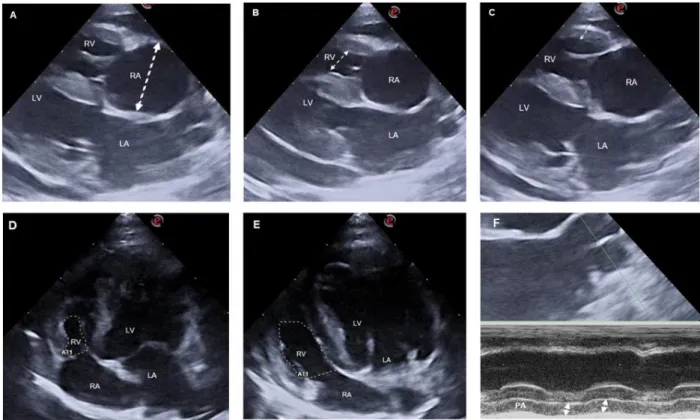
Figure 1. Representative measure of the RAD (A; dotted line), RVID (B; dotted line), RVFW (C; dotted line), RVAs (D; dotted dashed), RVAd (E; dotted dashed) and PA (F; dotted lines). 16 Figure 2. Box and whisker plot for body weight, age and heart rate in control cats, asymptomatic
HCM cats and cats with HCM+CHF. **P < 0.01. 18
Figure 3. Box and whisker plots for MAPSE IVS and TAPSE in control cats, asymptomatic HCM
cats and cats with HCM+CHF. **P < 0.01. 19
Figure 4. Box and whisker plots for LA size (LAD, LAD/Ao) assessment in control cats, asymptomatic HCM cats and cats with HCM+CHF. *P < 0.05; **P < 0.01. 19 Figure 5. Box and whisker plots of LV size and function indices in control cats, asymptomatic HCM cats and cats with HCM+CHF. *P < 0.05; **P < 0.01. 20 Figure 6. Box and whisker plots for aortic velocity and gradient and for pulmonary velocity gradient in control cats, asymptomatic HCM cats and cats with HCM+CHF. *P < 0.05. 21 Figure 7. Box and whisker plots for RV function indices (RVFWd, RVFS, RV FAC) in control cats,
asymptomatic HCM cats and cats with HCM+CHF. *P < 0.05. 22
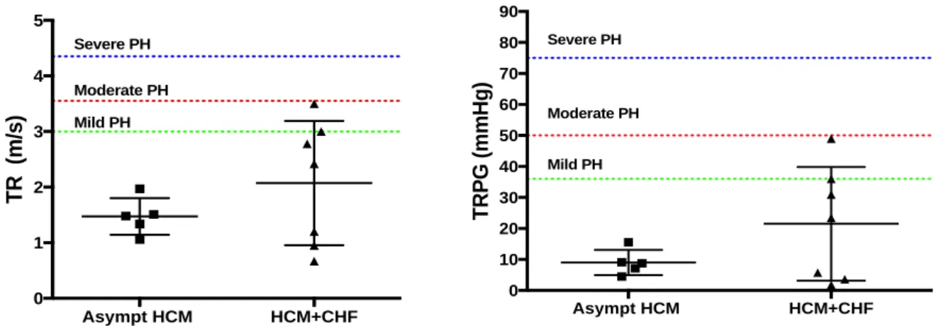
Figure 8. Box and whisker plots for TDI of RVFW myocardial systolic and diastolic velocities of RVFW in control cats, asymptomatic HCM cats and cats with HCM+CHF. 22 Figure 9. Scatter dot plots of TR and TRPG in the HCM group. For each group bars and error bars represent mean and standard deviation. The dotted lines represent the different reference value to classify the severity of PH (Diagnosis of PH: TRGP= 36 mmHg, mild PH: 36-50 mmHg,
moderate PH= 50-75 mmHg, severe PH ≥ 75 mmHg). 23
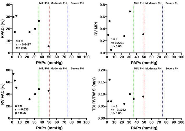
Figure 10. Scatter plots illustrating no significant differences (P>0.05) in correlations (r) between systolic pulmonary arterial pressures (PAPs) obtained by Doppler echocardiography of TRGP and the 4 indirect indices of pulmonary hypertension: FAC, RPADi, TDI RVFW S’ and RV MPI in
ix
LIST OF TABLES
Table 1. Clinical data of all studied cats (n=25). Bolded values denote statistical significance. 18 Table 2. Distribution of data for MAPSE IVS and TAPSE in 7 healthy cats. 19 Table 3. Distribution of data for RV MPI, RPADI and RVFWd in 7 healthy cats. 22 Table 4. Results of Pearson correlation for the prediction of HP severity via TRPG to estimate
1
I. INTRODUCTION
My curricular internship of the master degree in veterinary medicine was performed in Hospital Veterinário do Porto (HVP) and Davies Referrals, UK. In HVP, I could be part of the hospital clinical activity doing weekly rotations in first opinion and referral appointments, internment and critical care and surgery. I also did night shifts. In Davies Referrals I was in the anaesthesia department where I worked with specialists in the selection of anaesthetics to use in different situations, in monitoring of the anaesthesia parameters in different surgeries and while they performed imaging techniques. I was also in the cardiology department where I was part of their clinical activity, namely appointments, surgeries and complementary diagnostic exams like echocardiography and Holter monitoring. The objective of these internships was to improve the theoretical knowledge that I earned during my degree and to develop my practical skills in different areas of companion animal medicine.
Simultaneously, I integrated an investigation in which objective was to assess right ventricular function and pulmonary hypertension prevalence in cats with hypertrophic cardiomyopathy. The echocardiographic studies were performed in the HVP by Doctor Luís Lima Lobo according to the echocardiographic standards for transthoracic echocardiography of the American College of Veterinary Internal Medicine (Thomas et al., 1993). The echocardiographic indices were chosen based on literature from human and veterinary medicine (mainly cats and dogs) in the fields of hypertrophic cardiomyopathy, pulmonary hypertension and echocardiographic examination of heart function.
More specifically, the objective of this study was to estimate the number of cats with HCM that are affected with PH and to assess RV function and its involvement in this pathology. Considering the lack of studies in cats that relate both pathologies, as well as recognizing the importance of echocardiographic evaluation, this work highly contributes to the improvement of the knowledge about these pathologies and, also, to highlight the importance of right ventricular function assessment in cats with HCM.
2
II.
STATE OF THE ART
1. FELINE HYPERTROPHIC CARDIOMYOPATHY 1.1. Definition
A cardiomyopathy can be defined as a heterogeneous class of disorders in which there is a structural abnormality and functional impairment of the heart muscle. Therefore, it excludes hypertensive, vascular, valvular, pericardial, pulmonary, metabolic or congenital disorders (Fox
et al., 1999). Cardiomyopathies can be classified as primary (idiopathic) cardiomyopathy when
the heart disease results from an inherent problem in the myocardium and when the aetiology cannot be identified (Fox et al., 1999). In secondary cardiomyopathies, the myocardial involvement is a result of multiorgan systemic disorders (Smith et al., 2016).
Hypertrophic cardiomyopathy (HCM) is one of the four types of idiopathic heart muscle diseases and is characterized by a hypertrophied, nondilated (primarily left) ventricular myocardium in the absence of other cardiac, systemic or metabolic abnormalities that can produce the same magnitude of hypertrophy (Fox et al.,1999).
1.2. Aetiology
HCM is the most common heart disease in cats and although the disease is known to be inherited in some breeds, in most cases it is idiopathic (Abbott, 2010). HCM is a heritable disease in breeds like the Maine coon, Ragdoll, American shorthair (Côté et al., 2011) and has also been described in Persian, Norwegian Forest, Sphynx and mixed-breed cats. Although inherited in some breeds, HCM is not a congenital disease; it develops with age and can occur at any time during life span (Häggström et al., 2015).
In Maine Coon and Ragdoll cats, HCM is an autosomal dominant inherited disease and a myosin binding protein C gene (MYBPC3) mutation has been identified in some cats (Côté et al., 2011). However, some Maine Coon cats with myocardial hypertrophy do not have this mutation, suggesting that other causative mutations may exist or that there is a non-genetic cause of HCM in this breed (Häggström et al., 2015).
1.3. Pathophysiology
Understanding the genetic mutations leading to the development of hypertrophy has been a difficult task, since their precise mechanisms have not been fully described (Abbott, 2010). However, it is known that in this type of cardiomyopathy, some changes occur in sarcomeric proteins of cardiomyocytes responsible for cardiomyocyte dysfunction. This dysfunction induces cell stress responses, which increase cell transcription and result in the cardiomyocyte hypertrophy, increased collagen formation and myofiber disarray that typifies the final HCM phenotype. This phenotype is characterized by a concentric left ventricle (LV) hypertrophy,
3
myocardial fibrosis, and myofiber disarray, which leads to diastolic and possibly systolic dysfunction (Côté et al., 2011).
The main pathophysiologic feature of HCM is diastolic dysfunction, due to impaired diastolic filling of the LV caused by a non-uniform relaxation of the myocardium and increased stiffness (abnormal distensibility) of the ventricular muscle (Fox et al.,1999). The altered pattern of myocardial relaxation can be explained by increased myofilament sensitivity to calcium, intracytosolic calcium overload, changes in LV loading conditions and myocardial ischemia due to small coronary artery remodelling. The ventricular stiffness is augmented by concentric LV hypertrophy, myofiber disarray and myocardial fibrosis (Côté et al., 2011).
The diastolic dysfunction results in a ventricle that cannot properly fill with blood during diastole, leading to an increased LV diastolic filling pressure (White, 2015); in turn this is responsible for LA dilation, and elevation of its pressure, that consequently promotes elevated pressure in the pulmonary veins. When the LV diastolic filling pressure and pulmonary venous pressure exceed approximately 24 mm Hg (Côté et al., 2011), left sided congestive heart failure (CHF) occurs with the development of cardiogenic pulmonary oedema and/or pleural effusion (Smith et al., 2016). LA enlargement also increases the risk of thrombus formation (White, 2015) because it causes blood flow stasis, which results in erythrocyte aggregation and platelet activation (Côté et al., 2011). The thrombus can then break loose (become an embolus) and most commonly lodges in the terminal aorta, causing aortic arterial thromboembolism (ATE) (Smith et
al., 2016).
The diastole is the most affected phase of the cardiac cycle in cats with HCM, although in some cases, systolic abnormalities like systolic anterior motion (SAM) of the mitral valve and consequently LV outflow tract obstruction (LVOTO) may be present (Fox et al.,1999). The anterior motion of the mitral valve toward the interventricular septum (IVS) during systole occurs secondary to displaced, hypertrophied papillary muscles that pull the mitral valve into the LV outflow tract (LVOT) and thus causes LVOTO (Côté et al., 2011). Due to this displacement of the valve, mitral regurgitation (MR) can occur. The obstruction of the LV outflow tract caused by SAM leads to increased LV work, with impeded and turbulent ejection of blood flow throw the aortic valve, which may result in a murmur. It is important to mention that is not an intrinsic feature of HCM in cats, but when SAM is echocardiographically identified it is highly suggestive for the diagnosis of HCM (Luis Fuentes and Wilkie, 2017).
1.4. Clinical signs
Around half of the cats with HCM are asymptomatic and diagnosed incidentally on routine physical examination when a heart murmur or a gallop heart sound is auscultated (Côté et al., 2011). The most common auscultation finding in HCM is a systolic murmur (36–72% of cats)
4
(Smith et al., 2016), often associated with dynamic LVOTO (Luis Fuentes and Wilkie, 2017), followed by a gallop heart sound (33% of cats) (Smith et al., 2016). The gallop heart sound is a much more specific finding of HCM and generally reflects diastolic dysfunction (Luis Fuentes and Wilkie, 2017). Arrhythmias are a rare finding in cats with HCM (7%) (Smith et al., 2016).
Of those cats with CHF due to HCM, half had a triggering event such as fluid therapy, anaesthesia and surgery, or corticosteroid administration (Côté et al., 2011). There are some exceptions, as with early stages of decompensation when the owners may observe tachypnoea. However, cats with CHF usually exhibit peracute (in less than 24 hours) clinical signs associated with pulmonary oedema and/or pleural effusion (Fox et al.,1999). Dyspnoea is the most common sign (32–46% of cats) (Smith et al., 2016), but cough is rarely observed in cats with CHF. Lethargy, anorexia and vomiting may precede respiratory signs by 1 or 2 days in some cats. In the case of a significant pleural effusion, heart and lung sounds will be muffled (Fox et al.,1999). Syncope is less common (4%), and it may result from an arrhythmia, severe CHF, an intracardiac thrombus (Côté et al., 2011) or LVOTO (Fox et al.,1999). Sudden unexpected death as the first clinical manifestation of this disease may occur in few cats (Abbott, 2010).
ATE secondary to HCM occurs in approximately 12% to 17% of cats and causes a cessation of blood flow more commonly to the caudal legs (Smith et al., 2016). However occasionally paresis of one front leg (generally the right one) may occur (Fox et al.,1999). Cats with a thrombus in the aorta show acute clinical signs like acute paresis/paralysis, lameness and acute pain (Smith et al., 2016), while front leg paresis seems to be better tolerated (Fox et
al.,1999).
1.5. Diagnoses
Echocardiographic examination is the gold-standard method to diagnose HCM in cats (Abbott, 2010). It allows the assessment of systolic and diastolic myocardial function and chamber dimensions, quantification of concentric hypertrophy, identification of spontaneous echo contrast or an intracardiac thrombus and evaluation of the origin of a murmur (Côté et al., 2011). Cats with HCM can have different phenotypes of hypertrophy: generalized concentric hypertrophy, asymmetric (segmental) concentric hypertrophy of the septum or free wall or papillary muscle hypertrophy (Smith et al., 2016).
The diagnosis of HCM is established when there is an increased end-diastolic LV wall thickness in two-dimensional (B-mode) of 6 mm or greater in the absence of other secondary causes of concentric hypertrophy. The cut-off value (6 mm) is likely representative of a marker of HCM (Luis Fuentes and Wilkie, 2017) and there is an equivocal zone (cats suspected of HCM) from 5.5–5.9 mm. It is also important to mention that HCM can be classified, according with LV
5
wall thickness, as mild (6-6.5 mm), moderate (6.6–7.5 mm), and severe (>7.5 mm) (Côté et al., 2011).
LA size can be measured by B-Mode and Motion-mode (M-mode), and LA dilation is identified when the LA diameter to aortic diameter ratio on M-mode is >1.6 (Visser, 2017). SAM of the mitral valve can be identified on B-mode echocardiography from the right parasternal long-axis LVOT view. Therefore, continuous-wave Doppler may easily detect this abnormality because it shows a turbulent double jet of MR and turbulence in the LVOT arising from the common point of the anterior mitral leaflet obstruction (Côté et al., 2011). To quantify the severity of the obstruction, velocity of aortic blood flow can be estimated from the left apical parasternal 5-chamber view using continuous-wave Doppler (Smith et al., 2016).
Diastolic function can be evaluated using pulsed-wave Doppler to quantify mitral inflow velocity from the left apical four-chamber view that often, in cats with HCM, shows a delayed relaxation pattern: decreased early diastolic mitral velocity (E), increased late diastolic mitral velocity (A) which causes a reversed E/A ratio of <1, prolonged isovolumetric relaxation time, and prolonged deceleration time of the early diastolic mitral velocity. This delayed relaxation corresponds to the first level of diastolic dysfunction when there is an impaired early diastolic filling but normal LA pressures. As diastolic dysfunction worsens, LA pressure increases, leading to a pseudo-normal filling pattern of mitral inflow velocity. Then, when the dysfunction is severe, and the atrial pressure is significantly increased, a restrictive filling pattern is seen: increased early diastolic filling (E), decreased late diastolic filling (A), E/A ratio >2, decreased deceleration time, and shortened isovolumetric relaxation time (Smith et al., 2016).
Thoracic radiographs also provide valuable information about the size of the heart and pulmonary parenchymal and vascular changes (Fox et al.,1999), helping to identify the manifestations of CHF in cats with HCM, and to monitor response to treatment. Abnormalities like cardiomegaly, LA dilation, pulmonary venous distension, diffused patchy interstitial to alveolar pulmonary infiltrates (pulmonary oedema) and obscured cardiac silhouette (pleural effusion) are potential radiographic findings. However, sometimes the size of the heart may be normal in cats with mild HCM because of the concentric pattern of hypertrophy (Côté et al., 2011). An additional sensitive and accurate screening test to detect HCM in cats is the biomarker N-terminal pro-brain natriuretic peptide (NT-proBNP) that is released from the ventricles when the myocardium is stretched (White, 2015). It also helps to differentiate the origin of dyspnoea (CHF secondary to HCM or primary respiratory disease) (Côté et al., 2011) and to detect asymptomatic HCM, but there may be false negatives and positives (Côté et al., 2011).
6 1.6. Treatment
In asymptomatic cats with HCM and no CHF there is no consensus on when to start treatment and what is the most appropriate therapy (Côté et al., 2011). The main goals are to reduce LV hypertrophy, to improve diastolic function, to reduce the risk of ATE and to increase the time to heart failure. The decision to treat should be focused on severity of SAM of the mitral valve and left ventricular hypertrophy, size of the LA, presence of tachyarrhythmias, compliance of the owner to medicate daily to twice daily indefinitely and the cat’s temperament (Smith et al., 2016).
Beta-blockers (e.g. atenolol) and calcium channel blockers (e.g. diltiazem) are the drugs most commonly used in asymptomatic cats and they may reduce myocardial hypertrophy. Beta-blockers are more effective in reducing the severity of SAM than calcium channel Beta-blockers and help to prevent tachycardia. Administration of angiotensin converting enzyme (ACE) inhibitors or aldosterone antagonists in asymptomatic cats is not warranted. If there is evidence of spontaneous contrast, an intracardiac thrombus, or moderate to severe LA dilation, anticoagulant therapy such as clopidogrel is indicated (Smith et al., 2016).
The most common emergencies in symptomatic HCM cats include left-sided heart failure (pulmonary oedema and/or pleural effusion) and ATE (Smith et al., 2016). In cases of left-sided heart failure, the diuretic furosemide can be life-saving by reducing pulmonary oedema and slowing accumulation of pleural effusion. Parenteral furosemide should be administered and then the dose and frequency adjusted once the respiratory rate decreases to ≤50 breaths/minute and the respiratory effort decreases (Côté et al., 2011). Oxygen therapy is also beneficial (Fox et al., 1999), and in cats with respiratory distress due to severe pleural effusion, thoracocentesis is required to stabilize the patient (Fox et al., 1999). Once stabilized, the animal should be started on long-term treatment to maintain cardiac compensation, prevent ATE and improve myocardial function and quality of life (Fox et al., 1999).
Diuretics are the gold-standard treatment in chronic heart failure management and in cats stable enough oral furosemide can be administered; however, it is important to assess kidney function before starting long-term furosemide (Côté et al., 2011). The addition of an ACE inhibitor
(e.g. enalapril or benazepril) is also a standard approach once the cat is stable (Smith et al., 2016) because neurohormonal activation plays an important role in heart failure (Fox et al., 1999). Negative inotropic therapy (e.g. beta-blockers and calcium channel blockers) may be used in some cats with chronic heart failure. Also, prophylactic anticoagulant therapy may be started in cats with high risk of ATE(Côté et al., 2011).
7 1.7. Pulmonary Hypertension
1.7.1. Definition and Classification
Pulmonary hypertension (PH) can be defined as a pathologic condition resulting from an abnormally high pressure in the pulmonary circulation (Vezzosi et al., 2018) and can be classified as a primary disease (idiopathic) or more often a secondary disease (Pyle and Abbott, 2004). Then, depending on the anatomic location of the pulmonary vascular system affected it can be classified as pulmonary arterial hypertension (PAH) or pre-capillary or active if it affects the arterial side, or pulmonary venous hypertension (PVH) or post-capillary or passive if it occurs on the venous side (Kellihan and Stepien, 2012). In human medicine, PH can also be classified using a five-group system depending on the causative pathological process. The five groups are: I (PAH due to arteriolar vascular disease), II (PVH due to left heart disease), III (PH with chronic lung disease and/or hypoxia, IV (chronic thromboembolic pulmonary hypertension) and V (PH from unclear or multifactorial mechanisms) (Galiè et al., 2016).
1.7.2. Pulmonary Hypertension secondary to Hypertrophic Cardiomyopathy
In humans with HCM, PH can be a complication of elevated left-sided (LV and LA) diastolic pressures secondary to diastolic dysfunction (impaired relaxation and stiffness of the myocardium), LVOTO with MR or even systolic dysfunction that happens in end stages of HCM (Musumeci et al., 2017). This elevated LV filling pressure is passively back-transmitted to the pulmonary capillaries causing PH (Vezzosi et al., 2018). Although evidence of feline PH is limited to case reports (Ettinger et al., 2017), the same assumption may be extrapolated to cats with HCM.
Patients with left-sided heart disease may have concurrent PVH and PAH. PVH is caused by a combination of hypertension from increased LA pressures and reactive pulmonary arterial vasoconstriction due to acute or chronic hypoxia (caused by pulmonary oedema) (Kellihan and Stepien, 2012). PAH secondary to LA hypertension (LAH) occurs in the continuum of progression of heart disease to heart failure. LAH is associated with neurohormonal activation of the sympathetic nervous system, the renin-angiotensin-aldosterone system, and augmented activity of endothelin-1 (ET-1) (potent arterial vasoconstrictor), phosphodiesterase-5 (PDE 5) and natriuretic peptides (NP) that results in pulmonary arterial vasoconstriction (hypertension) (Stepien, 2009). In humans with HCM, PH is also associated with increased mortality (Ong et al., 2016).
8 1.7.3. Clinical signs and Diagnosis
The clinical signs of PAH can be similar to signs of left-sided CHF or may not be noticeable if signs of right-sided CHF are not present (Stepien, 2009). Murmurs of tricuspid or pulmonic insufficiency can be detected during systole or diastole respectively (Johnson, 2010).
In veterinary medicine, PH is more commonly diagnosed in dogs than in cats (Nelson and Couto, 2013) and echocardiography has replaced cardiac catheterization as a diagnostic approach for PH because it provides alternative values for many previously invasively measured parameters obtained by right heart catheterization. In combination with other tests it is possible to diagnose the presence and possible causes of PH (Stepien, 2009). There are multiple B-dimensional echocardiographic findings that are used to support the diagnosis of PH, including RV hypertrophy (due to acute and chronic RV pressure overload), septal flattening (when RV pressure approaches or exceeds LV pressure) and pulmonary arterial dilation (Kellihan and Stepien, 2012).
In dogs, a tricuspid regurgitation pressure gradient (TRPG) ≥ 36 mmHg (Vezzosi et al., 2018) or pulmonic insufficiency (PI) by Doppler echocardiography (Kellihan and Stepien, 2012) allows an estimation of pulmonary arterial pressure in systole (PAPs) and thus the identification of PH (Stepien, 2009). The TRPG is derived from the peak systolic regurgitation jet velocity using the modified Bernoulli equation: Pressure gradient (PG) = 4 × (peak TR velocity)2. Then, based on TRPG values, it is possible to classify dog’s PAH as mild (36-50 mmHg), moderate (51-75 mmHg) and severe ( 75 mmHg) (Vezzosi et al., 2018).
A study in dogs infected with heartworms and another that studied PH in dogs reported that right pulmonary artery distensibility index (RPADi) is a valuable method for early detection of the presence and severity of PH even when Doppler echocardiography does not show TR or PI (Visser et al., 2016; Venco et al., 2014). Venco et al. (2014) also demonstrated that RPADi has a strong correlation with invasive “gold standard” systolic PA pressures and it might be valuable to start applying this method in combination with TRPG to diagnose PH. To calculate RPADi, the right pulmonary artery (PA) is the one chosen because it is usually affected earlier and to a greater degree. Taking into account that the walls of PA distend when the blood pressure increases during systole and recoil when the blood pressure diminishes during diastole (Venco et al., 2014), RPADi is calculated by M-mode as the difference in diameter of the pulmonary artery in systole and diastole by the following formula: RPADi = (Pas - PAd)/Pas × 100 (Serrano-Parreño et al., 2017). In dogs, a normal pulmonary pressure is correlated with a RPADi ≥36% (Serrano-Parreño et al., 2017) and a RPADi < 35% is indicative of PH. Then, RPADi between 35%-28% is correlated with mild PH (30–55 mm Hg), between 27%-23% with moderate PH (56–79 mm Hg) and less than 22% with severe HP (>79 mm Hg) (Venco et al., 2014).
9 1.7.4. Treatment
The objectives of therapy for PAH due to left-heart disease are to improve haemodynamic status and clinical signs. These aims are achieved through reduction of PVH (managing LA hypertension) and reactive PAH that may be present in addition to PVH (using pulmonary vasodilators) (Stepien, 2009) and improving LV systolic and diastolic function (Kellihan and Stepien, 2012).
To reduce LA pressure combining diuretics (e.g. furosemide or torsemide) and optimal neurohormonal blockade (e.g. aldosterone blockade, ACE inhibition and beta-blockers) is a good approach. Reactive PAH can be diminished by using pulmonary vasodilators such as ET-1 antagonists, PDE-5 inhibitors and calcium-sensitizing phosphodiesterase-3 (PDE-3) inhibitors (Stepien, 2009). ET-1 antagonists (e.g., bosentan) promote pulmonary and systemic vasodilation but costs are prohibitive in veterinary medicine (Ettinger et al., 2017). PDE-5 inhibitors are pulmonary arterial vasodilators (mainly large arteries) and the most common drug of this class is sildenafil citrate (Stepien, 2009). Sildenafil is used in dogs since it diminishes clinical signs of PH and improves quality of life (Ettinger et al., 2017). In cats, there is little information available (Ramsey, 2017), with only one reported case of a cat with PH caused by a left-to-right shunt, where sildenafil was used (Novo-Matos et al., 2014). The PDE-3 inhibitors and also calcium sensitizing agents (e.g. pimobendan) differ from PDE-5 inhibitors because they vasodilate both large and resistance pulmonary arteries and the positive inotropic effects may result in decreased LA pressures (Stepien, 2009). The calcium-sensitizing 3 inhibitors (pimobendan) and PDE-5 inhibitors (sildenafil) are the most often used pulmonary vasodilators in dogs (Kellihan and Stepien, 2012), either separately or in combination, and are the most promising for therapy of PAH associated with left heart dysfunction (Stepien, 2009).
2. RIGHT VENTRICULAR FUNCTION
2.1. The Importance of Right Ventricle in Feline Hypertrophic Cardiomyopathy
In veterinary medicine, the quantitative assessment of RV function is not well studied and most of the time is not properly evaluated during routine clinical echocardiographic assessment (Visser, 2017). This might be explained by two main reasons: (i) the RV seems to be less commonly or obviously involved in cardiovascular diseases and (ii) its function is difficult to quantify compared with the LV due to the complexity of its three-dimensional shape, separate inflow and outflow regions, prominent endocardial trabeculations and marked load-dependence (Visser, 2017; Voelkel et al., 2006). In human medicine, the importance of quantitative RV function assessment as a predictor of clinical status, morbidity,and mortality is well recognized in cardiac diseases that also affect dogs and cats, including those regarded as left-heart specific, such as
10
mitral valve disease and HCM which enhance ventricular function as a single unit (Visser, 2017; Haddad et al., 2008).
Previous studies in cats with HCM that evaluated RV size and function documented that remodelling and dysfunction occurs in some cats with HCM and may be associated with clinical status and severity (Visser et al., 2017
;
Schober et al., 2016)
. Other recent studies in cats with HCM reported that increased RV wall thickness is common in cats with HCM and related to LV hypertrophy severity (Spalla et al., 2017). RV hypertrophy is also associated with increased risk of CHF, ATE, ventricular tachyarrhythmias and sudden death (Visser et al., 2017). The main mechanisms by which cats with LV dysfunction develop abnormal RV function and dilatation are: (i) PVH with subsequent PAH and (ii) as a consequence of the hypertrophic cardiomyopathic process involving the RV (Visser et all., 2017; Voelkel et al., 2006).2.2. Assessment of Right Ventricular Function
The most practical method for quantitative assessment of RV function in cats is echocardiography (Visser, 2017). There are several echocardiographic indices that can be measured and the ones acquired from the left apical 4-chamber view optimized for the right heart are: Tricuspid annular plane systolic excursion (TAPSE), RV-Myocardial performance index (RV-MPI), RV fractional area change (FAC) and Tissue Doppler imaging derived peak systolic (TDI RVFW S’), early diastolic (TDI RVFW E), late diastolic (TDI RVFW A) RV wall myocardial velocity of the lateral tricuspid annulus (Visser, 2017). Other important indices obtained from the right parasternal long-axis 4-chamber view are: Maximum right atrial diameter (RAD), RV internal dimension (RVID), RV fractional shortening (RVFS) and the thickest portion of the RV free wall at end-diastole (RVFWd) (Visser et al., 2017).
The TAPSE measurement consists of quantifying the maximal longitudinal displacement of the lateral tricuspid valve annulus towards the RV apex during systole (Visser, 2017) and is obtained from M-mode with the cursor over the tricuspid annulus (Boon, 2011). It is a marker of RV longitudinal systolic function and is lower in cats with HCM compared with healthy cats, confirming reduced systolic function possibly due to concomitant RV cardiomyopathy or PH secondary to left-sided disease (Spalla et al., 2017). In cats with HCM, reduced TAPSE is negatively associated with the severity and survival times. In most dogs, TAPSE is decreased with severe PH (Visser, 2017).
Measurement of the RV area to determine FAC is obtained by tracing the RV endocardial border at end-diastole (RVAd) and end-systole (RVAs). Then, FAC is calculated using the following formula: FAC = (RVAd - RVAs)/RVAd × 100 (Visser, 2017). FAC is a surrogate of RV ejection fraction and is decreased in dogs (Visser, 2017) and in humans with severe PH (Boon, 2011).
11
B-mode pulsed-wave TDI of the lateral tricuspid annulus allows the assessment of RV myocardial systolic and diastolic function. In humans, decreased TDI RVFW S’ is associated with decreased RV ejection fraction (Boon, 2011; Cincin et al., 2015). In cats with HCM, the TDI LVFW could detect LVFW dysfunction (Silva et al., 2013). TDI velocities can be reduced for reasons like PH and other causes of RV systolic dysfunction (Kellihan and Stepien, 2012).
The measurement of RAD is made at end-systole from the middle of the interatrial septum to the right atrial lateral wall in a cranial-caudal plane and parallel to the tricuspid valve annulus. RVID is measured at end-diastole (RVIDd) and end-systole (RVIDs) at the level of the RV where the tips of the opened tricuspid valve leaflets contact the endomyocardium and parallel to the tricuspid valve annulus. Then, RVFS is calculated as RVFS = (RVIDd - RVIDs)/RVIDd × 100. The RVFWd is measured at end-diastole from the inner edge of the RV endomyocardium to the outer edge of the RV epimyocardium, excluding the pericardium (Visser et al., 2017).
MPI or Tei index is an index of global (systolic and diastolic) myocardial function of the RV and LV (Boon, 2011). To evaluate RV function, this index is measured by pulsed-wave Doppler of the tricuspid and pulmonary inflow. Then, MPI is calculated: RV MPI = (IVCT + IVRT)/ET or RV MPI = (a-b)/b where “a” represents the time from closure to opening of tricuspid valve and “b” represents ejection time of pulmonary artery flow, “ET” is the ejection time, IVCT is isovolumetric contraction time and IVRT is isovolumetric relaxation time (Kellihan and Stepien, 2012). Augmented values of RV MPI are associated with RV myocardial dysfunction when changes in load are chronic (Boon, 2011; Visser, 2017). MPI is also a valuable indicator of PH and increased MPI may support the diagnosis of PH. In dogs, a value of >0.25 supports the diagnosis of PH (Kellihan and Stepien, 2012).
12
III. OBJECTIVES
HCM is the most common heart disease diagnosed in cats and there are many studies that focus on assessment of LV function. In human medicine, the involvement of the RV in HCM has also been described, and as a result there is an increase in the number of studies to assess RV function and its importance in HCM in veterinary medicine. In addition, PH has been studied in humans with HCM and in dogs with mitral valve disease and heartworm infections, but there is a lack of information in cats.
Therefore, the objective of the following prospective study was to focus on evaluating RV function and the prevalence of PH in cats with HCM. The specific objectives for this study were:
• Evaluate RV function by echocardiography in healthy cats and in cats with HCM (asymptomatic and symptomatic);
• Evaluate the prevalence of PH in cats by applying the echocardiographic indices used in dogs and humans;
• Compare the results obtained for different variables with different studied groups; • Compare the results obtained for the different evaluations with the reference intervals
for dogs and other species;
• Propose preliminary reference intervals for echocardiographic indices that in the literature were evaluated only in dogs (RV MPI and RPADi);
Learning the fundamentals of echocardiography, indices that can be measured, how to measure and to improve the knowledge about HCM and PH were skills also developed during this work. Finally, knowledge of statistical analysis and the discussion of the experimental results are an important step in the process of scientific learning and a complementary earned skill of my masters degree.
13
IV. MATERIALS AND METHODS
1. FELINE POPULATION CHARACTERIZATION
Cats were included in this prospective study if they underwent a complete standard echocardiographic examination according to human and veterinary guidelines (Thomas et al., 1993
;
Lang et al., 2015) with evaluation of left and right sides of the heart, pulmonary artery and related heart valves in M-mode, B-mode, and Doppler echocardiography. This echocardiographic study was performed in HVP, from September 2017 to March 2018, in 25 cats aged between 4 months and 20 years. To be included in the study, a complete case record (owner data, cat signalment (weight, breed, age, gender, body score), history, clinical signs, cardiac insufficiency classification if the animal had CHF (stands for International Small Animal Cardiac Health Council (ISACHC) and American College of Veterinary Internal Medicine (ACVIM)), complementary exams (type and evidence of pleural effusion (yes/no) or pulmonary oedema (yes/no)), concomitant diseases and current medications (type/dose) were required and recorded (see Appendix I).Cats were chosen only if they met the inclusion criteria and none of the exclusion criteria. Control cats had to be apparently healthy (with no clinical signs of systemic diseases), with a normal echocardiographic examination (normal myocardial structure and function) and not administered any medications that could affect the cardiovascular system. For cats diagnosed with HCM, the exclusion criteria included: any concomitant cardiac disease and any systemic disease that could affect LV wall thickness such as dehydration or hypovolemia, primary respiratory disease, hyperthyroidism, acromegaly, cardiomyopathies other than HCM, congenital diseases, neoplastic diseases and systemic hypertension (systolic blood pressure > 170 mmHg). In all cats diagnosed with HCM with an age superior to 6 years (high-risk) was dosed serum total thyroid hormone (T4) concentrations and measured blood pressures. Cats with MR were included only if it was secondary to SAM of the mitral valve. Cats with electrocardiographic alterations like sustained or clinically relevant tachy/brady-arrhythmia were also excluded.
Then, cats were allocated into 1 of 3 groups: (1) control group comprising apparently healthy cats with normal echocardiographic indices, (2) asymptomatic HCM group consisting of cats with HCM but with no clinical evidence of CHF (3) HCM + CHF group comprising cats with HCM and CHF. Cats were allocated to the control group if the end-diastolic LV wall thickness measured in B-mode was < 6 mm. Cats with a soft systolic heart murmur were also included in this group. Cats were diagnosed with HCM if the end-diastolic (diffused or segmental) LV wall thickness measured in B-mode was ≥ 6 mm. Cats with HCM that were not receiving any cardiac medication and had no signs or history of increased respiratory rate (tachypnoea) or effort (dyspnoea), syncope or ATE were classified as asymptomatic. The criteria used to diagnose CHF
14
were the presence of clinical signs compatible with CHF (tachypnoea, dyspnoea, distended jugular veins, ascites, abnormal thoracic auscultation), LA enlargement (diagnosed when LA dimension (LAD) >16 mm (Abbott and MacLean, 2006) or LA diameter to aortic diameter ratio on M-mode is >1.6 (Visser et al., 2017)), and radiographic or ultrasonographic evidence of pleural effusion or radiographic evidence of pulmonary oedema. In this third group of HCM + CHF, cats with more than mild pericardial effusion were excluded to ensure that the effects on RV size and function were originated only by HCM.
Cats were also classified based on reference values obtained in dogs (Visser et al., 2016): no PH if TRPG was < 36 mmHg and with PH if TRPG was ≥ 36 mmHg on echo-Doppler examination. The TRPG was measured based on the peak systolic TR jet velocity using the modified Bernoulli equation. The severity of PH was classified as mild if TRPG was between 36-50 mmHg, moderate if TRPG was between 36-50-75 mmHg and severe if TRPG was > 75 mmHg (Vezzosi et al., 2018; Visser et al., 2016).
2. ECHOCARDIOGRAPHIC EXAMINATION
All echocardiographic studies were performed by a single veterinary cardiologist. Cats received butorphanol (0.2 mg/kg IM or IV) before echocardiographic examination if sedation was required. The echocardiographic measurements were obtained by B-Mode, M-Mode and Doppler (pulsed-wave Doppler including color-flow Doppler and pulsed-wave TDI).
If the thickest portion of the interventricular septum (IVSd) and LV free wall (LVFWd) measured at end-diastole from both long-axis (Lx) and short-axis (Sx) images in B-Mode were ≥ 6 mm at any location, cats were diagnosed with HCM (if no exclusion criteria were met). These indices were also measured in end-systole (IVSs and LVFWs). LV internal dimension was measured at end-diastole (LVIDd) and end-systole (LVIDs) from the right parasternal Sx view. Then, LV fractional shortening (LVFS) was calculated as (LVIDd - LVIDs)/LVIDd × 100 (Visser et
al., 2017). LAD and left atrium-to-aorta ratio (LAD/Ao) were measured by M-mode from right
parasternal Lx view, LAD at end-systole and the aortic diameter at the end-diastole. Then, LAD/Ao was derived from these dimensions (Abbott and MacLean, 2006). A LAD/Ao >1.6 was used to determine LA dilation (Visser et al., 2017). LV ejection fraction (LV EF) was automatically measured by the ultrasound machine according to the Teichholz formula (Arora et al., 2010). The MAPSE IVS was measured by M-mode from the left apical 4-chamber view, with the cursor parallel to the IVS between the most basilar position of the mitral annulus in end-diastole and its most apical displacement at end-systole by the leading-edge method (Spalla et al., 2017).
Mitral valve and tricuspid valve peak flow velocity in early and late diastole were also measured using pulsed-wave and color-flow Doppler from the left parasternal 4-chamber view and the respective gradients were calculated using the simplified Bernoulli equation: PG = 4 ×
15
(peak velocity)2 (Boon, 2011). Aortic valve peak flow velocity (Ao) was also measured using the left parasternal apical 5-chamber view and pulmonary valve peak flow velocity (Pulm) was measured using the right parasternal 4-chamber view, both using pulsed-wave and color-flow Doppler and the respective gradients were calculated using the simplified Bernoulli equation: PG = 4 × (peak velocity)2 (Boon, 2011).
On the right side of the heart, several indices (RAD, RVID and RVFWd) were obtained from a right parasternal Lx 4-chamber view (Schober et al., 2016), and TAPSE, FAC and pulsed-wave TDI velocities of longitudinal RVFW myocardium were acquired from a left apical 4-chamber view (Visser et al., 2017). RAD was obtained at end-systole (RADs) and end-diastole (RADd) from the mid-point of the interatrial septum to the right atrial (RA) lateral wall in a cranial-caudal plane and aligned to the tricuspid valve annulus. RVID was measured at end-systole (RVIDs) and end-diastole (RVIDd) where the tips of the opened tricuspid valve leaflets contact with the RV endomyocardium and parallel to the tricuspid valve annulus. Then, RV FS was calculated as RVFS = (RVIDd-RVIDs)/RVIDd × 100. The presence of RV hypertrophy was estimated through the measurement of RVFW diameter at end-diastole (RVFWd) from the inner edge of the RV endomyocardium to the outer edge of the RV epimyocardium without including the pericardium (leading edge to trailing edge method) (Visser et al., 2017; Schober et al., 2016). A value higher than the maximum of RVFWd in healthy cats was used to identify the presence of RV hypertrophy. RVFWd was also indexed to body weight (iRVFW) in kilograms using the formula: iRVFWd = RVFWd/(body weight0.33). FAC was calculated tracing the RV area of the RV endomyocardial
border at end-diastole (RVAd) and end-systole (RVAs) excluding the papillary muscle and applying the following formula: FAC = (RVAd - RVAs)/RVAd × 100 (Visser et al., 2017). The TAPSE were measured by M-mode from the left apical 4-chamber view optimized for the RV, with the cursor as parallel as possible to the RVFW between the most basilar position of the tricuspid annulus in end-diastole and its most apical displacement at end-systole by the leading-edge method (Spalla et al., 2017; Visser, 2017) (Figure 1) (See Appendix V).
The pulsed-wave TDI velocities of longitudinal myocardial motion at the lateral tricuspid annulus were obtained by aligning the cursor as parallel as possible to the RVFW to measure peak systolic annular velocity (TDI RVFW S’) (Visser et al., 2015), and peak early (TDI RVFW E) and late (TDI RVFW A) diastolic annular velocities (Kellihan and Stepien, 2012). Then the respective gradients were calculated using the simplified Bernoulli equation: TRPG= 4 × (peak velocity)2 (See Appendix V). RV MPI was calculated using pulsed-wave Doppler (Kellihan and
Stepien, 2012), to measure the tricuspid inflow time (time between the opening and closure of tricuspid valve) and the pulmonary ejection time via pulsed-wave Doppler in order to apply the following formula: RV MPI = (x-ET)/ET where “x” represents the time from closure (cessation of
16
A wave) to opening (beginning of E wave) of the tricuspid valve and “ET” represents ejection time of pulmonary artery flow (Visser, 2017) (See Appendix V).
Peak systolic TR jet velocity was obtained using color-flow Doppler and continuous-wave Doppler (Vezzosi et al., 2018) using the left parasternal 4-chamber view optimized for the RV inflow tract (Kellihan and Stepien, 2012) and care was taken to align the cursor with the TR jet direction (Visser et al., 2016). Then, peak tricuspid regurgitation gradient (TRGP) was calculated using the simplified Bernoulli equation: TRPG = 4 × TR2. A peak TR velocity > 3 m/s
corresponding to a TRPG > 36 mmHg was used to diagnose PH (Borgarelli et al., 2015).
For RPADi, a M-mode echocardiography of the right PA using the Lx-4-chamber view was obtained with a fast speed in a way to acquire three cycles in a frame (Venco et al., 2014). Then, the minimum diastolic (PAd usually at the Q wave) and maximum systolic (PAs, usually at the T wave) diameter of right PA were measured using the edge to leading-edge technique with the cursor as perpendicular as possible with right PA. Then, RPADi was calculated by the following formula: RPADi = (PAs-PAd)/Pas × 100 (Visser et al., 2016) (Figure 1).
Figure 1. Representative measure of the RAD (Figure A; dotted line), RVID (Figure B; dotted line), RVFW (Figure C; dotted line), RVAs (Figure D; dotted dashed), RVAd (Figure E; dotted dashed) and PA (Figure F; dotted lines) (Images retrieved from the echocardiograms performed in HVP).
17 3. STATISTICAL ANALYSIS
Statistical analysis was performed using a commercially available statistical software package (GraphPad Prism, version 7.04, San Diego, CA, USA) and in all cases avalue of P < 0.05 was considered statistically significant. Descriptive statistics were generated, and the Shapiro-Wilk normality test was used to verify normal distribution of variables. Continuous data are reported as mean ± standard deviation (SD) and categorical data like age, weight, gender (coded as female= 0 male= 1) as a median and range (minimum-maximum).
Comparisons between the 3 studied groups were performed using one-way analysis of variance (ANOVA) or the Kruskal-Wallis test. One-way ANOVA was used to compare continuous, normally distributed data, with posthoc comparisons performed using Tukey’s test. The Kruskal-Wallis test was used to compare ordinal and continuous, non-normally distributed data, with posthoc comparisons performed using Dunn’s method.
Unpaired t-test was performed to compare between 2 groups of independent samples. In this study, we compared the TR in the Asymptomatic HCM and HCM+CHF groups. Pearson correlation was used to determine the strength of association between PAPs and RPADi, FAC and RV MPI after normality testing and both PAP and RPADi, FAC, RV MPI values from the study populations followed a Gaussian distribution.
From the cats in the control group, reference intervals for RVFWd, MAPSE IVS, TAPSE, RPADi and RV MPI were generated using the upper and lower limits on the distribution estimated with a 90% confidence interval (CI).
18
V. RESULTS
1. SAMPLE CHARACTERIZATION – CAT POPULATION 1.1. Gender, Age, Body Weight, Breed
From September 2017 to March 2018, 27 cats were included in the study, although 2 of these cats were subsequently excluded because they did not meet the defined inclusion criteria. Therefore, the population of cats evaluated (n=25) included: 7 cats in the control group, 9 in the asymptomatic HCM group and 9 in the HCM+CHF group. From this population, there were 12 males (most of them in the Asymptomatic HCM group (n=8)) and 13 females, with 6 in the control and HCM+CHF group and 1 in the asymptomatic HCM group.
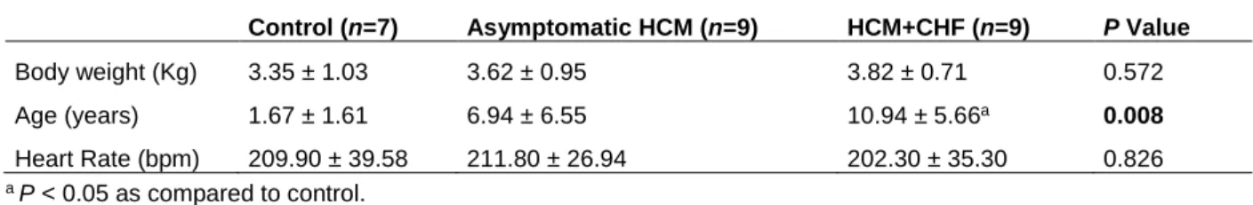
The median body weight of the population was 3.48 (2.3-5.35) kilograms, the median age was 5.00 (0.3-20) years, and the median heart rate was 214 (140-263) beats per minute. With respect to body weight and heart rate there was no statistically significant (P>0.05) difference between groups. Age was significantly greater (P<0.01) in the HCM+CHF group, as compared to control group (Figure 2 and Table 1).
Figure 2. Box and whisker plot for body weight, age and heart rate in control cats, asymptomatic HCM cats and cats with HCM+CHF. **P < 0.01.
The majority of cats were European shorthair (n=18), followed by Persian (n=4), Siamese (n=2) and Sphynx (n=1). The control group had 7 European shorthair, the asymptomatic HCM group had 5 European Shorthair, 1 Sphynx and 3 Persian and the HCM+CHF group had 6 European Shorthair, 2 Siamese and 1 Persian. Only one cat was sedated with butorphanol. A summary of the demographic data is presented in Table 1 and a summary of the clinical record results of the HCM group is presented in Appendix II.
Table 1. Clinical data of all studied cats (n=25). Bolded values denote statistical significance.
a P < 0.05 as compared to control. Control Asympt HCM HCM+CHF 0 2 4 6 B o d y W e ig h t (K g ) Control HCM Asympt HCM CHF 0 5 10 15 20 25 A g e ( y e a rs ) ** Control HCM Asympt HCM CHF 0 100 200 300 H e a rt r a te ( b p m )
Control (n=7) Asymptomatic HCM (n=9) HCM+CHF (n=9) P Value
Body weight (Kg) 3.35 ± 1.03 3.62 ± 0.95 3.82 ± 0.71 0.572
Age (years) 1.67 ± 1.61 6.94 ± 6.55 10.94 ± 5.66a 0.008
19 2. ECHOCARDIOGRAPHIC DATA
A summary of the echocardiographic data is presented in Appendix III. When comparing the 3 groups, cats with HCM+CHF had significantly (P<0.01) lower MAPSE IVS (a LV function index) than control and asymptomatic groups. However, when comparing MAPSE IVS of the asymptomatic HCM cats with the control group, values were not significantly different (P>0.05). TAPSE, a RV function index, was significantly decreased (P<0.01) in cats in the HCM+CHF group compared with asymptomatic HCM cats. Despite that, when comparing TAPSE of the HCM+CHF and asymptomatic HCM cats with the control group it was not significantly different (P>0.05) (Figure 3). Reference intervals for MAPSE IVS and TAPSE were generated from the control group (Table 2).
Figure 3. Box and whisker plots for MAPSE IVS and TAPSE in control cats, asymptomatic HCM cats and cats with HCM+CHF. **P < 0.01.
Table 2. Distribution of data for MAPSE IVS and TAPSE in 7 healthy cats.
MAPSE IVS TAPSE
Mean (mm) 4.70 6.56
Standard deviation 0.59 1.50
Reference interval (CI=90%) 4.27- 5.13 5.46 - 7.66
Minimum 4.2 4.6
Maximum 5.8 8.5
Left atrial size, as evaluated by LAD and LAD/Ao was significantly higher in cats in the HCM+CHF group compared with control (P<0.01) and asymptomatic HCM groups (P<0.05 and
P<0.01, respectively) (Figure 4).
Figure 4. Box and whisker plots for LA size (LAD, LAD/Ao) assessment in control cats, asymptomatic HCM cats and
cats with HCM+CHF. *P < 0.05; **P < 0.01. Control Asympt HCM HCM+CHF 0 2 4 6 8 10 12 M A P S E I V S ( m m ) ** ** Control Asympt HCM HCM+CHF 0 2 4 6 8 10 12 T A P S E ( m m ) **
20
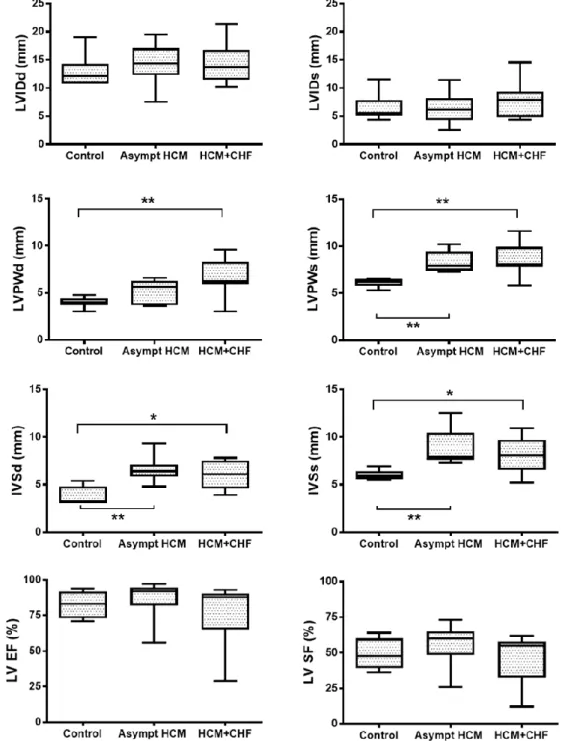
The LV size indices (LVIDd and LVIDs) did not differ significantly (P>0.05) among any of the groups. LVPWd was significantly (P<0.01) increased in cats with HCM+CHF compared with the control group. LVPWs was also significantly (P < 0.01) higher in HCM groups (Asymptomatic and CHF) compared with the control group. Both IVSd and IVSs were significantly increased in asymptomatic cats (P<0.01) and cats with HCM+CHF (P<0.05) compared with the control group. There were no statistically significant (P>0.05) differences for the LV function indices (LV EF and LV FS) among the groups (Figure 5).
Figure 5. Box and whisker plots of LV size and function indices in control cats, asymptomatic HCM cats and cats with
21
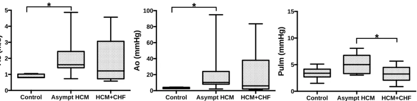
There were no significant (P>0.05) differences in the aortic flow between groups, except when comparing the asymptomatic HCM group with the control where there was a significative increase (P<0.01) in aortic velocity and velocity gradient. The pulmonary flow was not significantly (P>0.05) different between groups except for the HCM+CHF group, where there was a significative decrease (P<0.05) of the pulmonary velocity gradient compared with the asymptomatic HCM group (Figure 6).
Figure 6. Box and whisker plots for aortic velocity and gradient and for pulmonary velocity gradient in control cats,
asymptomatic HCM cats and cats with HCM+CHF. *P < 0.05.
For mitral inflow there were no significant differences (P>0.05) among the groups for the E wave (velocity and gradient), A wave (velocity and gradient) and E:A ratio. Tricuspid flow was also assessed and there were no significant differences (P>0.05) between groups for the E wave (velocity and gradient), A wave (velocity and gradient) and E:A ratio.
Concerning right heart size indices (RVIDd, RVDd, RAD), there were no statistically significant differences (P>0.05) between groups, except for RVFWd, which was significantly greater in the HCM+CHF group (P<0.05) than in the control group. RV function indices (RV FS, RV FAC) were significantly (P<0.05) decreased in cats in the HCM+CHF group compared with asymptomatic HCM cats. However, when comparing these RV function indices of HCM+CHF and asymptomatic HCM cats with the control group there were no significant differences (P>0.05). There was no statistically significative difference in RVIDs, between the different groups. When RVFWd was indexed to body weight (iRVFWd) to rule out an effect of body size on RVFWd, there were no statistically significant differences (P>0.05) between the 3 groups (Figure 7). The reference interval for RVFWd is presented in Table 3. From the HCM cats (asymptomatic and CHF) just one cat in the asymptomatic HCM group had an RVFWd > 3.2 mm. The value 3.2 mm corresponds to the maximum value of RVFWd in healthy cats, and above this threshold cats were diagnosed with RV hypertrophy.
Control Asympt HCM HCM+CHF 0 1 2 3 4 5 A o ( m /s ) * Control Asympt HCM HCM+CHF 0 20 40 60 80 100 A o ( m m H g ) * Control Asympt HCM HCM+CHF 0 5 10 15 P u lm ( m m H g ) *