Sofia Daniela da Silva Pereira
Adrenocortical tumors: Evaluation of immunohistochemistry
markers for their differential diagnosis and of the ERK signaling
pathway as a potential target in anti-tumor therapy
Tese de Candidatura ao grau de Doutor em
Ciências Biomédicas submetida ao Instituto de
Ciências Biomédicas Abel Salazar da Universidade
do Porto.
Orientador – Professor Doutor Duarte Pignatelli
Categoria – Professor Afiliado/ Investigador
Afiliação – Faculdade de Medicina da Universidade
do Porto & i3S
– Instituto de Investigação e
Inovação em Saúde, Instituto de Patologia e
Imunologia Molecular da Universidade do Porto
Coorientadora
– Professora Doutora Mariana P.
Monteiro
Categoria – Professora Associada
Afiliação
– Instituto de Ciências Biomédicas Abel
Salazar da Universidade do Porto
“People learn something every day, and a lot of
times it’s that what they learned before was wrong.”
Bill Vaughan
Aos meus pais,
Ao Bruno.
Fazer o doutoramento foi sem dúvida uma das decisões mais ambiciosas da minha vida. Apesar da exigência e do trabalho que este obrigou, eu não poderia ter sido mais feliz… Tudo porque tive sempre as pessoas certas ao meu lado. Que mesmo nos momentos mais complicados, quando tudo corria mal, sempre acreditaram em mim e me ajudaram a mim própria a acredita. A todas essas pessoas o meu muito obrigado do fundo do coração! Queria começar por agradecer à Professora Mariana Monteiro, minha orientadora de sempre e para sempre, minha amiga e uma inspiração para mim. Este doutoramento foi apenas possível com a sua orientação, ajuda e apoio. O meu muito obrigada pela sua confiança ao longo deste tempo e por me transmitido o gosto pela investigação científica. Por me ter permitido não só realizar este trabalho consigo, mas também integrar todos os seus restantes trabalhos.
Ao Professor Duarte Pignatelli, meu orientador, agradeço por todos os conhecimentos e entusiasmo que me transmitiu, por me ter contagiado o “bichinho” pela suprarrenal e suas patologias, por todo o apoio e disponibilidade demonstrados ao longo destes anos. Muito obrigada Professor.
Ao Professor Artur Águas, por todos os ensinamentos, palavras motivadoras e por acreditar em mim. Agradeço por me ter transmitido o gosto pela Anatomia. Foi sempre um prazer para mim assistir às suas aulas, e o Professor é sem dúvida um exemplo a seguir. Agradeço também por me ter deixado fazer parte do Departamento de Anatomia do ICBAS onde sou muito feliz.
À Madalena, pela amizade, ajuda, apoio, dedicação e por sempre acreditares neste trabalho. Contigo partilhei tudo durante estes anos e contigo tudo se tornou mais fácil e mais prazeroso. Esta tese é sem dúvida tanto minha como tua….
Ao Tiago, por toda a amizade, ajuda e partilha. Os nossos almoços tornaram sem dúvida estes anos mais leves. Apesar do contrassenso que isto possa parecer, obrigada…
À Ângela pela ajuda e amizade. És um exemplo de força e fizeste-me ver a vida de outra maneira…
À Professora Marta Guimarães, pela ajuda e por ser para mim um exemplo de entrega e dedicação à investigação científica.
Ao Prof. Duarte Monteiro, pelo carinho, pela amizade e toda a ajuda. Só mesmo o Professor para me convencer a ter aulas de desenho... E que feliz eu fui nas suas aulas apesar de não ter qualquer jeito para desenhar.
Às pessoas que orientei durante o meu doutoramento, Gilza, Joana, Rúben e António, convosco cresci e iniciei-me na tarefa de ensinar... Queria deixar um agradecimento especial ao António Palha, também ele uma pessoa muito especial, cuja alegria contagiante foi muito importante para mim.
Ao Sr. Costa e à Dona Manuela, por todas as palavras amigas, pela ajuda e ensinamentos. À Barbara pelo seu contagiante entusiasmo.
À Milaydis, um exemplo de resiliência. Obrigada por toda a alegria, ajuda e por nos últimos anos me ter tirado de cima tantos e tantos problemas burocráticos.
Ao restante Departamento de Anatomia do ICBAS, nomeadamente à Lúzia, Professora Maria João, Professora Paula e Professora Judite por terem feito parte desta fase da minha vida e me terem apoiado sempre.
Ao grupo de Cancer signaling and metabolism, o meu grupo do I3s/Ipatimup, em especial ao Professor Sobrinho-Simões, Professora Paula Soares e Professor Valdemar Máximo por toda a ajuda, ensinamentos e conselhos. Nas reuniões de grupo, aprendi e cresci muito convosco. O meu muito obrigada.
À Fundação para a Ciência e Tecnologia pelo financiamento da minha bolsa de doutoramento (SFRH/BD/89308/2012).
A todas as pessoas que apesar de não terem aqui o seu nome, participaram no trabalho e me ajudaram.
À minha família e amigos que sempre me apoiaram, motivaram e compreenderam a minha ausência muitas vezes “não justificada".
Ao Bruno, por tudo… Sem dúvida que nada disto seria possível se não me tivesses sempre apoiado, motivado e acarinhado. Por teres sempre tornado os meus obstáculos minúsculos com o teu otimismo e as minhas vitórias gigantes com o teu amor. Obrigada!
Aos meus pais, que nunca me deixaram desistir dos meus sonhos e a quem devo a realização de todos eles e de ser a pessoa que sou... A fé que me transmitiram é um pilar importante na minha vida, agora e sempre!
Acknowledgments ... IX Index of Contents ... XI Index of Tables ... XVII Index of Figures... XIX Publications ... XXIII Declaration of contributors ... XXV Abbreviations ... XXVII Abstract ... 1 Resumo ... 3 Chapter 1... 5 Introduction ... 5 1.1 Adrenal gland ... 7 1.2 Adrenocortical tumors ... 9 1.2.1 Adrenocortical carcinomas ...10
Adrenocortical carcinoma staging ...11
Adrenocortical carcinoma treatment ...12
1.3 Pathophysiology of Adrenocortical carcinoma ...14
1.3.1 Wnt Signaling pathway...14
Canonical Wnt signaling pathway ...14
β-catenin in adrenocortical carcinomas ...15
Alterations of other components of the canonical Wnt pathway found in ACT ...16
Wnt pathway as a treatment target for ACC ...17
1.3.2 IGF2 System ...19
IGF2 overexpression in adrenocortical tumors ...19
IGF receptors in adrenocortical tumors...21
IGFBPs in adrenocortical tumors ...21
IGF system as a treatment target for ACC ...23
1.3.3 Cell Cycle...24
G1-to-S phase transition in adrenocortical carcinomas ...25
G2-to-M phase transition in adrenocortical carcinomas ...34
Spindle assembly checkpoint regulation ...39
Cell Cycle regulators as a treatment target for ACC ...41
Chapter 2...43
Hypothesis and Aims of the study ...43
3.1 Abstract ...49
3.2 Introduction ...50
3.3 Aim ...51
3.4 Material and Methods ...52
Case Selection ...52
Immunohistochemistry (IHC) ...52
Immunofluorescence (IF) ...53
Computerized Image analysis ...53
Statistical analysis ...53
3.5 Results ...54
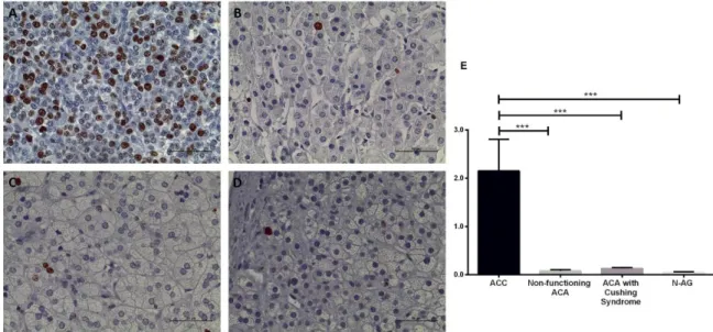
p27 is an excellent marker for the differential diagnosis between ACC and ACA ....54
Ki-67 is increased in ACC...58
No co-localization was observed between Ki-67 and p27 ...59
3.6 Discussion ...60
Chapter 4...63
CYP11B1 and CYP11B2 dual negativity is highly accurate for diagnosis of malignancy in functioning adrenocortical tumors ...63
4.1 Abstract ...65
4.2 Introduction ...66
4.3 Aim ...67
4.4 Material and Methods ...68
Case Selection ...68
Immunohistochemistry (IHC) and analysis ...68
Statistical analysis ...69
4.5 Results ...70
CYP11B1 expression in ACC is low ...70
CYP11B2 and CYP11B1 dual negativity is highly suggestive of malignant ACT ...70
StAR expression is decreased in ACC and ACAn ...72
4.6 Discussion ...75
Chapter 5...77
Angiogenesis and lymphangiogenesis in the adrenocortical tumors ...77
5.1 Abstract ...79
5.5 Results ...84
D2-40 expression ...84
CD31 expression ...84
Correlation between D2-40, CD31 expression with a steroidogenesis marker ...85
5.6 Discussion ...86
Chapter 6...89
Telomerase and N-cadherin differential importance in adrenocortical cancers and adenomas ...89
6.1 Abstract ...91
6.2 Introduction ...92
6.3 Aim ...95
6.4 Material and Methods ...96
Case Selection ...96
DNA extraction ...96
PCR and Sanger sequencing for TERT ...96
Telomerase, β-catenin and cadherins Immunohistochemistry (IHC) ...96
Statistical analysis ...97
6.5 Results ...98
No TERT promoter mutations were detected in ACT ...98
N-cadherin membrane expression is absent in the majority of ACC ... 100
β-catenin nuclear expression was present in both ACC and non-functioning ACA. ... 102
Nuclear telomerase and membrane N-cadherin expression were positively correlated in ACC... 103
6.6 Discussion ... 104
Chapter 7... 107
IGF2 role in adrenocortical carcinoma biology ... 107
7.1 Abstract ... 109
7.2 Introduction ... 110
7.3 Aim ... 111
7.4 Material and Methods ... 112
Adrenal tissue ... 112
IGF2 Immunohistochemistry (IHC) and data analysis ... 112
Cell Culture ... 112
Cell proliferation assay ... 113
Western Blot ... 114
Nuclear magnetic resonance (NMR) spectroscopy ... 114
Statistical analysis ... 116
7.5 Results ... 117
IGF2 in tumor and normal human adrenocortical tissues ... 117
The expression of IGF2 is significantly higher in ACC and ACAn ... 117
In vitro analysis of the influence of IGF2 in the H295R proliferation, viability, invasion and metabolism ... 118
A high IGF2 concentration increases H295R proliferation and viability ... 118
MEK inhibition reverts IGF2 triggered proliferation ... 118
IGF2 increases p27 expression ... 119
IGF2 does not influence cell invasion capacity ... 120
7.6 Discussion ... 122
Chapter 8... 125
MAPK/ERK pathway activation is a hallmark of malignancy and its inhibition is a promising treatment target for adrenocortical tumors ... 125
8.1 Abstract ... 127
8.2 Introduction ... 128
8.3 Aim ... 131
8.4 Material and methods ... 132
Adrenal tissue samples ... 132
Study of the adrenal hormonal secretion ... 132
Immunohistochemistry (IHC) procedures and analysis ... 132
Cell Culture ... 133
MEK Inhibitor treatment ... 133
Cell proliferation assay ... 133
Cell viability assay ... 134
Nuclear magnetic resonance (NMR) spectroscopy ... 134
Mitochondrial membrane potential assay ... 134
Western Blot ... 134
Steroids quantification ... 135
Statistical analysis ... 135
metabolism and steroidogenesis ... 139
MEK inhibitor significantly decreased H295R proliferation ... 139
Incubation of H295R cells with the highest concentration of MEK inhibitor increased glycolytic flux ... 140
Treatment with the lowest MEK inhibitor concentration increased acetate consumption while higher doses decreased it dose-dependently ... 141
Mitochondrial complexes analysis ... 142
MEK inhibition decreased steroids secretion by H295R ... 144
8.6 Discussion ... 145
Chapter 9... 149
Final Discussion ... 149
Molecular markers for differential diagnosis of adrenocortical tumors ... 151
Molecular targets for ACC treatment ... 152
New insights into the biology of adrenocortical carcinomas ... 153
What have we learned about steroidogenesis in the ACT? ... 155
Chapter 10 ... 157
Limitations and Future perspectives ... 157
References ... 161
Appendix 1 ... 193
Cell cycle regulators altered in adrenocortical carcinomas compared with adenomas ... 193
Appendix 2 ... 201
Publication 1 ... 201
Appendix 3 ... 213
Table 1- Modified Weiss system used for establishing differential diagnosis between
adrenocortical adenoma and adrenocortical carcinoma ... 10
Table 2 - Hereditary Tumor syndromes associated to adrenocortical tumors ... 11
Table 3 - World Health Organization (WHO)/ International Union Against Cancer (UICC) and European Network for the Study of Adrenal Tumors (ENSAT) classification of adrenocortical carcinoma... 12
Table 4 - Frequency of CYP11B1, CYP11B2, 17α-Hydroxylase and StAR immunostaining positivity in the different adrenocortical tumors ... 73
Table 5 - Correlation results between the CD31, D2-40 and the marker of steroidogenesis StAR ... 85
Table 6 - Telomerase reverse transcriptase’ nuclear expression in Adrenocortical carcinoma (ACC), Adrenocortical Adenoma (ACA) and Normal Adrenal Gland (N-AG). ... 99
Table 7 - N-cadherin membrane expression in Adrenocortical carcinoma (ACC), Adrenocortical Adenoma and Normal Adrenal Gland. ... 102
Table 8 - β-catenin staining localization distribution in the different study groups. ... 103
Table 9 – Antibodies used in this chapter ... 115
Figure 1 - Human adrenal gland stained by Masson tricromium ... 7
Figure 2 - Steroidogenesis in the different layers of the adrenal cortex. ... 8
Figure 3 - Flow chart for Adrenocortical carcinoma therapy ... 13
Figure 4 - Schematic representation of the canonical Wnt signaling pathway.. ... 18
Figure 5 - Alterations of chr11p15 in normal and malignant adrenocortical tissue ... 19
Figure 6 - IGF2 signaling ... 23
Figure 7- Schematic representation of the mammalian cell cycle. ... 25
Figure 8 - Schematic representation of p53 regulation ... 28
Figure 9 - Schematic representation of Rb regulation ... 31
Figure 10 - Schematic representation of CDC2/cyclin B regulation. ... 35
Figure 11 - Schematic representation of SAC regulation ... 40
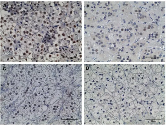
Figure 12 - Immunohistochemistry staining of p27 ... 54
Figure 13- Immunohistochemistry staining of p53 ... 55
Figure 14 - Immunohistochemistry staining of MDM2 ... 55
Figure 15 - Immunohistochemistry staining of p21 ... 56
Figure 16 - Immunohistochemistry staining of cyclin D1 ... 56
Figure 17 - Graphic representation of the percentage of p53, MDM2, p21, p27 and Cyclin D1 in the studied groups. ... 57
Figure 18 - ROC curves to assess the ability of the different molecular markers to distinguish between adrenocortical carcinomas from adrenocortical adenomas with the respective area under the curve. ... 57
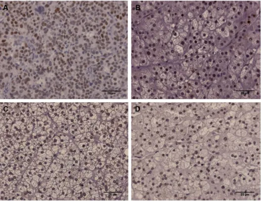
Figure 19 - Immunohistochemistry staining of Ki-67 and graphic representation of the percentage of the Ki-67 in the studied groups. ... 58
Figure 20 - ROC curves to assess the ability of Ki-67 to distinguish adrenocortical carcinomas from non-functioning adrenocortical adenomas, adenomas with Cushing syndrome and total adenomas with the respective area under the curve ... 58
Figure 21 - Immunofluorescence staining for Ki-67 and p27 in an ACC ... 59
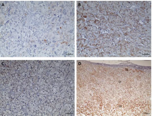
Figure 22 - Immunohistochemistry staining for CYP11B1 ... 70
Figure 23 - Immunohistochemistry staining for CYP11B2 ... 71
Figure 24 - Immunohistochemistry staining for 17α-Hydroxylase. ... 71
carcinomas adrenocortical adenomas with the respective area under the curve. ... 74
Figure 27 - Immunohistochemistry staining for D2-40 ... 84
Figure 28 - Immunohistochemistry staining of CD31 ... 85
Figure 29 - Immunohistochemistry staining of telomerase reverse transcriptase ... 99
Figure 30 - Immunohistochemistry staining of E-cadherin ... 100
Figure 31 - Immunohistochemistry staining of P-cadherin. ... 101
Figure 32 - Immunohistochemistry staining of N-cadherin ... 101
Figure 33 - Immunohistochemistry staining of β-catenin ... 102
Figure 34 - Relation between the N-cadherin and telomerase expression in the adrenocortical carcinomas. ... 103
Figure 35 - Immunohistochemistry staining for IGF2; graphic representation of the percentage of the area staining for IGF2 in the studied groups and ROC curves with the respective area under the curve to compare carcinomas and adenomas ... 117
Figure 36 - H295R cells proliferation (A) and viability (B) after incubation without or with IGF2 at the concentrations of 50 and 100ng/mL for 24 hours ... 118
Figure 37 - Relative phospho-ERK expression after IGF2 incubation at the concentrations of 50 and 100ng/mL for 5, 10 and 20 minutes (A). Cell proliferation (B) and viability (B) after IGF2 incubation (100ng/mL) with and without a MEK inhibitor (PD185352) at 10µM ... 119
Figure 38 - Relative p27 expression after 24 hours incubation with IGF2 at concentrations of 50 and 100ng/mL. ... 119
Figure 39 - Matrigel membrane invaded with H295R cells (A). N-cadherin expression after 24 hours incubation with IGF2 at the 50 and 100ng/mL concentrations evaluated by Western Blot (B) and immunofluorescence (C). ... 120
Figure 40 - Glucose (A) and glutamine (B) consumption; pyruvate (C), lactate (D) and alanine (E) production; lactate/glucose ratio (F) and lactate/alanine (G) after IGF2 incubation at the concentrations of 50 and 100ng/mL... 121
Figure 41 – MAPK/ERK Signaling Pathway. ... 129
Figure 42- Serum cortisol levels at 8.00h and 16.00h measured to assess the circadian rhythm and serum cortisol levels at 8.00h after the overnight 1mg Dexamethasone (Dxm) suppression test. ... 137
Figure 43 - Immunohistochemistry staining for phospho-ERK and the respective graphic representation of the phospho-ERKs staining score ... 138
Figure 44 - Immunohistochemistry staining for phospho-p38 and the respective graphic representation of the phospho-p38 staining score ... 139
incubation with a MEK inhibitor (PD184352) at concentrations of 0.1, 1 and 10 µM or with the vehicle (DMSO), during 12 and 24 hours ... 141 Figure 47 - JC1 ratio (A) and expression of the mitochondrial complexes III and IV (B and C) after the incubation with a MEK inhibitor (PD184352) at concentrations of 0.1, 1 and 10 µM or with the vehicle (DMSO), during 12 hours (B) and 24 hours (C) ... 143 Figure 48 - Cortisol, dehydroepiandrosterone sulfate (DHEA-S) and androstenedione secretion by H295R, after the incubation with a MEK inhibitor (PD184352) at concentrations of 1 and 10 µM or with the vehicle (DMSO), during 24 hours. ... 144
According with the Decreto de Lei nº115/2013 - Artigo 34º, I declare that in this thesis
were used results from the follow publications:
Pereira SS, Morais T, Costa MM, Monteiro MP, Pignatelli D (2013) The emerging role of the molecular marker p27 in the differential diagnosis of adrenocortical tumors. Endocrine connections 2 (3):137-145. doi:10.1530/EC-13-0025 (Publication 1 – Appendix 2)
Pereira SS, Máximo V, Coelho R, Batista R, Soares P, Guerreiro SG, Sobrinho‐Simões M, Monteiro MP, Pignatelli D (2016) Telomerase and N‐Cadherin Differential Importance in Adrenocortical Cancers and Adenomas. Journal of Cellular Biochemistry. doi:10.1002/jcb.25811 (Publication 2 – Appendix 3)
Publications submitted:
Pereira SS, Bourdeau I, Lacroix A, Monteiro MP, Pignatelli D, Cell cycle and apoptosis regulation in adrenocortical carcinoma, Submitted to Endocrine Reviews at March 2017 (Review proposal accepted)
Pereira SS, Costa MM, Guerrreiro SG, Monteiro MP, Pignatelli D, Angiogenesis and lymphangiogenesis in the adrenocortical tumors, Submitted to Pathology & Oncology Research at March 2017
The majority of the work presented in this thesis was performed by the author with supervision by Professor Mariana P. Monteiro and Professor Duarte Pignatelli. Any other collaboration is described below.
Chapter 3, 4 and 5: Immunohistochemistry studies and analysis were performed in collaboration with Tiago Morais and Madalena M. Costa.
Chapter 6: The screening of telomerase reverse transcriptase promoter mutations was performed in collaboration with Ricardo Coelho, Rui Baptista and Valdemar Máximo. The design of the experiments and the discussion of the results and implications were performed in collaboration with Valdemar Máximo, Paula Soares, Manuel Sobrinho-Simões and Susana Guerreiro.
Chapter 7: Immunohistochemistry studies were performed in collaboration with Madalena M. Costa; the cell culture studies were performed in collaboration with Ângela Moreira and Madalena M. Costa and the metabolites analysis were performed in collaboration with Marco G. Alves, Pedro F. Oliveira and Ivana Jarak.
Chapter 8: Immunohistochemistry studies were performed in collaboration with Jorge Ferreira; the cell culture studies were performed in collaboration with Madalena M. Costa; the metabolites analysis were performed in collaboration with Marco G. Alves, Pedro F. Oliveira and Ivana Jarak and the steroids quantification was performed in collaboration with Tiago Guimarães.
ACA Adrenocortical adenoma
ACAc Adrenocortical adenoma with Cushing syndrome ACAn Non-functioning adrenocortical adenoma
ACAt Total adrenocortical adenoma ACC Adrenocortical carcinoma
ACCc Adrenocortical carcinoma with Cushing syndrome ACT Adrenocortical tumor
ACTH Adrenocorticotropic hormone ALS Acid labile subunit
APC Adenomatous polyposis coli
APC/C Anaphase-promoting complex/cyclosome ASCT2 Alanine-Serine-Cysteine transporter AT1R Angiotensin II receptor type 1 ATM Ataxia telangiectasia mutated ATP Adenosine triphosphate ATR ATM and Rad3-related AUC Area under the curve AURK Aurora kinase
BrdU 5-bromo-2-deoxyuridine BUB Benzimidalozes
BUB1B Benzimidalozes homologue beta CAK Cdk-activating kinase
CAM Cell adhesion molecule Cdc2 Cell division cycle 2 Cdc25 Cell division cycle 25 CDK Cyclin-dependent kinase
CDKi Cyclin-dependent kinase inhibitor Chk1 Checkpoint kinase 1
Chr Chromosome
CK1α Casein kinase 1α CT Computed tomography
CYP11B2 Aldosterone synthase DAB 3,3’-diaminobenzidine DHEA Dehydroepiandrosterone
DHEA-S Ddehydroepiandrosterone sulfate DVL Dishevelled
Dxm Dexamethasone Ebp1 ErbB3 binding protein
EDP Etoposide, doxorubicin, cisplatin EDTA Ethylenediaminetetraacetic acid EMT Epithelial-mesenchymal transition ENC1 Ectodermal-neural cortex 1
ENSAT European Network for the Study of Adrenal Tumors ERK Extracellular signal-regulated protein kinase
ETS E26 transformation-specific
Fz Frizzled
GSK3-β Glycogen synthase kinase 3β HDAC Histone deacetylase
HPF High-power fields HRP Horseradish peroxidase HU Hounsfield units
IF Immunofluorescence
Ig Immunoglobulin
IGF1 Insulin-like growth factor 1
IGF1R Insulin-like growth factor 1 receptor IGF2 Insulin-like growth factor 2
IGF2R Insulin-like growth factor 2 receptor IGFBPs Insulin-like growth factor binding protein IHC Immunohistochemistry
IR Insulin Receptor
LPR Low-density lipoprotein receptor-related protein LYVE-1 Lymphatic endothelial hyaluronan receptor-1 MAD Mitotic arrest deficient protein
MAD2L1 Mitotic Arrest Deficient-Like 1 MAPK Mitogen-activated protein kinase Max MYC-associated protein X MCC Mitotic checkpoint complex MDM2 Murine doble minute-2 MPS1 Monopolar spindle 1
MRI Magnetic resonance imaging mTOR Mammalian target of rapamycin Myt1 Myelin Transcription Factor 1 N-AG Normal adrenal gland
NMR Nuclear magnetic resonance
PDE2A Phosphodiesterase 2A cGMP-stimulated
PHYHIP Phytanoyl-CoA 2-hydroxylase-interacting protein PI3K Phosphatidylinositol 3-kinase
PINK1 PTEN-induced putative kinase 1 Plk1 Polo-like kinase 1
pRb Retinoblastoma protein
PTEN Phosphatase and tensin homolog RALBP1 RalA-binding protein 1
Rbp1 ErbB3 binding protein
ROC Receiver operating characteristic ROS Reactive oxygen species
RPRM Reprimo
SAC Spindle assembly checkpoint SAS Sarcoma amplified sequence
SDS-PAGE Sodium dodecyl sulfate-polyacrylamide gel electrophoresis SE Standard Error
SHC Src homology 2 domain-containing transforming protein StAR Steroidogenic acute regulatory protein
TCA Tricarboxylic acid
TCF/LEF T cell-specific transcription factor/lymphoid enhancer-binding factor 1 TCGA The Cancer Genome Atlas
TERC Telomerase RNA component TERT Telomerase reverse transcriptase TGF-β Transforming growth factor β THS Tetrahydro-11-deoxycortisol
TK Tyrosine kinase
TNM Tumor–node–metastasis system TOP Topoisomerase
UICC Union for International Cancer Control VEGF Vascular endothelial growth factor WHO World Health Organization
ZF Zona Fasciculata
ZG Zona Glomerulosa
ZNRF3 Zinc RING finger 3 ZR Zona Reticularis
Adrenocortical tumors (ACT) are common neoplasms that are frequently found incidentally. Most of the tumors are benign while malignant adrenocortical carcinoma (ACC) is comparably more rare but often highly aggressive and with poor prognosis. The two major reasons for the clinical outcome of ACC are the difficulty of identifying malignant tumors at earlier stages and the non-existence of effective therapies.
The focus of this dissertation was the identification of the molecular patterns that characterize and are specific of adrenal malignancy, which could be useful in the clinical setting for the differential diagnosis of adrenocortical tumors. Once characterized the molecular features of ACC, our research efforts were then diverted towards the understanding of how the molecular fingerprints of ACC could be translated into different biological behaviors looking for insights to disclose potential therapeutic targets.
For that, we evaluated the expression pattern of a high range of molecular markers, involved in cell cycle regulation [p53, p21, murine doble minute-2 (MDM2), p27 and cyclin D1], cell proliferation (Ki-67), steroidogenesis [steroidogenic acute regulatory protein (StAR), 11β-hydroxylase (CYP11B1), aldosterone synthase (CYP11B2) and 17α-11β-hydroxylase], cell invasion (CD31 and D2-40), cell adhesion (N-, E- and P-cadherin and β-catenin), cell immortalization (telomerase) and cell signaling [insulin-like growth factor 2 (IGF2), phospho-ERK and phospho-p38] in tumor and normal adrenal tissues, to assess their possible usefulness for pathological diagnosis. By performing these studies, we showed that Ki-67 and p27 were the markers with the highest power to discriminate ACC from adrenocortical adenomas (ACA). Ki-67 is a well-stablish marker of malignancy in several cancers that has also been reported to be a useful tool for the differential diagnosis of ACT. In contrast, the utility of p27 for the pathological diagnosis of ACC had not been previously identified. So, we have demonstrated for the first time, that p27 could even be a more powerful diagnostic tool than 67, since it is able to exclude all ACA and diagnose more ACC when compared to Ki-67. Besides that our results suggested that p27 could possibly have an unknown role in adrenocortical tumorigenesis and thus represent a potential treatment target.
In addition, CYP11B1 was demonstrated to be very accurate for the distinction between ACC and adrenocortical adenomas with Cushing syndrome (ACAc), while CYP11B1 and CYP11B2 dual negativity was shown to be very specific for malignancy. Moreover, the incomplete pattern of the CYP11B1, CYP11B2 and 17α-hydroxylase expression in ACC can justify the increased secretion of steroid metabolites precursors witnessed in ACC which was observed in previous studies.
H295R, with IGF2 dose-dependently increased cell proliferation and viability, while IGF2 at different concentrations also modulates cell metabolism. So, we demonstrated that different IGF2 concentrations in ACC could be responsible for different biological behaviors of ACC and influence the response to treatment.
Another molecular fingerprint that was identified as potentially useful to distinguish different ACT is the pattern of expression of the adhesion molecule N-cadherin, due to the fact that the majority of the ACC depict a loss of cadherin membrane expression while all ACA retain cadherin membrane expression. Besides that, a positive correlation between the loss of the N-cadherin expression and the absence of telomerase expression was observed, suggesting the existence of a telomerase reverse transcriptase (TERT) non-canonical function in cell adhesion. We also found that TERT promoter mutations are infrequent in ACC and nuclear telomerase expression is only present in a minority of cases.
As ACC are often metastasized when first diagnosed, we decided to assess whether blood and lymph vessel density within ACT was correlated with malignancy or tumor functionality. Angiogenesis was shown to be increased in ACC, suggesting that this phenomenon may have an important role in ACT biological behavior, while lymph vascular density seems to be more closely related to the tumor functional status than malignancy.
Finally, once MAPK/MEK/ERK pathway activation is frequently dysregulated in the majority of human cancers, we decided to analyze the status of this pathway and to explore the potential of its inhibition as a therapeutic target in ACC. For that, we incubated H295R ACC cell line with different concentrations of a specific MAPK/MEK/ERK pathway inhibitor (PD184352). This inhibition lead to a significant decrease in cell proliferation as well as in steroidogenesis, besides increasing the redox state of the H295R cells. Overall, these findings suggest that MEK/MAPK/ERK signaling has a role in adrenocortical tumorigenesis that can be potentially targeted for ACC treatment, which if successfully achieved could lead to better clinical outcomes than the currently available therapy.
In summary, the results presented in this thesis are key to improve the pathological diagnosis of adrenocortical tumors and for driving the future development of novel anti-tumor therapies.
Os tumores adrenocorticais (ACT) são neoplasias comuns que frequentemente são detetados por acidente. A maioria destes tumores são benignos, enquanto que os tumores adrenocorticais malignos (ACC) são raros mas extremamente agressivos e com mau prognóstico. As duas principais razões para o desfecho clínico dos ACC são a dificuldade em identificar estes tumores malignos em fases precoces e a atual inexistência de terapias eficazes.
O objetivo desta dissertação foi a identificação de padrões moleculares que possam caracterizar e serem específicos da malignidade do ACT, e que possam ser úteis no cenário clinico para o diagnóstico dos tumores do córtex da suprarrenal. Uma vez identificados esses padrões moleculares nos ACC, focamos a nossa investigação na compreensão de como é que essas características moleculares poderiam ser traduzidas em diferentes comportamentos biológicos, levando à descoberta de potências alvos terapêuticos.
Para isso, avaliamos o padrão de expressão de uma variedade de marcadores moleculares envolvidos na regulação do ciclo celular (p53, p21, murine doble minute-2 (MDM2), p27 e ciclina D1), proliferação celular (Ki-67), esteroidogénese [steroidogenic acute regulatory
protein (StAR), 11β-hydroxylase (CYP11B1), aldosterone synthase (CYP11B2) and
17α-hidroxilase], invasão celular (CD-31 e D2-40), adesão celular (N-, E- e P-Caderina e β-catenina), imortalização celular (telomerase) e sinalização celular [insulin-like growth factor 2 (IGF2), fosfo-ERK and fosfo-p38] em tecido adrenocortical tumoral e normal, de modo a verificarmos a sua potencial utilidade no diagnóstico desta patologia. Realizando estes estudos, demonstramos que o Ki-67 e o p27 foram os marcadores com maior poder discriminativo no diagnóstico de ACC em relação aos adenomas adrenocorticais (ACA). O Ki-67 é um marcador de malignidade já bem estabelecido em vários cancros e já reportado anteriormente como útil no diagnóstico diferencial de ACT. Pelo contrário, a utilidade do p27 no diagnóstico patológico de ACC nunca tinha sido identificada. Assim, demonstramos pela primeira vez que o p27 pode ser uma ferramenta de diagnóstico mais poderosa que o Ki-67, uma vez que consegue excluir todos os adenomas e diagnosticar mais carcinomas do que o Ki-67. Além disso, os nossos resultados sugerem que o p27 tem possivelmente, um papel ainda desconhecido na tumorigénese de ACC e pode representar um potencial alvo terapêutico.
O CYP11B1 também demonstrou ser um marcador muito preciso na diferenciação entre ACC e adenomas adrenocorticais com síndrome de Cushing (ACAc). No que se refere à inexistência de marcação para ambos os CYP11B1 e o CYP11B2, esta demonstrou ser muito específico para definir malignidade. Adicionalmente, a baixa expressão do CYP11B1,
percussores de esteroides pelos ACC reportada em estudos anteriores.
O IGF2 também provou ser muito útil a distinguir ACC de adenomas adrenocorticais não funcionantes (ACAn). Por outro lado, a incubação da linha celular de carcinoma adrenocortical, H295R, com diferentes concentrações de IGF2, levou a um aumento dose-dependente da proliferação celular e viabilidade, ao tempo que diferentes concentrações de IGF2 modularam o metabolismo celular. Assim, demonstramos que diferentes concentrações de IGF2 nos ACC podem ser responsáveis por diferentes comportamentos biológicos e influenciar a resposta aos tratamentos.
Outra impressão molecular que foi identificada como potencialmente útil para o diagnóstico diferencial de ACT é a expressão da molécula de adesão N-caderina, devido ao facto da maioria dos ACC apresentar perda da expressão membranar da N-caderina ao passo que todos os ACA mantiveram essa expressão. Também foi observada uma correlação positiva entre a perda de expressão membranar da N-caderina e a ausência da expressão nuclear da telomerase, sugerindo a existência de uma função não canónica da telomerase reverse
transcriptase (TERT) na adesão celular. Também observámos que as mutações no promotor
da TERT são raras nos ACC e que a expressão nuclear da telomerase está presente apenas numa minoria dos casos.
Como a maioria dos ACC já metastizaram aquando do diagnóstico, decidimos avaliar a densidade de vasos sanguíneos e linfáticos dentro dos ACT e correlacionar essa densidade com a malignidade e com a funcionalidade tumoral. Observamos que a angiogénese estava aumentada nos tumores malignos enquanto que a densidade de vasos linfáticos parece estar mais associada com a funcionalidade dos tumores do que com a sua malignidade.
Finalmente como a via de sinalização MAPK/MEK/ERK se encontra frequentemente desregulada na maioria dos tumores humanos, nós decidimos avaliar a ativação desta via ACC e explorar o seu potencial como um alvo terapêutico. Para isso, incubamos a linha celular H295R com diferentes concentrações de um inibidor específico da via MAPK/MEK/ERK (PD184352). Esta inibição levou a uma diminuição significativa da proliferação celular bem como da esteroidogénese. Por outro lado, aumentou o estado redox das células H295R. No general, os nossos resultados sugerem que esta via tem um papel na tumorigénese adrenocortical e que pode ser um alvo potencial no tratamento de ACC, que pode levar a melhores resultados clínicos do que a terapia atualmente disponível.
Em sumário, os resultados presentes nesta tese são importantes tanto para a melhoria do diagnóstico patológico dos tumores adrenocorticais como também para impulsionar o
Chapter 1
1.1 Adrenal gland
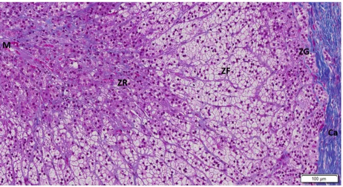
The adrenal glands are endocrine organs located above the superior pole of each kidney. Each gland has two distinct parts: an outer region, near the adrenal capsule, designated adrenal cortex that comprises 80% of the adrenal mass, and an inner region, so called adrenal medulla (Nussey and Whitehead 2001).
The adrenal cortex is responsible for adrenal steroid secretion and it is divided into three distinct morphological layers with different functionality, which are the glomerulosa, the fasciculata and the reticularis layers (Figure 1). These three layers present specific enzymatic features that are needed for the production of different steroids (Nussey and Whitehead 2001).
Figure 1 - Human adrenal gland stained by Masson tricromium (10x). Costa MM (not published): Ca-
capsule; ZG- zona glomerulosa; ZF- Zona fasciculata; ZR-Zona reticularis; M- medulla.
The outer layer is the glomerulosa layer that is responsible for mineralocorticoids production, predominantly aldosterone. Mineralocorticoid secretion is mainly regulated by angiotensin II but it can also be influenced by adrenocorticotropic hormone (ACTH) and potassium. The intermediate layer is fasciculata that produces glucocorticoids, mainly cortisol, having ACTH as the major regulator. The inner layer is reticularis that produces adrenal androgens, namely dehydroepiandrosterone (DHEA) and dehydroepiandrosterone sulfate (DHEA-S), which can be converted into testosterone or aromatized to estrogens in peripheral organs, such as the adipose tissue (Figure 2) (Mulrow and Franco-Saenz 1996, Nussey and Whitehead 2001).
The adrenal medulla, located in the center of the adrenal gland, is considered to be a modified sympathetic ganglion, as it produces catecholamines, such as norepinephrine and epinephrine (McCorry 2007).
Adrenal tumors can arise both from the adrenal cortex or medulla. Tumors originating from the cortex are named adrenocortical tumors, while those originating from the medulla are designated pheochromocytomas (Lee and Duh 2009).
Figure 2 - Steroidogenesis in the different layers of the adrenal cortex. In red are represented the
enzymes located in the mitochondria and at blue the enzymes located in the smooth endoplasmic reticulum.
1.2 Adrenocortical tumors
Adrenocortical tumors (ACT) are common tumors of the adrenal cortex, affecting 3% to 10% of the human population (Else, Kim et al. 2014). These can be classified according to their biological behavior in benign or malignant, and according their functionality in non-functioning or functioning tumors (Lee and Duh 2009). The majority of the ACT are benign, non-functioning and discovered incidentally during imaging for unrelated clinical reasons (Pignatelli 2011). The detection of non-functioning adrenocortical tumors (both benign and malignant) increased significantly over the last years, due the widespread use of computed tomography (CT), magnetic resonance imaging (MRI) and abdominal ultrasonography (Low, Dhliwayo et al. 2012, Audenet, Mejean et al. 2013).
Functioning ACT can secrete steroids autonomously and independently of ACTH or renin-angiotensin system regulation, leading to various clinical syndromes depending on the secreted steroids, namely Conn’s syndrome when aldosterone secreting, Cushing’s syndrome for cortisol secreting or virilizing syndrome in result of androgen production (Audenet, Mejean et al. 2013, Else, Kim et al. 2014). Some malignant tumors are able to secrete precursor steroids or even inactive steroids and hence in spite of being hormonally functioning do not produce a clinical syndrome (Arlt, Biehl et al. 2011). The symptoms related with excessive hormone secretion can occasionally accelerate the adrenal tumor finding. Thus, due to the elevated possibility of apparently asymptomatic patients harboring functional tumors, according to the European Network for the Study of Adrenal Tumors (ENSAT), all patients with identified adrenal masses should undergo endocrine function tests for glucocorticoid (minimum 3 of 4 tests), sexual steroids and steroid precursors production, mineralocorticoid excess and catecholamine excess (Fassnacht, Johanssen et al. 2009, Berruti, Baudin et al. 2012).
The prognosis of adrenocortical carcinomas is very different from adrenocortical adenomas which stresses the importance of differential diagnosis between the two entities (Soon, McDonald et al. 2008, Lafemina and Brennan 2012). However, distinguishing benign from malignant adrenocortical tumors is not always simple. So far, the size of the lesion on the imaging studies are the strongest predictor of the adrenocortical malignancy and according to recent guidelines, ACT with more than 4 cm should be treated surgically. (Nieman 2010, Kapoor, Morris et al. 2011). Although the tumors size needs to be taken into account, it should not be used alone in order to avoid disregarding small sized malignant tumor masses at a stage where these could have a better prognosis (Lafemina and Brennan 2012). In addition to size, a threshold value of 10 Hounsfield units (HU) of unenhanced CT scan was shown to have a high specificity and sensibility for the differential diagnosis of adrenocortical malignant lesions, particularly when associated with decreased contrast washout (Allolio and Fassnacht 2006, Mazzuco, Durand et al. 2012).
After surgical removal, the pathological diagnosis is based on the tumor macroscopic characteristics, such as size, presence of an intact capsule, areas of necrosis or hemorrhage, adjacent organ invasion and lymph node metastasis. At the microscopic level, there are several morphological features used to assess malignancy whenever metastasis have not been identified. The most widely used method for pathological diagnosis is the Weiss scoring system that relies in criteria such as, nuclear grade, mitotic rate, abnormal mitosis, proportion of clear cells, necrosis, diffuse architecture, invasion of the tumor capsule, sinusoid and venous invasion (Weiss 1984, Allolio and Fassnacht 2006, Lafemina and Brennan 2012). More recently, a modified Weiss scoring system has been proposed, based on the five strongest and more reliable criteria (mitotic rate, abnormal mitosis, proportion of clear cells, necrosis, and capsular invasion), thus eliminating the parameters that were considered to be subjective or difficult to interpret in the original Weiss system (Table 1) (Lau and Weiss 2009, Tissier 2010).
Table 1- Modified Weiss system used for establishing differential diagnosis between adrenocortical
adenoma and adrenocortical carcinoma [adapted from (Lau and Weiss 2009)].
Histological Criteria 0 1
Mitotic rate <5 per 50 high-power fields (HPF) >5 per 50 HPF
Abnormal mitoses Absent
Presence of abnormal distribution of chromosomes or
excessive number of mitotic spindles
Necrosis Absent Present
Clear Cells Clear cells comprising > 25% of the tumor
Clear cells comprising 25% or less of the tumor
Capsular invasion Absence of capsular invasion
Nests or cords of tumor extending into or through tumor
capsule
Weiss score calculation: 2x mitotic rate criterion + 2x clear cells criterion + abnormal mitoses + necrosis + capsular invasion (score of 3 or more suggests malignancy)
1.2.1 Adrenocortical carcinomas
ratio of 1.5:1 (Roman 2006, Low, Dhliwayo et al. 2012, Chagpar, Siperstein et al. 2014). The tumors are even rarer in children although there is a region in South Brazil where their incidence is ten times higher than the rest of the world (Ribeiro, Michalkiewicz et al. 2000). The majority of the ACC are functional (50-60%), with Cushing’s syndrome alone being the most frequent presentation among adults (45%), followed by Cushing’s syndrome in association with a virilization syndrome (25%) (Ng and Libertino 2003, Allolio and Fassnacht 2006, Pignatelli 2011).
ACC may be also associated with hereditary syndromes (Table 2) such as Li-Fraumeni syndrome, Beckwith-Wiedemann syndrome, and multiple endocrine neoplasia type 1, Lynch syndrome, familial polyposis coli syndrome or more rarely even with congenital adrenal hyperplasia or Carney complex (Else, Kim et al. 2014). It must be stressed however, that the majority of ACC are in fact sporadic (Else 2012, Mazzuco, Durand et al. 2012, Chagpar, Siperstein et al. 2014).
Table 2 - Hereditary Tumor syndromes associated to ACT [adapted from (Soon, McDonald et al. 2008)].
Hereditary tumor syndrome
Gene (locus) Prevalence of ACT
Li-Fraumeni syndrome TP53 (ch17p13),
hCHK2 (ch22q12.1) ACC: 3%–4%
Beckwith-Wiedemann syndrome IGF2, H19, CDKN1C,
KCNQ1 (ch11p15) ACC: 5%
Multiple endocrine neoplasia 1 MEN1 (ch11q13) ACA: 55%; ACC: rare
Congenital adrenal hyperplasia CYP21B (ch6p21.3) ACA: 82%; hyperplasia: 100%; ACC: rare
ACA: adrenocortical adenoma; ACC: adrenocortical carcinoma; ACT: adrenocortical tumor
Adrenocortical carcinoma staging
The disease stage and margin-free resection are the majors’ prognostic factors in ACC (Fassnacht, Johanssen et al. 2009, Berruti, Baudin et al. 2012). There are two main tumor– node–metastasis (TNM) classifications to evaluate ACC, the one proposed by the International Union Against Cancer (UICC) in 2004 and more recently the TNM classification proposed by the ENSAT that seems to have an improved prognostic accuracy (Table 3) (Fassnacht, Johanssen et al. 2009, Fassnacht, Libe et al. 2011).
According the ENSAT classification, the 5-year disease-specific survival rate is approximately 82% for stage I, 61% for stage II, 50% for stage III, and 13% for stage IV (Fassnacht, Johanssen et al. 2009). The majority of the ACC are diagnosed in an advanced stage leading
to a poor prognosis (Else, Kim et al. 2014). The most common metastatic sites of the ACC are lung (46-79%), liver (44-93%), lymph nodes (18-73%) and peritoneum (16-79%) (Allolio, Hahner et al. 2004).
Table 3 - World Health Organization (WHO)/ International Union Against Cancer (UICC) and European
Network for the Study of Adrenal Tumors (ENSAT) classification of adrenocortical carcinoma [adapted from (Fassnacht, Johanssen et al. 2009) and (Lacroix 2016)].
Stage WHO/UICC (2004) ENSAT (2008)
I T1; N0; M0 II T2; N0; M0 III T3; N0; M0 T3-4; N0; M0 T1-2; N1; M0 T1-4; N1; M0 IV T3; N1; M0 Any M1 T4; N0-1; M0 Any M1
T1: tumor ≤5 cm; T2: tumor >5 cm; T3: tumor infiltration in surrounding tissue; T4: tumor invasion in adjacent organs; N0: no positive lymph nodes; M0: no distance metastases; N1: positive lymph nodes; M1: presence of distant metastasis
Adrenocortical carcinoma treatment
Surgery is the most important treatment for ACC, while complete surgical resection (R0) is the only potential curative approach (Figure 3) (Fassnacht, Johanssen et al. 2009, Libe 2015). Even though there is a high rate of ACC recurrence after R0 surgery, thus adjuvant therapy is mandatory (Libe 2015). Open adrenalectomy is the most consensual operation type, since laparoscopy carries a greater risk of malignant cell dissemination (Leboulleux, Deandreis et al. 2010, Libe 2015). In addition, Reibentanz et al reported a significantly reduced risk for tumor recurrence and disease related death if the lymph nodes were resected during the adrenalectomy (Reibetanz, Jurowich et al. 2012).
In patients with incomplete resection (R1) or undefined resection (Rx), adjuvant mitotane therapy is recommended to reduce the risk of recurrence and control the hormone excess, which can be conjugated with tumor irradiation (Allolio and Fassnacht 2006, Terzolo, Angeli et al. 2007, Fassnacht, Libe et al. 2011, Berruti, Baudin et al. 2012). However, the benefits of mitotane as adjuvant therapy have been questioned due to the lack of data from controlled clinical trials or from large prospective studies with consistent assessments of mitotane dosing (Kopf, Goretzki et al. 2001, Pignatelli 2011, Berruti, Baudin et al. 2012). Furthermore, the
Mitotane is also associated with significant toxicity, such as, adrenal insufficiency requiring compensation with hydrocortisone; vertigo; central nervous system disorders and gastro-intestinal side effects (Kroiss, Quinkler et al. 2011, Libe 2015).
Figure 3 - Flow chart for Adrenocortical carcinoma therapy [adapted from (Fassnacht, Libe et al. 2011,
Else, Kim et al. 2014)].
In metastatic ACC when total resection is not technically possible, surgery can only be recommended to control excess hormone production, in slow progressing tumors or in tumors where 80% of their mass can be resected (Libe 2015). Still, the first line therapy for advanced ACC is mitotane alone or in the combination with other drugs, such as etoposide, doxorubicin, cisplatin (EDP) or with streptozotocin (Sz) (Fassnacht, Libe et al. 2011, Berruti, Baudin et al. 2012). Mitotane in combination with EDP is associated with a better progression-free survival as compared to the mitotane with Sz combination, however no difference was observed on overall survival (Fassnacht, Libe et al. 2011, Libe 2015).
Similarly to what is routinely used for other types of tumors, molecular and immunohistochemistry markers are now being considered of great potential as diagnostic and prognostic tools in adrenocortical tumors (Wachenfeld, Beuschlein et al. 2001, Allolio and Fassnacht 2006, Tissier 2010). Overexpression of Ki-67 proliferation marker and Insulin-like
growth factor 2 (IGF2) has been repeatedly demonstrated to be good predictors of malignancy and prognosis (Wachenfeld, Beuschlein et al. 2001, Tissier 2010, Lafemina and Brennan 2012). However, no single or combination of molecular markers has yet been validated for use in the clinical practice. While more recently, the Helsinki Score has been proposed as diagnosis and staging tool for adrenocortical carcinomas, based not only on morphological features, such as the Weiss system, but also on the Ki-67 index (Helsinki Score: 3 × mitotic rate +5 × necrosis + Ki-67 index). Duregon et al, verified that this score was better compared to the Weiss system in predicting ACC prognosis (Duregon, Cappellesso et al. 2016).
1.3 Pathophysiology of Adrenocortical carcinoma
The key molecular mechanisms that seem to be involved in the ACC pathophysiology are the activation of the Wnt pathway, IGF2 system and cell cycle alterations, which include those occurring in the well-studied tumor gene suppressor, p53.
1.3.1 Wnt Signaling pathway
Wnt signaling pathway is a highly conserved molecular cascade that requires glycoproteic extracellular ligands of the Wingless family (Wnts) for activation, which regulates a diversity of cellular processes essential for embryonic development and homeostatic systems (Komiya and Habas 2008).
The majority of the early studies on the Wnt signaling pathway focused on the canonical β-catenin-dependent pathway. However, in the past few years, other Wnt pathways have been disclosed including the non-canonical planar cell polarity pathway involved in ciliogenesis and the non-canonical Wnt/calcium pathway involved in cell movement (Komiya and Habas 2008). The β-catenin-dependent Wnt signaling role in the normal adrenocortical tissue development, homeostasis and tumorigenesis is well-stablished (Berthon, Martinez et al. 2012), and thus described here in further detail.
Canonical Wnt signaling pathway
β-catenin is a molecule associated with cadherin membrane proteins, which plays a structural role in the cell adhesion, as well as in the Wnt signaling pathway (Wijnhoven, Dinjens et al. 2000, Komiya and Habas 2008) (Figure 4).
targeted for degradation by the proteasome (Figure 4) (Komiya and Habas 2008). The bind of Wnt ligands to Frizzled (Fz) receptors with low-density lipoprotein receptor-related protein (LPR) coreceptor, results in the inhibition of the regulatory complex Axin-APC-GSK3-β, leading to β-catenin accumulation in the cytoplasm and nuclear translocation. In the nucleus, β-catenin regulates target gene expression by binding to the T cell-specific transcription factor/lymphoid enhancer-binding factor 1(TCF/LEF) (Figure 4) (Komiya and Habas 2008).
In normal adrenal gland development, Wnt signaling activity is restricted to the zona glomerulosa and it is involved in cortex zonation (Berthon, Drelon et al. 2014, Walczak, Kuick et al. 2014). Besides that, β-catenin activation is associated with an upregulation of Angiotensin II receptor type 1 (AT1R) and aldosterone synthase (Berthon, Sahut-Barnola et al. 2010, Berthon, Drelon et al. 2014).
β-catenin in adrenocortical carcinomas
Tissier et al was the first author to demonstrate that Wnt signaling activation was frequent in adrenocortical tumors. Cytoplasmic and/or nuclear accumulation of β-catenin was found in the majority of the analyzed adrenocortical tumors, being higher in ACC (77%). Activating somatic mutation in CTNNB1, the gene that encodes β-catenin, was shown in a similar percentage of adrenocortical adenomas (ACA) (27%) and ACC (31%) and these mutations were observed only in ACT with abnormal β-catenin accumulation and most were point mutations on exon 3 that corresponds to a GSK3-β/CK1 phosphorylation site. These mutations prevent β-catenin phosphorylation and induce accumulation of the protein. In ACA, β-catenin alterations were more frequent in nonfunctioning ACA, suggesting that β-catenin pathway activation might be involved in the development of non-functioning ACT adrenocortical adenomas and adrenocortical carcinomas (Figure 4) (Tissier, Cavard et al. 2005).
This study was soon followed by others that showed CTNNB1 mutation to be present with equal prevalence in benign and malignant ACT, as well as associated steroidogenesis dysfunction and thus more prevalent in non-functioning tumors (Masi, Lavezzo et al. 2009, Bonnet, Gaujoux et al. 2011, Parviainen, Schrade et al. 2013, Kovach, Nucera et al. 2015). Through the comparison of ACC with different outcomes, CTNNB1 mutation and abnormal β-catenin immunostaining was shown to be present predominantly in ACC with poor-outcome and ENSAT stages III and IV, thus with locally advanced or metastatic disease (Ragazzon, Libe et al. 2010, Gaujoux, Grabar et al. 2011). The presence of β-catenin nuclear staining was positively correlated with the presence of tumor necrosis and high mitotic count (Gaujoux, Grabar et al. 2011)
Non-functioning ACA harboring CTNNB1 mutations were found to be significantly larger and heavier than those ACA without these mutations (Bonnet, Gaujoux et al. 2011). Durand et al, using microarray analysis identified the genes that are differentially expressed in ACT with
CTNNB1 mutations and high nuclear β-catenin immunostaining compared with wild-type CTNNB1 ACT without nuclear accumulation of β-catenin. Isthmin1, zebrafish homolog (ISM1),
RalA-binding protein 1 (RALBP1), phosphodiesterase 2A cGMP-stimulated (PDE2A) and ectodermal-neural cortex 1 (ENC1) were found to be overexpressed in the mutated tumors, while phytanoyl-CoA 2-hydroxylase-interacting protein (PHYHIP) was found to underexpressed (Durand, Lampron et al. 2011).
Using a transgenic mouse model with constitutive β-catenin activation in the adrenal cortex (ΔCat), Berthon et al observed a progressive sub-capsular cell hyperplasia of steroidogenic cells due to proliferation and ectopic expansion of sub-capsular cell populations resulting in cortical and medullary dysplasia. 10-month-old mice developed primary hyperaldosteronism and over a 17 months’ time course, ΔCat mice adrenal glands developed malignant characteristics such as neovascularization and local invasion, demonstrating β-catenin role as an adrenal oncogene, leading to the development of benign aldosterone-secreting ACT and promoting malignancy (Berthon, Sahut-Barnola et al. 2010).
H295R is a human adrenocortical cancer cell line that harbors an activating CTNNB1 mutation on the GSK3β phosphorylation site. Gaijoux et al silenced the CTNNB1 gene in H295R cells leading to a specific Wnt signaling pathway inactivation with significant reduction of CTNNB1 and AXIN2 expression, which was responsible for a decrease in cell proliferation, accumulation of cells in the G1 phase and increased apoptosis in vitro. CTNNB1 gene silenced H295R cell xenografts in athymic nude mice completely prevented tumor cells growth as compared to what was observed using unsilenced H295R cells of the control group (Gaujoux, Hantel et al. 2013).
Alterations of other components of the canonical Wnt pathway found in ACT
Since abnormal β-catenin immunostaining is more frequent in ACC than in ACA, despite the similar rate of CTNNB1 mutations, it has been hypothesized that other components of Wnt signaling pathway could also be involved.
Wnt pathway co-receptors LRP5 and LRP6 expression is more frequently observed in ACA that in ACC, with LPR5 expression being more frequent in virilizing ACT, while LRP6 expression is similar regardless ACT subtype (Parviainen, Schrade et al. 2013).
4 is one of the Wnt ligands that is a critical component of the reproductive system. WNT-4 gene expression was found to be significantly higher in aldosterone producing ACA, and lower in cortisol secreting ACA with Cushing syndrome and virilizing ACC as compared to normal adrenal glands, ACC with Cushing syndrome and virilizing ACA (Kuulasmaa,
observed 12-bp deletion in exon 7 of AXIN2 gene in 7% of ACA and 17% of ACC (Chapman, Durand et al. 2011). Guimier et al observed this AXIN2 genetic change only in 2% of ACC analyzed and no AXIN1 alterations were found in ACC (Figure 4) (Guimier, Ragazzon et al. 2013). APC mutations were found in 2-3.3% of ACC cases (Gaujoux, Grabar et al. 2011, Assie, Letouze et al. 2014, Zheng, Cherniack et al. 2016).
Zinc RING finger 3 (ZNRF3) is also associated with the Wnt receptor complex that inhibits Wnt signaling by promoting Fz and LRP6 receptors degradation (Hao, Xie et al. 2012). Assié et al reported the finding of ZNRF3 gene somatic alterations in 21% of ACC, which included homozygous deletions and mutations. In their ACC series, CTNNB1 and ZNRF3 alterations were found to be mutually exclusive. ACC with altered ZNRF3 presented β-catenin gene targets activation, but it was weaker than in ACC with CTNNB1 mutations (Assie, Letouze et al. 2014). Another research group has only observed ZNRF3 gene alterations to be present in 12.2% of the analyzed ACC cases (Juhlin, Goh et al. 2015). The Cancer Genome Atlas (TCGA) study found ZNRF3 homozygous deletion (chr22q12.1) and non-silent mutations of this gene in 16% and 19.3% of ACC, respectively (Zheng, Cherniack et al. 2016). ZNRF3 role as tumor suppressor gene in ACC has been confirmed by Hanin et al after observing that ZNRF3 overexpressing H295R cells showed decreased cell proliferation and an increased apoptosis, while ZNRF3 silenced H295R cells were protected against apoptosis (Figure 4) (Hanin, Marthe et al. 2016).
Wnt pathway as a treatment target for ACC
The accumulated evidence supporting the role of the Wnt pathway in ACT tumorigenesis, compounds that inhibit β-catenin transcription, such as PKF115-584 and PNU74654, were tested in vitro to evaluate their therapeutic potential for ACC.
PKF115–584 dose-dependently inhibited H295R proliferation, even in the presence of increased steroidogenic factor-1 levels, a protein well-known to be involved in this cell line proliferation; while it also decreased the percentage of H295R cells in S-phase and increased apoptosis (Doghman, Cazareth et al. 2008). PNU-74654 also decreased H295R proliferation and increased early and late apoptosis. Besides that PNU-74654 inhibited the H295R steroidogenesis, decreasing cortisol, testosterone, androstenedione production levels, SF1 and CYP21A2 gene expression and protein levels of steroidogenic acute regulatory protein (StAR) and aldosterone synthase. This study confirmed the role of Wnt pathway in the control of initial and late steps of steroidogenesis (Leal, Bueno et al. 2015). Therefore, this data supports that these inhibitors may become useful for treatment of tumors with activated Wnt signaling pathway.
Figure 4 - Schematic representation of the canonical Wnt signaling pathway. In the absence of Wnt
ligands, β-catenin is rapidly phosphorylated by GSK3-β, a member of a regulatory complex composed by Axin, APC and CK1α. β-catenin phosphorylation results in its ubiquitination and degradation. ZNRF3 is associated with the Fz receptor complex that inhibits Wnt signaling pathway by promoting Fz receptor
1.3.2 IGF2 System
IGF2 is a growth factor secreted mainly in the liver but also in the majority of the tissues where it can act in an autocrine or a paracrine way. IGF2 is described to regulate the cell growth, differentiation and metabolism that is mostly expressed during embryogenesis to promote fetal growth (Livingstone 2013).
IGF2 gene is located at chr11p15 and it is only expressed from the paternal allele, while the
maternal one is imprinted by promoter methylation (DeChiara, Robertson et al. 1991). The chr11p15 region is organized in two different clusters: a telomeric domain that includes the
IGF2 gene and the H19 gene and a centromeric domain that includes the CDKN1C gene
(Figure 5) (DeChiara, Robertson et al. 1991, Ribeiro and Latronico 2012). The CDKN1C encodes a cyclin dependent kinase inhibitor that regulates the G1-S phase of the cell cycle and its role in the ACT that will be further detailed under the “Cell Cycle” topic of this Thesis.
IGF2 overexpression in adrenocortical tumors
Loss of the maternal allele or loss of the imprinting with the duplication of the paternal allele leads to increased IGF2 expression and decreased H19 and CDKN1C expression. Alterations in the chr11p15 region are frequently observed in sporadic ACT (Gicquel, Raffin-Sanson et al. 1997, Ribeiro and Latronico 2012) (Figure 5).
Figure 5 - Alterations of chr11p15 in normal and malignant adrenocortical tissue. The imprinted chr11p15 locus contains the genes CDKN1C, IGF2, and H19. In normal adrenocortical tissue, only the paternal allele of the IGF2 gene is expressed and the maternal alleles of CDKN1C and H19 are expressed. In adrenocortical carcinoma, paternal isodisomy with loss of the maternal allele is frequently observed, which leads to IGF2 overexpression.
Gicquel et al described that loss of heterozygosity (LOH) in chr11p15, characterized by the loss of the maternal allele and duplication of paternal one, and/or IGF2 gene overexpression were found in 93.1% of ACC and in only 8.6% of ACA (Gicquel, Raffin-Sanson et al. 1997). LOH in chr11p15 was correlated with an increase of the Weiss score and associated with higher risk of ACC recurrence. It also showed to be a strong predictor of shorter disease-free survival (Gicquel, Bertagna et al. 2001).
Many studies have demonstrated that IGF2 gene expression was 10 to 20 fold higher in ACC when compared to ACA and normal adrenal glands (Ilvesmaki, Kahri et al. 1993, Gicquel, Raffin-Sanson et al. 1997, Boulle, Logie et al. 1998, Gicquel, Bertagna et al. 2001, de Fraipont, El Atifi et al. 2005, Giordano, Kuick et al. 2009, Soon, Gill et al. 2009, Ragazzon, Assie et al. 2011, Guillaud-Bataille, Ragazzon et al. 2014, Nielsen, How-Kit et al. 2015). Higher IGF2 expression levels were associated with more aggressive phenotype and risk of ACC recurrence (Boulle, Logie et al. 1998, Gicquel, Bertagna et al. 2001). Chr11p15 LOH was found to have a greater prognostic value when compared to IGF2 overexpression, suggesting that the loss of genes expressed from the maternal allele are important for ACC tumorigenesis (Gicquel, Bertagna et al. 2001)
The IGF2 protein expression has also been reported to be 8 to 80 fold higher in ACC than in ACA or normal adrenal glands (Ilvesmaki, Kahri et al. 1993, Boulle, Logie et al. 1998, Erickson, Jin et al. 2001, Schmitt, Saremaslani et al. 2006, Soon, Gill et al. 2009), although no differences in IGF2 plasma levels have been described between patients with ACA, ACC or healthy volunteers (Patel, Ellis et al. 2014). The utility of IGF2 combined with Ki-67 for the differential diagnosis between ACC and ACA was found to be highly sensitive (96-100%) as well as specific (95.5-100%) (Schmitt, Saremaslani et al. 2006, Soon, Gill et al. 2009).
When Guillaud-Bataille et al compared ACC with high and low mRNA IGF2 levels, these were found to present similar Weiss scores, Ki-67 indexes, ENSAT stages, overall and event-free survival rates and occurrence of metastasis, thus the authors concluded that IGF2 status correlates with malignancy but it is not a good prognosis marker (Guillaud-Bataille, Ragazzon et al. 2014).
Transgenic mice overexpressing human IGF2, despite having increased IGF2 serum levels and moderate expression in the adrenal cortex, only depict mild adrenocortical hyperplasia due to the increased fasciculata volume and do not develop ACT (Weber, Fottner et al. 1999). In addition, in another study using two different transgenic mouse models, one overexpressing IGF2 specifically in the adrenal cortex and other overexpressing IGF2 and constitutively active β-catenin in the adrenal cortex, have shown that IGF2 was able to recruit adrenal progenitor cells but not to induce adrenocortical tumor development. In mice with constitutive β-catenin expression, IGF2 overexpression did not seem to influence benign β-catenin-induced tumors, although a mild increase of Weiss score and tumor proliferation was observed for ACC in a late stage (Drelon, Berthon et al. 2012). These studies suggest that IGF2 overexpression does not has a role in adrenocortical tumor development but could be involved in malignant progression (Weber, Fottner et al. 1999, Drelon, Berthon et al. 2012).
![Table 1- Modified Weiss system used for establishing differential diagnosis between adrenocortical adenoma and adrenocortical carcinoma [adapted from (Lau and Weiss 2009)]](https://thumb-eu.123doks.com/thumbv2/123dok_br/18903282.935548/40.892.116.777.548.934/modified-establishing-differential-diagnosis-adrenocortical-adenoma-adrenocortical-carcinoma.webp)