UNIVERSIDADE DE LISBOA
Faculdade de Medicina Veterinária
CHARACTERIZATION OF IN VITRO MODELS FOR THE STUDY OF CANDIDATE G-QUADRUPLEX LIGANDS TARGETING THE HUMAN C-KIT PROTO-ONCOGENE
PROMOTER
LARA ZORRO SHAHIDIAN
CONSTITUIÇÃO DO JÚRI ORIENTADOR
CO-ORIENTADORA
2013 LISBOA
Doutora Maria Inês Sanches Falcão da Fonseca
Doutor Mauro Dacasto
Doutora Berta Maria Fernandes Ferreira São Braz
Doutor António José de Freitas Duarte Doutor Mauro Dacasto
Doutor Fernando António da Costa Ferreira
UNIVERSIDADE DE LISBOA
Faculdade de Medicina Veterinária
CHARACTERIZATION OF IN VITRO MODELS FOR THE STUDY OF CANDIDATE G-QUADRUPLEX LIGANDS TARGETING THE HUMAN C-KIT PROTO-ONCOGENE
PROMOTER
LARA ZORRO SHAHIDIAN
DISSERTAÇÃO DE MESTRADO INTEGRADO EM MEDICINA VETERINÁRIA
CONSTITUIÇÃO DO JÚRI ORIENTADOR
CO-ORIENTADORA
2013 LISBOA
Doutor Mauro Dacasto
Doutora Berta Maria Fernandes Ferreira São Braz
Doutor António José de Freitas Duarte Doutor Mauro Dacasto
Doutor Fernando António da Costa Ferreira
i Inscription
“After years of having a dog, you know him. You know the meaning of his snuffs and grunts and barks. Every twitch of the ears is a question or statement, every wag of the tail is an exclamation.”
― Robert R. Boy's Life
- I dedicate this work to my sister, who taught me the meaning of true friendship! -
iii Acknowledgements
To my supervisor, Prof. Dr. Mauro Dacasto, for allowing me to develop the work necessary for this thesis with his research group. I am deeply thankful for all the insides, support and guidance given to me during and after my training period. It was a privilege I will never forget.
To my co-supervisor, Prof. Dr. Berta São Braz, for being a constant source of support and encouragement. For all the trust and time dedicated to guarantee the success of this thesis.
To Dr. Eleonora Zorzan, without whom this work could not be possible. For all the hours we spent together in the lab and the incomparable dedication and friendship given to me.
To Dr. Vanessa Zancanella, with whom I learned so much. For all the knowledge, guidance and friendship given to me.
To Prof. Dr. Mery Giantin, for being an example of what a researcher should be. Dedicated, hard-worker and humble, three characteristics of an exceptional professional.
To Dr. Rosa Loparelli, for teaching me everything is possible with determination and persistence.
To the remaining staff of the Faculty of Veterinary Medicine of the University of Padua, who made my time in Italy so pleasant and joyful.
To all my friends who always encouraged me and gave me the energy to move forward. For helping me be a better person every day.
And finally, to my family. To my mother and father, who taught me to always follow my dreams and supported me in every decision I ever made. To my sister, to whom I dedicate this work, for being the steadiest rock in my life. To my brother, for reminding me every day we are responsible for our own future.
v Abstract
Characterization of in vitro models for the study of candidate G-quadruplex ligands targeting the human c-KIT proto-oncogene promoter
Proto-oncogene c-KIT has been implicated in the development and growth of several tumors, e.g. mast cell tumors (MCT) in dogs and gastrointestinal stromal tumors (GIST) in humans. Several therapeutic approaches directed to the blocking of receptor tyrosine kinases (RTK), such as c-KIT, have been created. However, after a short period of recovery, these drugs lose efficiency and the tumor relapses. A new approach, aiming to control c-KIT’s transcription, is being tested. This approach relies on the use of small molecule inhibitors (SMI) that specifically block DNA secondary structures, G-quadruplexes, located on the promoter regions of many proto-oncogenes, including c-KIT.
The main goal of this work is the development of in vitro models through which the study of candidate SMIs for human c-KIT is possible.
An in vitro model, composed by cytotoxicity tests aimed for the determination of the SMI’s inhibitory concentration 50 (IC50) on two human cell lines and by real time quantitative PCR (qPCR) for the study of gene expression alterations, has been developed and validated. The cytotoxicity tests were also used to identify the IC50 of three candidate ligands for c-KIT.
vii Resumo
Caracterização de modelos in vitro para o estudo de possíveis ligandos de G-quadruplex dirigidos à região promotora do proto-oncogene c-KIT no Homem
O proto-oncogene c-KIT tem sido relacionado com o desenvolvimento e crescimento de vários tumores, incluindo mastocitomas em cães e tumores do estroma gastrointestinal no Homem. Várias abordagens terapêuticas têm vindo a ser desenvolvidas tendo como objetivo bloquear os recetores de tirosina quinase, tais como c-KIT. No entanto, após um curto período de recuperação, estes fármacos perdem eficácia e o tumor reaparece. Está a ser testada uma nova abordagem, que visa controlar a transcrição de c-KIT. Esta abordagem recorre ao uso de pequenas moléculas inibidoras que bloqueiam, de forma específica, estruturas secundárias de ADN, G-quadruplex, localizadas na região promotora de vários proto-oncogenes, incluindo
c-KIT.
O principal objetivo deste trabalho é o desenvolvimento de modelos in vitro que possam ser utilizados para estudar possíveis moléculas inibidoras para o c-KIT humano.
Assim foi desenvolvido e validado um modelo in vitro, composto por testes de citotoxicidade, que visam determinar a concentração inibitória 50 dos ligandos, em duas linhas celulares humanas, e por métodos de PCR quantitativo em tempo real, para o estudo das alterações na expressão génica. Os testes de citotoxicidade foram também utilizados para identificar a concentração inibitória 50 de três possíveis ligandos para c-KIT.
Palavras-chave: c-KIT, citotoxicidade, G-quadruplex, HGC27, MCF7, pequenas moléculas inibidoras
viii
ix Table of contents Inscription ... i Acknowledgements ... iii Abstract ... v Resumo ... vii Table of contents ... ix Table of figures ... x Table of abbreviations ... xi
1. Training period activities ... 1
2. Introduction ... 3
2.1. Receptor tyrosine kinases (RTK) ... 3
2.2. The c-KIT receptor ... 3
2.3. SCF and c-KIT regulation ... 4
2.4. c-KIT’s functions ... 5
2.5. Mutations in human and canine c-KIT ... 6
2.6. MCTs ... 8
2.6.1. MCs and MCTs ... 8
2.6.2. c-KIT and MCTs ... 9
2.6.3. MCTs – diagnostic and clinical presentation ... 9
2.6.4. MCTs – treatment ... 10
2.7. New therapeutic approach for cancer treatment ... 10
2.7.1. Approved TKIs for human medicine ... 11
2.7.2. Approved TKIs for veterinary medicine ... 12
2.8. Resistance to TKIs ... 14
2.9. G-quadruplex ... 15
2.9.1. G-quadruplex: structure ... 15
2.9.2. Studies on G-quadruplex ... 17
2.9.3. G-quadruplex in biological systems ... 18
2.9.3.1. G-quadruplexes in the promoter region of human c-KIT... 20
2.9.4. G-quadruplex structures as potential anticancer drug targets ... 21
2.10. SMIs ... 22
2.11. MCF7 and HGC27 cell-lines ... 23
3. Aims of the present study ... 25
6. References ... 26
Appendix 1. Poster taken to the Forth International G-quadruplex Convention ... Error! Bookmark not defined. Appendix 2. Abstract of the article co-written by the student ... 37
x Table of figures
Figure 1 - Basic structure of c-KIT (modified from Gilfillian & Rivera, 2009). ... 4 Figure 2 - Activation of c-KIT by SFC binding (modified from Lennartsson & Rönnstrand, 2012). ... 5 Figure 3 - Comparison between mutation in canine and human GIST (modified from Gregory-Bryson et al., 2010). ... 7 Figure 4 - Schematic representation of G-quadruplex structures (modified from Chen & Yang, 2012). ... 16 Figure 5 - Classification of unimolecular G-quadruplexes found in promoter regions
(modified from Brooks, Kendrick & Hurley, 2010). ... 19 Figure 6 - Sequences involved in the formation of the two G-quadruplexes present in the promoter region of c-KIT: c-KIT 1 and c-KIT 2 (original). ... 20 Figure 8 - Schematic representation of c-KIT 1 (modified from Qin & Hurley, 2008). ... 21 Figure 7 - NMR structure of c-KIT 1 (taken from Todd, Haider, Parkinson & Neidle, 2008).21 Figure 9 - Schematic representation of the transcription regulation of proto-oncogenes caused by G-quadruplex formation mediated by SMI (modified from Ma et al., 2013)... 22 Figure 10 - MCF7 cell-line (original). ... 24 Figure 11 - HGC27 cell-line (original). ... 24
xi Table of abbreviations AgNOR ALT AQ ATP BCL2 BCR-ABL bp B2M CD CD117 cDNA c-KIT CML CSF1R Ct DMSO DNA dNTP DLFP e.g. EGFR EMEM FADH FBS FLT1 FNA FRET g g G GAPDH GIST GUSB
Argyrophilic nuclear organizing region Alanine aminotransferase
Anthraquinones derivatives Adenosine-5'-triphosphate B-cell CLL/lymphoma 2
Breakpoint cluster region-abelson Base pairs
Beta-2-microglobulin Circular dichroism
Cluster of differentiation 117 Complementary DNA
v-kit Hardy-Zuckerman 4 feline sarcoma viral oncogene homolog Chronic myelogenous leukemia
Colony stimulating factor 1 receptor Cycle threshold
Dimethyl sulfoxide Deoxyribonucleic acid
Deoxyribonucleotide triphosphate Dual Labeled Fluorescent Probes Exempli gratia/ for example
Endothelium growth factor receptor Eagle’s Minimal Essential Medium
Flavin adenine dinucleotide hydroquinone form Fetal bovine serum
FMS-related tyrosine kinase 1 Fine needle aspirate
Förster resonance energy transfer Grams
Relative centrifugal force Guanine
Glyceraldehyde 3-phosphate dehydrogenase Gastrointestinal Stromal Tumor
xii Gy HER2 HGF HKG HMBS HPRT1 IC50 Ig-like ITD KRAS L LDH m M MC MCT MET Min mRNA MTT MYC NAD+/ NADH NADPH NCBI ND NEAA NMR NSAID P/ S PBS PCR PDGFA PDGFRA PDGFRB Primer F Gray
Human epidermal growth factor receptor 2 Hepatocyte growth factor
Housekeeping gene
Hydroxymethylbilane synthase
Hypoxanthine phosphoribosyltransferase 1 Inhibitory concentration 50
Immunoglobulin-like Internal tandem duplication
v-Ki-ras2 Kirsten rat sarcoma viral oncogene homolog Liter
Lactase dehydrogenase Meter
Molar Mast cell Mast cell tumor Met proto-oncogene Minutes
Messenger RNA
Tetrazolium dye colorimetric
v-myc myelocytomatosis viral oncogene homolog Nicotinamide adenine dinucleotide
Nicotinamide adenine dinucleotide phosphate National Center for Biotechnology Information Naphthalene diimide
Non-essential amino acids Nuclear magnetic resonance
Non-steroidal anti-inflammatory drugs Penicillin/streptomycin
Phosphate buffered saline Polymerase chain reaction
Platelet-derived growth factor alpha
Platelet-derived growth factor receptor alpha Platelet-derived growth factor receptor beta Primer forward
xiii Primer R q.a.d. qPCR r2 RNA Rnase RPL13A Rq RT RTK SCF Sec SMI SRB STK-1 TAE T/ E TCA TERT TKI Tm TSS UPL UV w/ v V VEGFR ΔGº ΔH ™ ® ≥ < ºC % Primer reverse Every other day Real time quantitative
Coefficient of determination
Ribonucleic acid Ribonuclease
Ribosomal protein L13a Relative quantification Retrotranscription Receptor tyrosine kinase Stem cell factor
Seconds
Small molecule inhibitor Sulforhodamine B
Serum thymidine kinase 1 Tris-acetate-EDTA Trypsin-EDTA Trichloroacetic acid
Telomerase reverse transcriptase Tyrosine kinase inhibitor
Melting temperature Transcription starting site Universal ProbeLibrary Ultraviolet
Mass/ volume Volts
Vascular endothelium growth factor receptor Gibbs free energy
Enthalpy Trademark
Registered trademark symbol Higher or equal than
Lower than Celsius Percentage
1 1. Training period activities
In the last year of her Integrated Masters degree, the student took a training period, of approximately 9 months, in the Department of Comparative Biomedicine and Food Science, University of Padua, Italy. The training period, which took place under the LLP/ Erasmus Program, was supervised by Prof. Dr. Mauro Dacasto and co-supervised by Prof. Dr. Berta São Braz.
The Department where the author had her training is deeply involved in research on basic sciences applied to Veterinary Medicine, genetic variability in animal species, food safety and comparative pathology and medicine. Within this Department, the student was engaged in the activities peculiar of the research group in Veterinary Pharmaco- and Toxicogenomics. This one is composed by:
Mauro Dacasto (Associate Prof, DVM, PhD, Dipl. ECVPT); Mery Giantin (Assistant Prof., DVM, PhD);
Rosa Maria Lopparelli (Technician, MSc in Agricultural Sciences, PhD);
Vanessa Zancanella (Post-doctoral researcher, MSc in Biotechnology for Food Science, PhD);
Eleonora Zorzan (PhD student, MSc in Biotechnology for Food Science).
The student was involved, both, in the activities directly related to the thesis work - described into detail over the following chapters – and in other concurrent research projects. These ones were mostly related to Veterinary oncology and anti-cancer chemotherapy. Overall, the student had the opportunity to learn and practice basic biomolecular techniques. Among these ones: the extraction of nucleic acids from tissue, blood and cell samples; agarose gel electrophoresis; spectrophotometric determination of extracted nucleic acids quali-quantitative traits; reverse transcription (RT); polymerase chain reaction (PCR); real time quantitative PCR (qPCR); cloning, and a number of techniques associated with thawing, maintenance, growth, splitting, and cryopreservation of established cell lines.
As a result of her involvement, the student was inserted as a co-author in an abstract entitled “Characterization of the promoter region of proto-oncogene c-KIT in canine mast cell tumour”. This abstract was accepted as a poster and presented at the 4th
International Meeting on Quadruplex Nucleic Acids, which was held in Singapore on July 1st-4th, 2013. A copy of both poster and abstract of the work can be found in the “Appendix” chapter.
Besides laboratorial activities, the student also had the opportunity to participate to classic Journal Club lab meetings. These ones consisted in weekly scientific discussions, during which a member of the research group presented a paper related with the active research
2
projects and coordinated a critic discussion with other members of the research group. The author not only had the opportunity to participate in the discussions, but she played an active role, by presenting articles and coordinating the resultant discussion.
3 2. Introduction
2.1. Receptor tyrosine kinases (RTK)
The RTKs are the main mediators involved in the transmission of extracellular signals into the cell. These transmembrane receptors play an important role in processes such as cellular growth, proliferation, differentiation, metabolism and motility. Most RTKs bind to growth factors, though some bind to other polypeptide ligands (Bennasroune, Gardin, Aunis, Crémel & Hubert, 2004; Hubbard & Miller, 2007). It is possible to classify RTKs according to 20 different subfamilies; each subfamily shares a homologous domain that specifies its catalytic tyrosine kinase function (Zwick, Bange & Ullrich, 2001). Type III RTKs are characterized by five extracellular immunoglobulin-like (Ig-like) domains and an intracellular kinase domain, which includes a hydrophilic insertion sequence of 70-100 amino acids. Members of this subfamily include the platelet-derived growth factor receptor alpha and beta polypeptides (PDGFRA and PDGFRB), serum thymidine kinase 1 (STK-1) and v-kit Hardy-Zuckerman 4 feline sarcoma viral oncogene homolog (c-KIT) (Small et al., 1994).
2.2. The c-KIT receptor
The c-KIT proto-oncogene codes for a RTK which is found to be deregulated in many diseases, including cancer (Lennartsson & Rönnstrand, 2012). In 1986 the v-KIT oncogene, the viral homolog of c-KIT, was first identified with the discovery of the Hardy-Zuckerman 4 feline virus, a retrovirus isolated from a feline fibrosarcoma (Besmer et al., 1986). One year later, c-KIT was cloned and sequenced (Yarden et al., 1987).
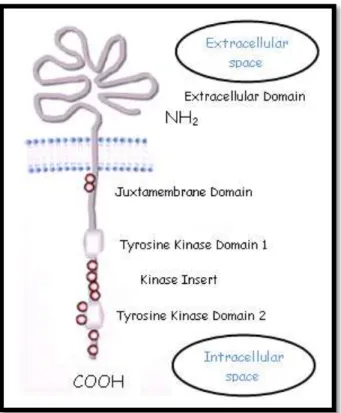
The c-KIT proto-oncogene is localized in the chromosome 4 of the human genome (Yarden et al., 1987), while on the dog genome it is mapped on chromosome 13 (Reimann-Berg, Murua & Nolte, 2012). In both species the gene is composed of 21 exons of which: exon 1 encodes the translational initiation codon and the signal peptide, exons 2-9 the remainder of the extracellular part of the protein, exon 10 the transmembrane region and the remaining exons, exons 11-21, encode the intracellular part of the receptor (Lennartsson & Rönnstrand, 2012). The c-KIT receptor, also known as cluster of differentiation 117 (CD117), is a type III RTK which binds to the stem cell factor (SCF), a cytokine responsible for the stimulation of mast cell growth and differentiation (Preziosi, Morini & Sarli, 2004). As illustrated in figure 1, the c-KIT receptor is composed by the five extracellular immunoglobulin-like domains and by two intracellular kinase subdomains, tyrosine kinase domain 1 and 2, separated by an
4
approximately 80 amino acids long kinase insert sequence and ends with a COOH-terminal tail. Between the extracellular and the intracellular portion there is a single transmembrane domain (Yamamoto, Tojo, Aoki & Shibuya, 1993; Lennartsson & Rönnstrand, 2012).
Figure 1 - Basic structure of c-KIT (modified from Gilfillian & Rivera, 2009).
The red circles represent the major tyrosine phosphorylation sites.
2.3. SCF and c-KIT regulation
The SCF, also known as mast cell growth factor, steel factor or KIT ligand, is a growth factor expressed by fibroblasts and endothelial cells in various tissues throughout the body, including brain, endothelium, gametes, heart, kidney, lung, skin, liver, thymus and bone marrow stromal cells. The gene coding for SCF maps to chromosome 12 in humans and 15 in dogs. In humans, the gene is composed of 9 exons. SCF can exist as a shorter, membrane-bound, or a longer, soluble, form. Both are able to bind c-KIT and activate its intrinsic tyrosine kinase activity (Lennartsson & Rönnstrand, 2012).
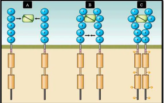
The activation of c-KIT begins when a SCF homodimer simultaneously binds two c-KIT monomers (figure 2A). The interaction between SCF and each c-KIT monomer only involves the first three Ig-like domains of c-KIT, which have a complementary shape and charge, allowing a tight binding with SCF (figure 2B). As a result of this interaction, the two c-KIT monomers involved are drawn closer to each other, to the point where a conformational
5
change in each monomer takes place, enabling homotypic interactions between Ig-like domains 4 and 5 of the two adjacent c-KIT molecules (figure 2C). This new proximity, between the two receptors involved, also affects the intracellular domain, facilitating its activation and subsequent transphophorylation (Zhang, Zhang, Joachimiak, Schlessinger & Kong. 2000; Yuzawa et al., 2007). As the phosphorylation process continues in an orderly manner along the receptor, the phoshorylation of the juxtamembrane domain is followed by the phosphorylation of the kinase insert and, finally, the phosphorylation of the activation loop (Lennartsson & Rönnstrand, 2012).
Figure 2 - Activation of c-KIT by SFC binding (modified from Lennartsson & Rönnstrand, 2012).
A - The SCF protein leads the formation of c-KIT homodimers by interacting with its Ig-like domains 1, 2 and 3; B - As a result of their interaction, SFC brings two c-KIT monomers close to each other, allowing interactions between Ig-like domains 4 and 5 of adjacent monomers; C - The new homodimeric state of c-KIT permits an efficient trans-phosphorylation along the c-KIT receptor.
2.4. c-KIT’s functions
Once activated, c-KIT is involved in a wide range of biologic activities, including cell proliferation, migration, maturation, and survival (Heissig, Werb, Rafii & Hattori, 2003; Metcalfe, 2008; Gregory-Bryson, Bartlett, Kiupel, Hayes & Yuzbasiyan-Gurkan, 2010). The c-KIT receptor is, mainly, expressed in stem and precursor cells and its expression is usually downregulated upon terminal differentiation, suggesting that signaling through the SCF/ c-KIT axis might be a key element in conferring and maintenance of stemness. There are,
6
however, some types of cells that even at their final stage of differentiation continue to express high levels of c-KIT. Mast cells (MC), melanocytes and interstitial cells of Cajal might be some of the most important examples (Pittoni, Piconese, Tripodo & Colombo, 2011). In mast cells, adding to the previously mentioned functions, c-KIT has been associated with fibronectin adhesion, chemotaxis and degranulation (London, 2013). The c-KIT protein plays an important role during both embryogenesis and adulthood (Webster et al., 2006).
2.5. Mutations in human and canine c-KIT
There are many diseases associated with an aberrant c-KIT expression, including Piebald – a disease that affects mammals, resulting in a pattern of non-pigmentation and white spotting –, atopic dermatitis, allergic rhinitis, asthma, rheumatoid arthritis (Jeong, Choi, Kim, Kim & Kim, 2011) and gastrointestinal motility disorders (Breuer et al., 2010). Furthermore, several mutations have been reported in c-KIT, in both human and canine species, all of which can potentially lead to the activation of c-KIT in the absence of SCF binding. Aberrant autophosphorylation of c-KIT, enabling the cell to develop independently from growth and survival signals, is one of the key features of malignance (Hanahan and Weinberg, 2000). According to different authors, mutations in human c-KIT occur in 50-80% of all gastrointestinal stromal tumors (GIST). It is estimated, however, that up to 35% of the remaining GIST cases exhibit mutations in PDGFRA. Since GISTs associated with c-KIT or
PDGFRA mutations have similar downstream signaling pathways, it is thought that PDGFRA
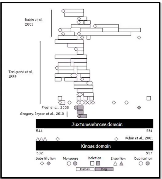
mutations serve as an alternative tumorigenic mechanism to c-KIT in GISTs (Gregory-Bryson et al., 2010; London, 2013). In GIST, the majority of c-KIT mutations occur in exons 9, 11, 13 and 17. Exon 11 by itself comprises 90% of all c-KIT mutations. In figure 3, the main mutations in exon 11 are reported. Typically, these mutations consist in deletions resulting in perturbation of the negative regulatory function of the juxtamembrane domain, with consequent ligand independent c-KIT activation (Khanna & Gordon, 2009).
Besides GIST, also acute myelogenous leukemia, pediatric mastocytosis, small cell lung cancer and prostate cancer have been linked to aberrant c-KIT expression. In fact, 70% of all small cell lung cancer cases present overexpression of c-KIT (Webster et al., 2006; Bodemer et al., 2010; London, 2013). In some melanoma types, specifically acral melanomas – which occur on the foot soles and on the palm of the hands – and mucosal melanoma, it has been reported the existence of activating c-KIT mutations within exons 11 and 13 (Lennartsson & Rönnstrand, 2012).
7
Figure 3 - Comparison between mutation in canine and human GIST (modified from Gregory-Bryson et al., 2010).
Codon number is based upon the human amino acid sequence [Genbank: NP_00213].
In dogs, mutations in c-KIT occur in 15-50% of mast cell tumors (MCT), and have been associated with tumors showing a more aggressive phenotype (Downing, Chien, Kass, Moore & London, 2002; Webster et al., 2006), possibly due to an increased proliferation and resistance to apoptosis (Gleixner et al., 2007; Letard et al., 2008). The most common type of mutation found in canine MCTs are internal tandem duplications (ITD) for the most part involving exon 11 (Webster et al., 2006). Zemke et al. (2002), as part of a study which analyzed 88 canine MCTs from selected canine breeds, identified c-KIT ITDs in 9% of tumors. However, in intermediate and high grade MCTs mutations can be present in up to 30-50 % of all cases (Zemke, Yamini & Yurbasiayan-Gurkan, 2002; Webster, et al., 2006). Other types of mutations in MCTs include deletions and point mutations in exons 8, 9, 11 and 12 of the
8 2.6. MCTs
2.6.1. MCs and MCTs
Mast cells are found in most organs and tissues of the body, it is, however, in areas that interface the environment, such as the skin, the lungs and the gastrointestinal tract, that MCs are present in higher numbers. One of the distinctive characteristics of MCs is the existence of granules in their cytoplasm, where inflammation mediators, including histamine, proteases, chemotactic factors, cytokines and metabolites of arachidonic acid are stored. These granules can be dyed through the use of cationic dyes that bind the granule proteoglycans, resulting in metachromasia, and therefore permitting the identification of mast cells (Welle, Bley, Howard & Rüfenacht, 2008). Mast cells play an important role in immunological, inflammatory and immediate-type allergic reactions. When a physical or chemical trauma occurs, or when stimulated by immune mechanisms, MCs respond by releasing the contents of these granules to the extracellular compartment (Galli, Nakae & Tsai, 2005).
Mast cells are known to suffer neoplastic transformation, originating solitary and multiple tumors. The biological behavior of MCTs is highly variable, and their etiology is still unknown; however, likewise other tumors, MCTs are believed to have multifactorial causes (Gross, Ihrke, Walder & Affolter, 2005).
In dogs, MCTs are one of the most common tumor types. When considering cutaneous tumors alone, MCTs account for up to 20% of all cases. Most MCTs occur in the dermis and subcutaneous tissue; nevertheless, visceral forms, originated in the gastrointestinal tract, urethra, spine bone marrow, conjunctiva, larynx, liver, oral cavity, salivary gland nasopharynx and spleen, can also be found (London & Seguin, 2003; Ohmori et al., 2008; Takeuchia et al., 2010). No differences in prevalence between male and female have been identified, whereas it is known that some dog breeds, such as Boxers and Labrador Retrievers, are more prone to develop MCTs (Webster, Yuzbasiyan-Gurkan, Miller, Kaneene & Kiupel, 2007; Warland & Dobson, 2013). Interestingly, in Boxers and Pugs, MCTs are usually considered - from a histological point - of low or intermediate grade, while Shar-Peis and particularly young individuals are predisposed to develop poorly differentiated MCTs, which are biologically more aggressive (Blackwood et al., 2012). The mean age of the affected dogs is 8.5 years (Passantino et al., 2008), but occasionally MCTs are found in animals as young as 4 to 6 months.
9 2.6.2. c-KIT and MCTs
Since it was first identified, c-KIT has been considered a proto-oncogene due to the close connection between alterations in its expression or activity and the development of certain types of tumors. The involvement of c-KIT in tumorigenesis can be of two types: one in which the activation of c-KIT plays the main role in the initiation of the development of the neoplasm and another in which c-KIT has no substantial role in tumor initiation, but it acquires one upon tumor progression (Pittoni et al., 2011).
As explained before, once activated c-KIT initiates a signaling cascade resulting in a wide array of biological activities including cell proliferation, migration, maturation and survival. The c-KIT receptor is expressed in both normal and neoplastic MCs presenting, however, a higher expression in poorly differentiated MCTs. Several c-KIT mutations have been identified in MCTs. Even though the complete understanding of the consequences of such mutations has not been achieved, it is known that c-KIT mutations and aberrant c-KIT localization are associated with increased expression of Ki67 and argyrophilic nuclear organizing regions (AgNOR), both of which are considered markers of increased cellular proliferation (Webster et al., 2007).
2.6.3. MCTs – diagnostic and clinical presentation
Upon the suspicion of a MCT, the definitive diagnosis can be achieved through cytology, most commonly using fine needle aspirate (FNA), and/or histopathology. Cytology of a MCT consists on the predominant presence of MCs. The clinical presentation of the lesions varies according to the tumors grade: well-differentiated cutaneous MCTs consist of slow growing, hairless and solitary lesions, while poorly differentiated cutaneous MCTs are characterized for being rapidly growing, ulcerated and pruritic lesions, sometimes surrounded by small “satellite lesions” (Blackwood et al., 2012).
Clinical signs depend on both the localization and the stage of the tumor. Systemic signs, including anorexia, vomiting, melaena, widespread erythema, edema and gastrointestinal ulceration, are more frequent in visceral forms of MCTs and are associated with a poorer prognosis (Mullins et al, 2006).
10 2.6.4. MCTs – treatment
The therapeutic approach to each MCT case depends on different aspects, including clinical grading and tumor location. There are now several treatment algorithms aiming to guide oncologists on treatment choice (Blackwood et al., 2012). In localized, non-metastatic MCTs, surgical excision of the tumor is the preferable treatment choice. A 2 cm margin for grade I and II or a 3 cm margin for grade III, and a deep margin, including at least one fascial layer, should be respected (Chaffin & Thrall, 2002; Weisse, Shofer & Sorenmo, 2002).
Surgical excision should be considered the best local approach for MCTs, and radiation should be restricted to those cases where surgery did not achieve total local control (Blackwood et al., 2012). In intermediate grade MCTs, adjuvant radiation therapy results in a 1-2 years disease-free interval in 81–95% of cases (Poirier et al., 2006).
Chemotherapy, in general, is used in three different situations; (a) in cases of high-grade tumors where systemic therapy is required to treat, delay or prevent the dissemination of metastases; (b) as a preliminary treatment, prior to surgery or radiation, aiming to reduce tumor size and improve the likelihood of achieving complete surgery excision or tumor elimination through irradiation; (c) in those cases where residual microscopic disease remains and surgical excision and radiation are not possible (Blackwood et al., 2012).
Several drugs and protocols are used to treat canine MCTs, reflecting the limitations with this approach. Prednisolone and vinblastine are most commonly used as first-line therapy and lomustine as part of a second-line of therapy. When choosing a chemotherapeutic agent, the potential drug toxic effects should always be taken in consideration. Among the aforementioned chemotherapeutic agents, vinblastine is known to cause perivascular irritation, while both vinblastine and lomustine are potentially myelosuppressive; hence, checking patients’ haematology, prior to each dose, is mandatory. Lomustine is also hepatotoxic and monitoring alanine transaminase (ALT) levels is therefore recommended (Blackwood et al., 2012).
2.7. New therapeutic approach for cancer treatment
Over the last years, many experimental settings, aiming to develop drugs capable of specifically blocking proteins that act as drivers of uncontrolled cancer cell growth and survival, have been undertaken. In general, two approaches are most commonly used: the use of monoclonal antibodies and tyrosine kinase inhibitors (TKI).
11
It is now possible to engineer antibodies capable of recognizing specific epitopes on a variety of proteins. With the development of these techniques, antibodies that recognize and bind the extracellular domain of RTKs or circulating growth factors, resulting on the functional inhibition of these proteins, have been created (London, 2013).
A successful example of monoclonal antibody is trastuzumab (Herceptin; Genentech, South San Francisco, CA, USA). Trastuzumab is a monoclonal antibody targeting human epidermal growth factor receptor 2 (HER2), a human epidermal growth factor receptor over-expressed in approximately 30% of all breast cancers and other epithelial tumors (Harris, 2004). Results obtained are very encouraging: in women with metastatic HER2-positive breast cancer, the response rate is as high as 25% when used alone (Vogel et al., 2001) and when combined with chemotherapy, the response rate improves to 50 % (Slamon et al., 2001). As a consequence of these results, trastuzumab is now part of the routine therapeutic approach for women with HER2-positive breast cancer (Arteaga et al., 2012).
Nevertheless, except for trastuzumab and few other cases, this strategy has not presented good results. Failure in therapy response might be due to the location of the receptors, since a considerable proportion of the expressed receptors reside inside the cell and, therefore, are not accessible to the antibodies in the extracellular space (Lennartsson & Rönnstrand, 2012). The second approach mentioned above, which also aims to inhibit specific proteins, is based upon the use of TKIs. These drugs can act either as competitive or allosteric inhibitors, blocking protein-protein interactions (Zhang, Yang & Gray, 2009). Overall, as a result of interrupting the survival and growth signals, these molecules prevent adenosine-5'-triphosphate (ATP) binding to the kinase domain, inducing cell death. Comparing to monoclonal antibodies, TKIs are often easier to be synthesized in large and orally bioavailable quantities that can readily enter cells and bind the intended target (London, 2013).
2.7.1. Approved TKIs for human medicine
The first TKI approved for humans was imatinib (Gleevec, Novartis Oncology US, East Hanover NJ, USA). Imatinib is an orally administered drug, initially developed to inhibit the breakpoint cluster region-abelson (BCR-ABL) fusion protein and PDGFRA. Later studies revealed that imatinib inhibited also c-KIT (Lennartsson & Rönnstrand, 2012), presenting substantial action against GIST (Heinrich et al., 2003). The kinase inhibitory effect of imatinib is caused by its capacity to bind the ATP pocket of ABL, c-KIT and PDGFRA, blocking the kinase phosphorylation process and preventing, therefore, its signaling. In patients with chronic myelogenous leukemia (CML), in which fusion proteins are present in
12
more than 95% of the cases, treatment with imatinib results in a remission rate close to 95% in the chronic phase and 20-50% for patients with blast crisis. In GIST patients, response rates of 50-70% have been reported, far better than the 5% response rate observed with the use of standard chemotherapy alone (London, 2013).
Another TKI approved for the use in humans is sunitinib (Sutent; Pfizer). Similarly to imatinib, sunitinib acts on several RTKs, such as vascular epithelium growth factor receptor (VEGFR), PDGFRA/B, c-KIT, FMS-related tyrosine kinase 1 (FLT1), colony stimulating factor 1 receptor (CSF1R) and RET receptor. It shares with imatinib its mechanism of action, as it sits in the ATP-binding pocket of the RTK. As a result of its multitargeted nature, sunitinib presents activity over a wide range of cancers, including GIST. In 2006, sunitinib was approved for the treatment of imatinib-resistant GIST and renal cell carcinoma patients. A surprising 61% of imatinib-resistant GIST patients, when treated with sunitinib demonstrated disease regression or stable disease for more than 4 months. Moreover, 65% of renal cell carcinoma patients, in which the treatment with interleukin-2 and/or interferon failed, once treated with sunitinib showed partial response or stabilization of the disease (London, 2009).
2.7.2. Approved TKIs for veterinary medicine
In veterinary medicine, two TKIs have been approved. The first one, toceranib phosphate (Palladia; Pfizer Animal Health, Madison, NJ, USA), is an orally available TKI blocking several RTKs such as VEGFR2, PDGFRA and c-KIT. Since toceranib is structurally very similar to sunitinib, it may have activity over other receptors too (Papaetis & Syrigos, 2009). In the first evaluation of toceranib in dogs, a phase I clinical trial, the TKI was tested in animals suffering from different types of cancer. When considering the results in patients suffering from MCTs, 10 of 11 dogs with c-KIT mutations responded to the therapy. In this first study the maximum tolerated dose was established as 3.25 mg/ kg every other day (q.a.d.). The main adverse effects included anorexia, diarrhea, vomiting and gastrointestinal bleeding. Toceranib has also been shown to induce a protein-losing nephropathy and/or hemolytic anemia in some patients (London, 2009). Concomitant medications proved to control these toxicities (London et al., 2003). A consequent trial, consisting of a placebo-controlled randomized study with 86 dogs suffering from grade 2 and 3 MCTs, revealed a 37.2% response rate in animals treated with toceranib, comparing to a 7.9% rate in animals treated with placebo. Also, of 58 animals receiving toceranib after the placebo trial, 41.4% experienced an objective response. Interestingly, the response rate was higher in animals with
13 c-KIT mutations (69 versus 37%: London et al., 2009).
Toceranib was approved in 2009 and since then it has been used to treat various canine solid tumors, mainly after failure of primary therapy or in case of metastatic disease. High response rates have been registered, with clinical benefits reaching 74% in anal gland sac adenocarcinoma, osteaosarcoma, thyroid carcinoma and head, neck and nasal carcinomas (London et al., 2012).
Several studies in which different combinations of toceranib with non-steroidal anti-inflammatory drugs (NSAID), other chemotherapeutic drugs or radiation have been tested in animals suffering from MCTs revealed very encouraging results. The association of vinblastin and toceraninb, for instance, with a maximum dose of vinblastine at 1.6 mg/ m2 every other week and toceranib at 3.25 mg/ kg q.a.d., showed a 71% over all response rate (Robat et al., 2012). Even the combination of toceranib with radiation showed interesting results. In a study in which dogs with non-reseactable MCTs received prednisolone, omeprazole, diphenhydramine and toceranib for a week before starting coarse fraction radiation therapy (6 Gy once a week, for a total of 4 weeks), the response rate was 76.4% (58.8% of patients achieved full response and 17.6% partial response). As important as the response rate achieved, in this study there were no evidence of enhanced radiation-induced toxicities (Carlsten et al., 2012).
The second TKI approved for veterinary use was masitinib (Kinavet; AB Science, Paris, France). Masitinib has shown activity over c-KIT, PDGFRA, PDGFRB and the cytoplasmic kinase Lyn. During a phase II study on masitinib, which included 13 animals suffering from grade II and III MCTs, six patients responded to treatment, two achieved complete response, two partial responses and in the remaining two disease stabilization was achieved. In a subsequent placebo-controlled phase III study, involving more than 200 dogs with non-metastatic grade II and III MCTs, the response rate was not significantly different between the two groups: the response rate in the placebo group was 15%, while on the masitinib-treated group it was 16%. However, there was a significant difference regarding the progression time: the progression time in the placebo-treated group was 75 days against 118 days in the masitinib treated group. Masitinib has been proven to be well tolerated by both dogs and cats suffering from MCTs, with mild vomiting and diarrhea being the most common side effect (Hahn et al., 2008). Masitinib is also known to induce in some patients a mild neutropenia that does not predispose dogs to bacterial infection and it is often time-resolving, and the development of localized muscle cramping, which can be readily treated with NSAIDs (London, 2009). Masitinib appears to have action against both mutant and wild-type c-KIT (Isotani et al., 2006; Isotani et al., 2009).
14 2.8. Resistance to TKIs
The immediate results obtained by treating tumor cells with TKIs have been very positive, unfortunately, in most cases the tumor relapses within 6 to 18 months. Resistance mechanisms are known only for few TKIs, i.e. imatinib, while remain only partly understood for the great majority of them (Rosenzweig, 2012). It has been hypothesized that several cellular alterations contribute to drug resistance, hindering the search for strategies that can prevent or circumvent this issue.
Probably, the most studied mechanisms of drug resistance are the ones associated with imatinib treatment of BCR-ABL-positive CML. In these patients, drug resistance due to the development of point mutations in the kinase domain of BCR-ABL, preventing imatinib binding, has been described. Other patients develop resistance, through up-regulation of
BCR-ABL messenger ribonucleic acids (mRNA), resulting in an over-expression of the protein,
beyond imatinib capacity to block protein function (Mahon et al., 2000). Other mechanisms that have been reported include elevated P-glycoprotein expression, enhanced multidrug efflux and activation of other growth factor pathways (London, 2013).
Another well studied example regards patients with mutations in the endothelium growth factor receptor (EGFR) that respond to erlotinib, an EGFR inhibitor. Three different mechanisms have been described: (a) the generation of a second mutation in the EGFR ATP binding pocket that delays drug binding; (b) the amplification of the gene encoding met proto-oncogene (MET) and (c) the over-expression of hepatocyte growth factor (HGF), the ligand for MET (Kosaka, Yamaki, Mogi & Kuwano, 2011).
In a recent study, Gao et al. (2013) identified the development of secondary mutations in GIST patients after treatment with imatinib. In 65.8% of cases where drug resistance was developed, secondary mutations were noticed.
As mentioned above, mechanisms of resistance to other TKIs are not so well characterized. It should be mentioned that drug resistance to tyrosine inhibitors can occur through epigenetic alterations of genes responsible for the regulation of the cellular response to kinase inhibitors that may follow alterations in histone acetylation/ deacetylation and chromatin and histone methylation (Rosenzweig, 2012).
All the aforementioned TKIs, act as inhibitors of the kinase protein after it has been expressed. As a result, it has been reported that the same drug can present different response rates which depend from the presence of a wild-type or a mutated c-KIT genotype. For this reason recent researches aiming to develop drugs capable of controlling c-KIT expression have been undertaken. In particular, it has been hypothesized that the promote region of c-KIT could be
15
the best target of these new drugs, and two structures have been considered particularly attractive targets, the so-called G-quadruplex structures (Fernando et al., 2006).
2.9. G-quadruplex
2.9.1. G-quadruplex: structure
In 1953, Watson and Crick described for the first time deoxyribonucleic acids (DNA) double helix structure. For almost a decade, it was thought the entire genome presented this structure, but in 1962 Gellert and his co-workers collected fiber X-ray diffraction data on guanylic acid that suggested the existence of tetrameric units assembled into large helical structures in guanine (G) rich DNA sequences (G-quadruplexes: Bryan & Baumann, 2011).
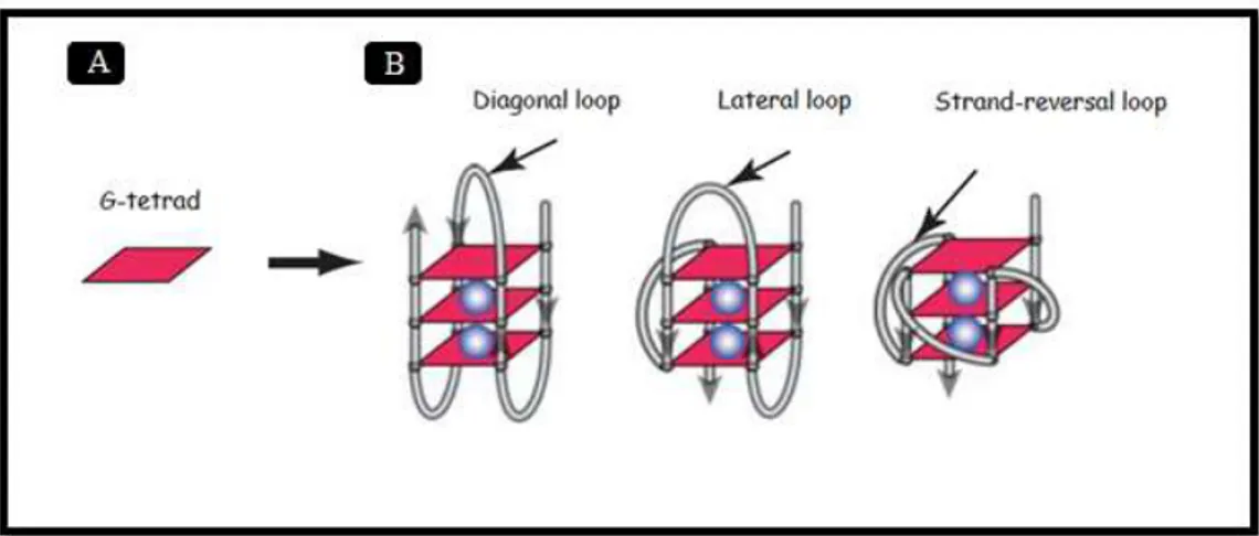
G-quadruplexes are DNA secondary structures formed by stacked G-tetrads. Each G-tetrad is composed by four Gs connected by cyclic Hoogsteen hydrogen bonding in which each of the four bases is the donor and the acceptor of two hydrogen bonds, thereby forming a square planar platform (Bryan & Baumann, 2011). The connective bond involved in the formation of G-tetrads is one of the many differences between G-tetrads and the normal double helix DNA. In the latter, nucleotides are connected by the Watson-Crick hydrogen bond, in which only two or three bonds exist (Yang & Okamoto, 2010).
The ability to form stable and extensive self-connections is limited to G; no other nucleotide can mime a similar association. The unique G hydrogen donor and acceptor sites allow G nucleotides to establish such strong bonds among themselves (Bryan & Baumann, 2011). The guanine residues in each G-tetrad can adopt either syn or anti glycosidic conformation, while the residue in the antiparallel strand necessarily adopts the opposite conformation. G-quadruplex structures can be formed by one or more DNA strands, to a maximum of four (Yang & Okamoto, 2010).
Intramolecular G-quadruplexes, formed by one single DNA molecule, are usually composed by three tetrads, with four or more G-tracts involved in the tetrad formation, three or more loops and two flanking segments. The G-strands of intramolecular G-quadruplexes can be parallel, anti-parallel or hybrid and the loops, as represented in figure 4, can exhibit three possible conformations: strand-reversal, lateral and diagonal. Tetramolecular G-quadruplexes, on the other hand, tend to adopt a well-defined structure, in which all Gs display an anti-glycosidic conformation and all DNA strands are parallel (Yang & Okamoto, 2010; Chen & Yang, 2012).
Evidence suggests that the structure developed by the G-quadruplex is strictly dependent on its respective DNA sequence. In fact, small changes in the sequence can dramatically modify
16
the structure of the G-quadruplex or even prevent its formation. Rankin et al. (2005) have proved how the substitution of a single nucleotide in a four-base loop of a c-KIT G-quadruplex sequence resulted in inhibition of the G-G-quadruplex formation. The formation and stabilization of G-quadruplexes is also dependent on the presence of monovalent cations, specifically K+ and Na+ which, as reported in figure 4, occupy the central cavity of the G-quadruplex and neutralize the electrostatic repulsion between the O6 atoms of adjacent stacked G-tetrads. Both K+ and Na+ ions are the principal cations found in vivo, thus implying that G-quadruplex structures are favored in physiological conditions. Between these two ions, K+ is considered to be the most relevant, due to its high intracellular concentration (K+ presents a intracellular concentration ~140 mM, while Na+ intracellular concentration is ~10 mM: Chen & Yang, 2012). These two cations exhibit differences in their location within the G-quadruplex structures: the sodium ion can present various geometries, while K+ is always equidistant between each tetrad plane, featuring a better coordination with the O6s and lower dehydration energy (Hud & Plavec, 2006). The ionic radius seems to be a key factor for G-quadruplexes stability (Bryan & Baumann, 2011).
Figure 4 - Schematic representation of G-quadruplex structures (modified from Chen & Yang, 2012).
A – Schematic representation of a tetrad; B – Three schematic representations of intramolecular G-quadruplexes. The blue balls represent the stabilizing cations (K+ or Na+).
Although the primary nucleotide sequence determines the quadruplex structure, some G-rich sequences can, in the presence of different cations, give raise to different G-quadruplex structures. In fact, highly conserved human telomeric sequences, which only consist of d(TTAGGG)n are able to form various G-quadruplex structures with low energy differences (Chen & Yang, 2012).
17 2.9.2. Studies on G-quadruplex
Various techniques have been developed to study the formation of G-quadruplexes. These, simple to complex, techniques allow studying the G-quadruplex formation both ex vivo and in
vivo. One of the earliest used was a simple non-denaturating acrylamide gel electrophoresis,
through which it can be observed how G-rich sequences present aberrant migration along the gel. This observation is compatible with the theory that G-rich sequences can form secondary structures, with consequent variations in migration.
Even though native gel electrophoresis is still used in G-quadruplex studies, more elaborate techniques have been created to verify the G-tetrads structure. Circular dichroism (CD) is a technique in which the sample is exposed to circularly polarized light. The structure of the possible G-quadruplex is evaluated through the interaction between the structure and the light. Variations in the structure can be perceived and extrapolated according to the wavelength of the CD spectrum. When carried out over a certain range of temperatures, CD spectroscopy can also be used to determine the melting temperature (Tm), the change in enthalpy (ΔH) and Gibbs free energy (ΔGº) - thermodynamic parameters providing information about the stability of the G-quadruplex structure (Bryan & Baumann, 2011).
A recent method, which has rapidly gained increasing importance for studying G-quadruplexes conformation and dynamics, is the single-molecule Förster resonance energy transfer (FRET) spectroscopy. In this technique, the oligonucleotide is labeled with a donor and an acceptor fluorophore. When the oligonucleotide folds the donor fluorophore transfers its energy to the acceptor fluorophore. The efficiency of the donation depends on the distance between the two parties involved. This technique permits not only to evaluate the formation of DNA secondary structures, but also to analyze dynamic variations in the G-quadruplex structure (Bryan & Baumann, 2011).
More detailed information on the precise molecular structures can be obtained through high-resolution techniques, such as nuclear magnetic resonance (NMR) and X-ray crystallography. With NMR it is possible to determine the strands’ orientation and loops’ configuration. X-ray crystallography can be used to generate high-resolution structures (Bryan & Baumann, 2011). All the above mentioned techniques study the formation and conformation of G-quadruplex
ex vivo. By contrast, the existence of secondary DNA structures is inherently difficult to prove in vivo. At present, three techniques have been created to study the existence of G-quadruplex in vivo. The first one is the use of specific antibodies against parallel and anti-parallel
G-quadruplexes. Through the use of this method, the existence of G-quadruplexes in ciliates telomeres has been demonstrated. The main concern about this approach is that the reagents
18
used for the detection of the structures may be responsible for the formation of the G-quadruplexes, by shifting the equilibrium towards the folded form (Bryan & Baumann, 2011). Other techniques used in in vivo studies of G-quadruplexes include the use of either the radiolabeled G-quadruplex ligand 360A (2,6-N,N’-methyl-quinolinio-3-ylpyridine dicarboxamide) or the fluorescent G-quadruplex ligand BMVC 3,6-bis(1-methyl-4-vinylpyridinium) carbazole diiodide (Yang & Okamoto, 2010).
2.9.3. G-quadruplex in biological systems
Bioinformatic analyses have identified within the human genome about 375000 sequences where the formation of G-quadruplex structures is possible. A better understanding of the functions of these structures has nowadays gained increasing interest among researchers. Biologically relevant G-quadruplexes were first discovered in telomeric DNA. In several species, it was proved the existence of guanine-guanine base-paired hairpin structures in single-stranded telomeric terminal sequences. As an example, in humans, telomeres contain a 5- to 10- kb d(TTAGGG) tandem sequence with a 50- to 600 nt- single stranded 3’ overhang at the chromosome end, with the ability to form DNA G-quadruplexes (Chen &Yang, 2012). The G-quadruplex structures formed from human telomeric sequences have been the center of intense investigation, as, in vitro, these structures block the telomere elongation by the cancer-associate enzyme telomerase. However, long single-strandedness is not a prerequisite for G-quadruplex formation.
Studies characterizing the distribution of rich sequences concluded that possible G-quadruplex forming sequences are not found randomly throughout the genome. Besides their location in telomeric DNA, possible G-quadruplexes have been found in noncoding regions of the genome - especially gene promoters -, immunoglobulin switch regions, ribosomal DNA and certain regions of ribonucleic acid (RNA: Chen &Yang, 2012). But even their distribution in gene promoters is not uniform: these structures are frequently found in the promoter regions of proto-oncogenes, such as v-myc myelocytomatosis viral oncogene homolog (MYC),
c-KIT and B-cell CLL/lymphoma 2 (BCL2); however, their formation in tumor suppressor
genes is much inferior (Bryan & Baumann, 2011).
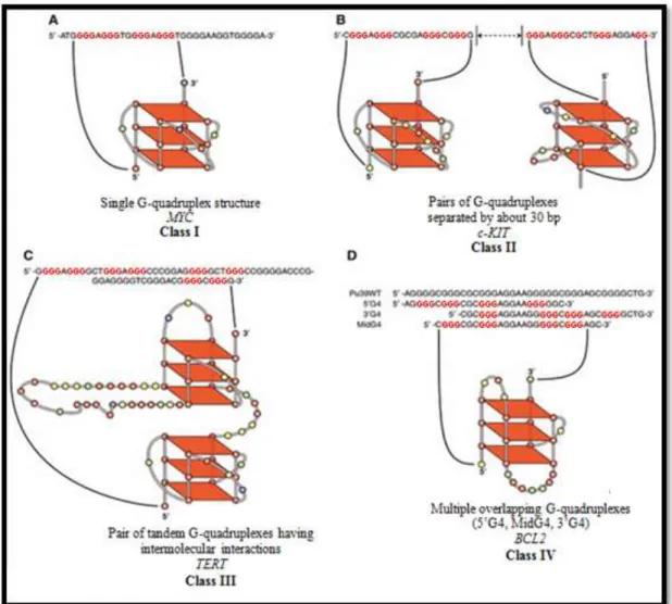
Promoter G-quadruplexes can be classified into 4 different classes, according to the number of G-quadruplexes and their relative location. Class I is composed by those cases where a single quadruplex structure exists. The most studied member of this class is MYC’s quadruplex (figure 5A). Class II is characterized by the presence of two distinctly different G-quadruplexes, separated each other by about 30 base pairs (bp). The only known members of
19
this family are c-KIT’s G-quadruplexes (figure 5B). Class III is formed by those cases where two G-quadruplex structures are so closely positioned that they form tandem structures, which are more stable than individual G-quadruplexes. The G-quadruplexes present in the promoter region of human telomerase reverse transcriptase (TERT) are members of this class (figure 5C). Members of class IV present multiple overlapping G-quadruplexes. Both, PDGFRB and
BCL2 are members of this class (figure 5D: Brooks, Kendrick & Hurley, 2010).
Figure 5 - Classification of unimolecular G-quadruplexes found in promoter regions (modified from Brooks, Kendrick & Hurley, 2010).
Due to the high number of possible G-quadruplexes within promoter regions, these structures have been associated with the process of gene regulation. It was hypothesized that the formation of G-quadruplex structures could block gene transcription, since, when comparing to duplex DNA, they are more stable - presenting a higher Tm (Lipps & Rhodes, 2009). In
MYC, for instance, a single mutation which destabilizes the formation of a G-quadruplex
located in its promoter region, results in a threefold increase in basal transcription activity of the MYC gene (Bryan & Baumann, 2011).
20
Besides the aforementioned evidences on the existence of G-quadruplex in vivo, there is accumulating evidence that many proteins are capable of interacting with G-quadruplexes and in some cases can even promote their unfolding (Bryan & Baumann, 2011).
Contrary to what happens in telomeres, where repeating tandem sequences not only exist but are also a key element in the quadruplex forming sequences, in the promoter regions the G-quadruplex forming sequences are diverse, varying in number and length of G-tracts and intervening bases (Qin & Hurley, 2008). The G-quadruplexes in the promoter regions often contain more than four G-tracts, suggesting they can form more than one G-quadruplex structure through the combination of different four G-tracts, and with each G-quadruplex a potential combination of loop isomers due to the unequal number of Gs in each G-tract. It is thought that the different G-quadruplex arrangements may not be permanent, but be in a dynamic equilibrium with each other (Dailey, Miller, Bates, Lane & Trent, 2010). Another particular feature of these G-quadruplexes, lays on the fact they differ greatly in energy, due to the many possible arrangements of stable structures and minor unstable structures.
A short nucleotide sequence motif, G3NG3, known to form a robust parallel-stranded structure motif with a 1-nt double-chain-reversal loop, has been found in all types of promoter G-quadruplexes. In fact, due to its prevalent occurrence, it has been proposed that the G3NG3 motif has been evolutionarily selected to serve as a stable foundation for the promoter intramolecular G-quadruplex structures to build upon (Yang & Okamoto, 2010).
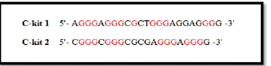
2.9.3.1. G-quadruplexes in the promoter region of human c-KIT
Within the promoter of the human c-KIT proto-oncogene two G-quadruplex forming sequences have been identified (figure 6): c-KIT 1 and c-KIT 2. These two sequences, both located in a nuclease hypersensitive region, are separated by only 31 bases. The c-KIT 1 sequence is located between 87- and 109- bp and c-KIT 2 between 140- and 160- bp relative to the transcription starting site (TSS: Balasubramanian, Hurley & Neidle, 2011).
Figure 6 - Sequences involved in the formation of the two G-quadruplexes present in the promoter region of c-KIT: c-KIT 1 and c-KIT 2 (original).
21
Comparatively, c-KIT 2 seems to form a standard unimolecular parallel G-quadruplex by using a K+ ion; on the other hand, the structure suggested to be formed in c-KIT 1 (figure 7), is not as simple. As expected, due to their proximity, similarly to c-KIT 2, c-KIT 1 also uses K+ as the stabilizing cation. NMR studies of the c-KIT 1 G-quadruplex (figure 8), revealed that even though its sequence includes four three-guanine runs, the backbone of one G-strand corner of the G-tetrad core is interrupted by a single guanine from the linker and two consecutive guanines from the forth three-guanine G-tract. One of the particular features of this G-quadruplex, lays on the existence of a snapback parallel-stranded G-quadruplex. The
c-KIT 1 is a unique structure composed by three double-chain-reversal loops; this arrangement
is observed in normal intramolecular parallel-stranded G-quadruplex, and by an additional lateral loop. The c-KIT 1 G-quadruplex also includes a G3NG3 sequence, which, as explained above, serves as a core structure (Yang & Okamoto, 2010).
Figure 8 - Schematic representation of c-KIT 1 (modified from Qin & Hurley, 2008).
2.9.4. G-quadruplex structures as potential anticancer drug targets
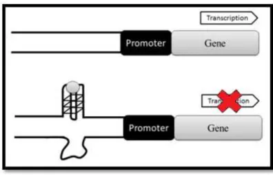
With the recognition of the biological importance of G-quadruplexes, the research and development of compounds capable of interacting with these structures has intensified. The goal is to discover compounds capable of stabilizing G-quadruplex structures and therefore repressing gene transcription (figure 9). The concept of using G-quadruplex targeting drugs is a revolutionary approach in cancer treatment (Balasubramanian et al., 2011).
Sun et al. (1997) reported for the first time the results of the interaction between G-quadruplex ligands and telomeric G-G-quadruplexes. In this study, the use of G-G-quadruplex ligands resulted in the inhibition of telomerase, an enzyme which is active in most cancer cells and not in non-cancerous cells (Sun et al., 1997; Yang & Okamoto, 2010).
Figure 7 - NMR structure of c-KIT 1 (taken from Todd, Haider, Parkinson & Neidle, 2008).
22
Since then, several families of small molecules capable of acting as inhibitors (small molecule inhibitors, SMIs) have been identified or created. Information about their crystal structures and NMR data are available providing, to some extent, information on the way by which they interact with each quadruplex structure. Different ligand families bind differently to the G-quadruplex (Bates, Mergny & Yang, 2007).
Figure 9 - Schematic representation of the transcription regulation of proto-oncogenes caused by G-quadruplex formation mediated by SMI (modified from Ma et al., 2013).
The conformational diversity among DNA G-quadruplexes indicates these structures can be targeted by specific ligands. However, a completely specific drug towards a determined G-quadruplex structure has not yet been found nor developed. In fact, it is thought such specificity might not be necessary. When considering tyrosine kinases for instances, SMIs targeting several G-quadruplexes might result in an increased inhibitor effect. The drawback of these drugs consists in a larger number of associated side-effects (Chen & Yang, 2012).
2.10. SMIs
Several small molecule families capable of interacting with G-quadruplex structures and stabilizing their structures have been identified (di Antonio, et al., 2009). Efficient SMIs are usually composed by an extended planar system, capable of interacting with guanine-tetrads, and two to three positively-charged side chains, which confer an additional stability to the complex by binding DNA loops (Zagotto et al., 2007). In fact, ligands capable of interacting with G-quadruplex structures, both at the G-tetrad and loops levels, can show selectivity, not only for quadruplex over duplex DNA, but also for the target quadruplex over other G-quadruplexes. Understanding how a SMI interacts with its G-quadruplex receptor is fundamental in the design and optimization of G-quadruplex ligands (Hou et al., 2012).
23
Naphthalene diimides (ND) are a SMI family which has been used in several studies as the quadruplex-binding core motif, due to the combination of two factors: its chemical accessibility and their extensive planar surfaces in its structure. These two characteristics, together with the presence of a cationic group within its structure, suggest NDs are predisposed to bind quadruplex over duplex DNA (Micco et al., 2013). Several SMI members of the ND family have been described so far, not only towards telomere G-quadruplexes, but also to promoter G-quadruplexes, namely c-KIT 1 and c-KIT 2 (Hampel et al., 2012).
Anthraquinones derivatives (AQ) represent another important SMIs family, which has been studied for its DNA-interacting properties. Several AQs have been used as chemotherapeutic agents in anticancer treatment. Doxorubicin, for example, an anthracycline antibiotic, is being used as an anticancer drug against a wide range of cancer types, for over 30 years. Disubstituted amido-anthraquinones are considered particularly relevant SMIs, as, depending on their pattern of disubstituition, they can bind specifically to quadruplex over duplex DNA. Several studies on telomere G-quadruplexes have used AQs as ligands (Huang, 2008; Zagotto, 2008; Huang, 2009).
Bisantrene, a known intercalating agent, possess a structural motif characteristic of effective G-quadruplex ligands. Several bisantrene-analogues have been studied as possible SMIs. Depending on the number, location and nature of their side chains, these molecules present different affinity with DNA molecules (Folini et al., 2010).
2.11. MCF7 and HGC27 cell-lines
The use of human and animal cell-cultures in in vitro studies has become an important tool in biotechnology and biomedical research, since immortalized cell-lines provide a simple model for complex biological systems. Cell cultures are now being used in several scientific areas, such as toxicity testing, virology, drug screening and development, cancer research and gene therapy. In this project, two different cell-lines, the MCF7 and the HGC27 cell-lines, were used. Both cell-lines had already been used in studies aiming to develop and test SMIs capable of interacting with c-KITs G-quadruplexes (Bejugam et al., 2007; Gunaratnam et al., 2009; McLuckie et al., 2011).
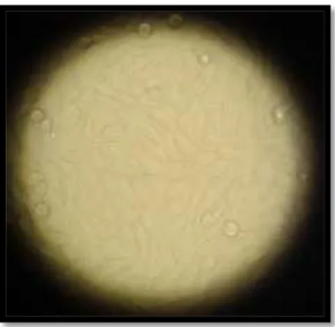
The MCF7 cell-line was established from a breast adenocarcinoma human female patient, in 1970. MCF7 cells are characterized by their epithelial-like form, for growing in monolayer, being capable of forming domes (see figure 10) and for presenting a doubling time of about 38 hours (Cell Biolabs inc., 2010; ATCC, 2012). Several studies have relied on MCF7 for
24 Figure 10 - MCF7 cell-line (original).
Figure 11 - HGC27 cell-line (original).
The HGC27 is a stable cell-line, established in 1976 through the culture of the metastatic lymph node of an undifferentiated gastric carcinoma human patient. The HGC27 cell-line is characterized by its polygonal shaped cells (figure 11), for presenting a doubling time of, proximately, 17 hours and for adhering to glass surfaces in monolayer (Akagi & Kimoto, 1976). The HGC27 is known for expressing both c-KIT and SCF genes (Hassan et al., 1998)
25 3. Aims of the present study
The c-KIT proto-oncogene is known to play an important role in the development of MCTs in dogs. Several approaches aiming to decrease the protein expression have been developed. Less than fifteen years ago, TKIs were first approved for the treatment of human cancers in which c-KIT was over expressed and, more recently (2009), for the treatment of canine MCTs. Despite the encouraging results initially obtained with these drugs, it is now known they present limitations, namely due to the development of resistance (Rosenzweig, 2012).
Thus, the discovery of new molecular targets controlling c-KIT transcription represents nowadays a subject of interest within the scientific community. In this respect, the two G-quadruplex structures present in the promoter region of c-KIT may represent interesting molecular targets for drugs aiming to modulate c-KIT expression.
The work developed on this thesis was inserted in a collaborative research project between the Department of Comparative Biomedicine and Food Science and the Department of Pharmaceutical and Pharmacological Sciences of the University of Padua. The main goal of the project was the development in vitro of new SMIs capable of interacting with human and canine c-KIT’s G-quadruplex structures and, therefore, modulating its expression. The work performed for this thesis was specifically aimed to develop a valid in vitro model for the study of SMIs for humans. For the achievement of this goal, the work was divided into three parts: a) the selection of in vitro human cellular models (established cell lines) for the testing of SMIs; b) the execution of cytotoxicity tests, namely the measurement of the inhibitory concentration 50 (IC50) value, by using three candidate SMIs, the aforementioned cell lines and a battery of cytotoxicity assays and c) the optimization and validation of qPCR assays to study the gene expression of c-KIT and other proto-oncogenes presenting G-quadruplex structures within their promoter regions.