Original Research
Distinct temporal patterns of electrical stimulation in
fl
uence neural
recruitment during PTZ infusion: An fMRI study
Michel Bernanos Soares Mesquita
a,b, Daniel de Castro Medeiros
a, Vinícius Rosa Cota
a,c,
Mark P. Richardson
b, Steven Williams
b, Márcio Flávio Dutra Moraes
a,b,*aNúcleo de Neurociências (NNC), Departamento de Fisiologia e Biofísica, ICB, UFMG, Brazil bKing
’s College London, London, UK
cUniversidade Federal de São João Del Rei, Brazil
a r t i c l e
i n f o
Article history:
Available online 31 October 2010
Keywords: Basolateral amygdala Pentylenetetrazole Synchronization Desynchronization Electrical stimulation Synfire chains
a b s t r a c t
Our working hypothesis is that constant inter-pulse interval (IPI) electrical stimulation (ES) would resonate with endogenous epileptogenic reverberating circuits, favoring seizure, while random inter-interval ES protocol would promote desynchronization of such neural networks, interfering with the abnormal recruitment of neural structures. Male Wistar rats were stereotaxically implanted with a monopolar ES carbon-fiber electrode (minimizing fMRI artifact) in the amygdala. A 7T fMRI scanner was used to evaluate brain activity during ES,fixed four pulses per second ratio, using either a periodic IPI (ES-P) or random IPI (non-periodic ES-NP) stimulation paradigm. Appropriate imaging protocols were used to compare baseline BOLD (blood oxygen level dependent) MRI with scans during ES. A second series of experiments, both without stimuli and under the same ES paradigms, were evaluated during continuous infusion of pentylenetetrazole (PTZ, 4 mg/ml/min) through an i.v. catheter. Our results show that temporal lobe activation during ES-P or ES-NP did not present any statistical differences during ES. However, during PTZ infusion, PTZ-P facilitated recruitment of the temporal lobe ipsilateral to ES while PTZ-NP showed significantly less activation ipsilateral to ES and, in turn, less inter-hemispheric differ-ences. Altogether, our results support the hypothesis of reverberating circuits being synchronized by ES-P and desynchronized by ES-NP. Time-coded low frequency stimulation may be an interesting alternative treatment for patients with refractory epilepsy.
Ó2010 Elsevier Ltd.
1. Introduction
Epilepsy is a very common and serious primary brain disease, with an incidence of 24e53/100,000 and prevalence of 0.4e0.8% in
developed countries (Hauser et al., 1998), characterized by recur-rent and spontaneous seizures caused by hyperexcitable and hypersynchronous underlying neural networks (Engel, 1996). The suppression of seizures is the main therapeutic goal for neurolo-gists treating people with epilepsy, even when lacking a better etiological understanding of the disease. Despite the great advances of drug development over the last decades (Loscher and Schmidt, 2002), pharmacological treatment is still unable to satisfactorily
control seizures in about one third to one fourth of epilepsy patients (French, 2007; Loscher and Schmidt, 2002; Wuttke and Lerche, 2006). In addition, many of these patients, with refractory epilepsy, are not eligible for ablative surgery, which, in most cases, requires a readily identifiable epileptogenic focus (Centeno et al., 2006; Spencer, 2002; Wiebe et al., 2001). Thus, investigation of alternative methods of treatment, such as electrical stimulation (ES) of neural structures (Theodore and Fisher, 2004), is of great interest in current epileptology. ES may be applied peripherally [e.g. Vagus Nerve Stimulation e VNS (Ben-Menachem, 2002; Binnie, 2000; Valencia et al., 2001) or Trigeminal Nerve Stimula-tioneTNS (DeGiorgio et al., 2003; Fanselow et al., 2000)] or
tar-geted to a variety of structures within the central nervous system [Deep Brain StimulationeDBS (Benabid et al., 2002; Hodaie et al., 2002; Vonck et al., 2005), such as: the anterior nucleus of the thalamus (Hamani et al., 2004; Mirski et al., 1997), subthalamic nuclei (Benabid et al., 2002; Chabardes et al., 2002) or the epilep-togenic focus itself (Vonck et al., 2002)]. Classically, ES is believed to work either by suppressing or inhibiting epileptogenic structures, *Corresponding author at: Núcleo de Neurociências, Department of Physiology
and Biophysics, Institute of Biological Sciences, Federal University of Minas Gerais, Av. Antonio Carlos, 6627, CEP 31270-901 Campus Pampulha, Belo Horizonte, MG, Brazil. Tel.:þ55 31 3499 2930; fax:þ55 31 3499 2924.
E-mail address:[email protected](M.F.D. Moraes).
Contents lists available atScienceDirect
Progress in Biophysics and Molecular Biology
j o u r n a l h o m e p a g e : w w w . e l s e v i e r . c o m / l o c a t e / p b i o m o l b i o
0079-6107Ó2010 Elsevier Ltd.
doi:10.1016/j.pbiomolbio.2010.10.005
Open access under the Elsevier OA license.
analogous to surgical ablation (McIntyre and Grill, 2001; McIntyre et al., 2004a; Volkmann, 2004), or by activating or stimulating neural networks that would modulate seizure-like activity. Never-theless, this simplistic view does not explain the complex under-lying mechanisms by which ES is effective (Theodore and Fisher, 2004) on suppressing seizures (McIntyre et al., 2004a; McIntyre et al., 2004b).
Elegant numerical simulations of neural network dynamics have guided epileptologists in better understanding the relationship between excitability and synchronicity (Kudela et al., 2003), leading to paradigm changing ideas on how to suppress the mass recruitment of neural substrates during ictogenesis. In a neural modeling study,Tass and Hauptmann (2007)showed that it would be theoretically possible to desynchronize neural masses with appropriate spatialetemporal ES protocols without the need for
chronic high frequency (HF) stimulation, which could lead to undesirable complications (Freund, 2005; Kumar et al., 2003; Rodriguez-Oroz et al., 2005; Volkmann et al., 2004). The impor-tant change in paradigm of these new approaches, especially when comparing to anticonvulsant drug therapy, is to target the synchronization aspect of epileptogenic neural networks instead of their excitability. Our laboratory tested time-coded ES patterns, designed to either resonate or desynchronize epileptogenic rever-berant circuits, in the PTZ animal model of epilepsy (Cota et al., 2009). In this work we showed that afixed 4 stimuli per second ES could either facilitate or interfere with the behavioral manifes-tation of the seizure depending on how the stimulus was tempo-rally coded. Such novel stimulation techniques may selectively desynchronize neuronal populations involved in epilepsy, thus being the biological validation of the“network desynchronization” therapeutic approach.
Although Cota et al. (2009)presented behavioral evidence to support the anticonvulsant effect of desynchronizing ES (dES), there is still no direct biological validation regarding how, and to what extent, ES interferes with neural mass recruitment during the transition to the ictal state. Understanding how abnormal activity propagates throughout the epileptic brain requires the acquisition and analysis of data containing anatomic, temporal and magnitude components. Electrophysiological measures could be used to determine seizure propagation (Durand, 1993; Gale, 1992; Moraes et al., 2005b; Snead, 1992; Timofeev and Steriade, 2004) however; this technique presents inherent insensitivity with respect to anatomic localization. Functional magnetic resonance imaging (fMRI) is much better suited to this purpose and has been applied to quantifying seizure propagation in the PTZ animal model of epilepsy (Keogh et al., 2005). Once the methodological problems regarding physical compatibility between intracranial ES and fMRI imaging were resolved, our objective was to provide biological evidence for theoretical modeling data suggesting that low frequency desynchronizing ES (non-periodic/random temporally coded stimuli, anti-resonant with epileptogenic oscillators) would disrupt PTZ seizure propagation while synchronizing ES (periodic stimuli resonant with epileptogenic oscillators) would promote or facilitate neural mass recruitment. By applying ES paradigms to only one side of the brain (amygdaloid complex in the temporal lobe), the contra-lateral hemisphere could be considered as a paired control.
2. Materials and methods
All experiments were done in accordance with United King-dom’s Animals (Scientific Procedures) Act 1986, under Home Office approval, with respective project and personal licenses, for researchers involved with procedures.
2.1. Electrical stimulation
We designed and built an electrical stimulator composed of a constant-current isolation unit (Digitimer DS3 Isolated Constant Current Stimulator) driven by a PC-programmable clocking system. Cþþsoftware was developed to program the stimulator with two patterns of temporally coded stimuli, all delivering a total of four pulses per second (to guarantee the same energyflow): 1) constant inter-pulse intervals (IPIs) of 250 ms (periodic stimulus, P); 2) randomized IPI (non-periodic stimulus, NP). Each single stimulus consisted of a 500
m
A square wave pulse of 100m
s duration. Thetemporal patterns used were chosen based on previous report from our laboratory (Cota et al., 2009) that showed an optimal pro-convulsive effect for P-IPI and anti-pro-convulsive action of the NP-IPI.
2.2. MRI compatible electrodes
The electrode used for stimulation had to be compatible with fMRI imaging. Three kinds of electrodes were tested, using a monopolar montage, in order to choose the one with least imaging artifact: stainless steel (model #791400, A-M Systems, California, USA), tungsten (California Fine Wire Company) and glass-coated carbon fiber. The construction of the carbon-fiber electrode is described in detail elsewhere (Moraes and Garcia-Cairasco, 1997). In summary, a bundle of 10e20
m
m carbon-fibers is placed insidea glass micropipette while attached to a microelectrode puller (Glass 2BBL, World Precision Instruments, Inc., New Haven, CT). In addition, after pulling the pipette, a stereomicroscopic guide is used to heat the micropipette tip in order to melt the glass over the carbonfiber and, thus,firmlyfixing it to the micropipette.
2.3. Animal preparation
Male Wistar rats were randomly assigned to two experimental groups: ES group (n¼7) and ES with PTZ (n¼21). All animals underwent a surgical procedure for implantation of a monopolar stimulation electrode in the amygdaloid complex. Animals were anesthetized via systemic i.p. urethane injection (1.5 g/kg at 1.5 g of urethane in a 10 ml saline solution) and locally with lidocaine chlorohydrate plus epinephrine (2%) and then positioned in a stereotaxic frame (Stoelting). Coordinates for the right basolateral amygdala (AP¼2.8 mm; ML¼5.0 mm referenced from the bregma suture and 7.2 mm from dura-mater) were derived from the Paxinos and Watson Atlas for Rats (Paxinos and Watson, 1998). The elec-trode,fixed in the skull with acrylic, was connected to the positive lead of the stimulator. The negative lead was connected to a carbon pad electrode attached to the left hind paw of the rat. After the surgical procedure, animals were positioned inside the MRI scanner while temperature, pulse oxymetry and respiratory frequency were monitored. A constantflow of air/oxygen (80/20%) was provided by means of connecting polyethylene tubing to the animal nose.
2.4. Groups
The ES group was designed to identify differences in activated areas due to the stimulation pattern itself. Thus, stimulation para-digms of 40 s of silence followed by 20 s of each kind of ES pattern were applied continuously until a total of 24 repetitions were made for each ES pattern. Due to interlaced distribution of ES patterns, each animal was its own control when comparing the effect of ES-P and ES-NP against silence (noES).
vein for continuous PTZ infusion. An infusion rate of 0.1 ml/min using a PTZ concentration of 40 mg/kg was optimally chosen to avoid significant changes in blood volume while promoting abnormal neural mass recruitment (seizure propagation) within an average period of 15 min. Animals were sacrificed with an anesthetic over-dose before any behavioral ictal manifestation was evident. Animals from the ES with PTZ group were divided into three subgroups: without stimulation (PTZ-noES, n¼7), periodic stimulation (PTZ-P,
n¼7) and non-periodic stimulation (PTZ-NP, n¼7).
2.5. Image acquisition
MR images were acquired using a 7.0 T horizontal small bore magnet (Varian, Palo Alto, CA, USA) and a custom built transmit-receive bird-cage RF coil with an inner diameter of 40 mm, linked to a LINUX-based control console running VnmrJ acquisition software (v2.3, Varian, Palo Alto CA, USA). Structural T2-weighted images
were acquired using a fast spin-echo multi-slice sequence (FSEMS), with the following scanning parameters: TR¼4000 ms; echo train length¼4; effective TE¼60 ms; 4 averages; FOV¼3232 mm;
matrix¼128128 voxels; slice thickness¼1 mm; whole brain
coverage with 15 axial slices (interleaved); total scan dura-tion¼8:32 min. Functional images were acquired with a single
shot gradient-echo echo-planar sequence (GE-EPI), with the following scanning parameters: TR¼2500 ms; TE¼14 ms;
FOV¼3232 mm; matrix¼6464 voxels; slice
thick-ness¼1 mm; whole brain coverage with 15 axial slices
(inter-leaved); 1152 volumes were acquired in the ES experiments, with a total scan duration of 48 min; 400 volumes were acquired in the PTZ experiments, with a total scan duration of 16.6 min.
2.6. Image analysis
Pre-processing and statistical analyses were performed using the Jim image analysis package, Version 5.0 (Xinapse Systems Ltd., Northants, UK,www.xinapse.com; 2010) and SPM5 (http://www. fil.ion.ucl.ac.uk; 2010).
For each subject, fMRI time series were realigned using a least squares approach and a 6-parameter (rigid body) spatial trans-formation (SPM5), where each image was aligned to thefirst image of the time series. A brain mask was then created manually (Jim), in order the remove non-brain tissue from the images, and applied to all realigned images using a matlabÒ
script. The realigned/masked images were then spatially normalized to a standard in-house template using a 12-parameter spatial transformation (SPM5). Finally, normalized images were Gaussian smoothed using a full-width and half-maximum 3D kernel of 0.750.751.5 mm (150%
of the voxel size).
Pre-processed images were statistically analyzed using a voxel by voxel, mass-univariate approach based on a general linear model (GLM). This process estimates a GLM according to the experimental protocol (i.e. resting period and type of electrical stimulation, for the ES group and pre and post PTZ infusion in the PTZ group) convolved with a hemodynamic response function (HRF) followed by interrogation of results using contrast vectors to produce group statistical parametric maps (SPM5). Data from the ES study were high-pass-filtered in time, using a cut-off period of 180 s and conditioned for temporal autocorrelations (due to aliased biorhythms) using AR1 correction. Contrast images (between-condition contrast) from each subject were used to produce a random-effects group-level t-test analysis. Data from the PTZ study were analyzed using a group-level fixed-effect approach. Since the temporal evolution of PTZ-induced changes was relatively slow, time series were not convolved with a HRF, temporally filtered or conditioned for temporal autocorrelations. Group
statistical parametric maps were overlaid to FSEMS anatomical images (co-registered to the standard template). All data are dis-played with an uncorrected threshold of p<0.05.
Finally, SPM results from the PTZ study were analyzed for acti-vation laterality. MatlabÒ
scripts, written in house, were used to determine the mean value of supra-threshold T values, from statistical parametric maps from each subject, for ipsi and contra-lateral hemisphere in each of the 15 slices acquired (T threshold corresponding to a p<0.001; given by SPM5). Mean T values for
ipsi and contra-lateral hemisphere of each individual slice, within each group, were compared with paired t-test (Prism 4.0c, Graph-Pad Software Inc).
3. Results
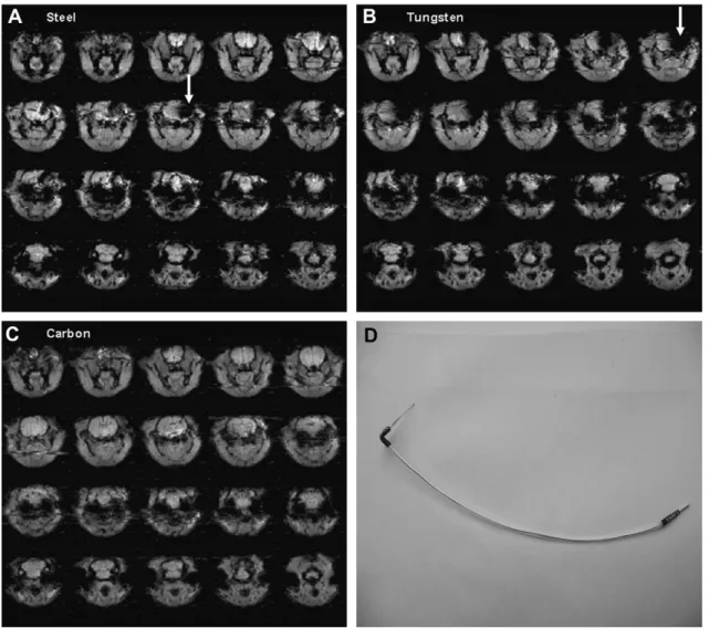
Fig. 1depicts the image artifacts generated by the three elec-trodes (stainless steel, tungsten and carbonfiber), implanted in the right basolateral amygdala. We can observe significant loss of signal surrounding the steel and tungsten electrodes (dark areas in the right hemisphereewhite arrows) rendering the images unsuitable
for analysis. However, no noticeable image artifact is present in the images acquired with carbon-fiber electrodes.Fig. 1also shows an example of the carbon-fiber electrode chosen for experimental procedures. The electrode depicted inFig. 1was used throughout the experiments, even when no stimulation was applied.
As expected, the ES group (with no PTZ) showed a statistically significant recruitment of the area surrounding electrode place-ment for both ES paradigms (Fig. 2A and B slices 0 andþ10), which
corresponds to the amygdaloid complex. In this experiment, stim-ulation paradigms (i.e. NP and P) were applied to the same animal, preceded by a control silent period (without stimulation), in order to allow paired statistics, see methods for details. Although we could observe a trend of fewer activated voxels in the ES-NP group, within a region of interest (ROI) involving the temporal lobe, paired t-test did not show significant difference between ES-NP and ES-P. In addition to the areas around the electrode, ES-P produced contra-lateral activation spreading along thalamic and hippo-campal structures, whereas ES-NP activated more anterior/frontal structures, mainly nucleus accumbens. The effect of NP stimulation recruiting frontal areas (Fig. 2B, sliceþ60) reinforces the idea that
the amygdaloid complex is able to differentiate temporally coded processes even when the same overall stimulating frequency is used (4 stimuli per second).
The ES with PTZ group showed a bi-hemispheric recruitment for the PTZ-noES animals, in agreement with previous reports found in literature (Keogh et al., 2005). In general, a similar acti-vation/inactivation pattern was also observed in the groups submitted to ES (Fig. 3). Nevertheless, the PTZ-P showed a more pronounced activation of the side ipsilateral to electrode implan-tation (slices þ30,þ40, þ50 and þ60 of Fig. 3B) while PTZ-NP
protected the ipsilateral hemisphere against abnormal recruitment (slices 20, 10, 0 andþ10 ofFig. 3C).Fig. 3also shows that
PTZ-NP had less activated posterior areas when compared to either PTZ-noES or PTZ-P.
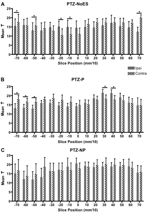
The mean value of T was calculated for the right and left hemispheres, for each animal from the parametric maps of each slice, in order to determine lateralization during the PTZ seizure progression. We can observe that the PTZ-NP abolishes the later-alization in all the slices, whereas PTZ-P significantly enhances lateralization in slicesþ30 andþ40 (Fig. 4). The mean T analysis did
not statistically confirm the diminished ipsilateral activation of slices 20, 10, 0 andþ10 (Fig. 3C). The PTZ-noES showed
signif-icant ipsilateral lateralization in slices 20 and 10 (þ0, although
blood brain barrier (BBB). Both PTZ-noES and PTZ-P showed contra-lateral contra-lateralization for more posterior slices.
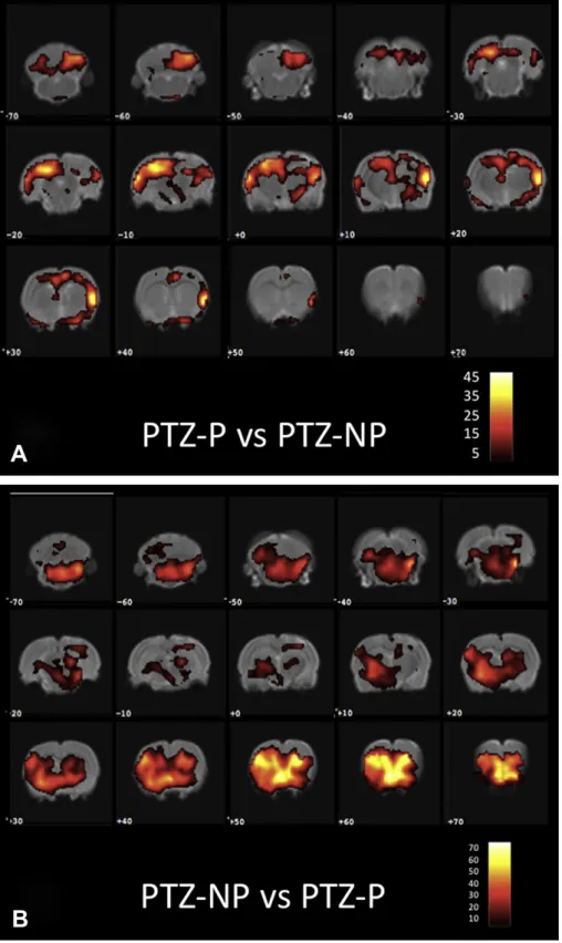
Fig. 5 depicts a voxel by voxel analysis of PTZ infused rats comparing PTZ-P (periodic stimulation) against PTZ-NP (non-periodic stimulation) in panel A, and PTZ-NP against PTZ-P in panel B.Fig. 5A clearly shows an increased ipsilateral activity, regarding electrode placement, when compared to Fig. 5B (slices varying from 20 to þ20 are clearly less compromised in panel B). The
same activation observed in Fig. 2B (slice þ60) is evident
throughout the whole frontal area ofFig. 5B. It is important to highlight that not onlyFigs. 2 and 5come from different experi-ments, using different animals, but thatFig. 2uses paired statistics whileFig. 5A and B is from respectively two different groups of animals continuously infused with PTZ. Thus, altogether, these results raise an interesting hypothesis that NP stimulation may not only play a direct desynchronizing effect in slices neighboring electrode placement (slices 20 to þ20 from Fig. 5A when compared toFig. 5B), but could also indirectly recruit frontal areas (Fig. 5B slicesþ40 toþ70) that may contribute inhibiting seizure
propagation. Nevertheless, the direct hypothesis (through desynchronizing effect) and indirect hypothesis (through activa-tion of PTZ seizure-inhibitory frontal areas) are not exclusive.
In addition, when comparing SPM analysis of slices 20 toþ20 from Figs. 3B and C, with 5A and B, the lateralization effect of
suggested inFig. 4 is even clearer.Fig. 5A presents pronounced activation (slicesþ10 toþ40) the same regions ofFig. 3B, which
neighbor electrode placement of PTZ-P animals. In contrast, for PTZ-NP maps, Figs.3C and5B show diminished activity in regions neighboring electrode placement (slices 20 toþ30).
4. Discussion
The results show that distinct temporal patterns of ES, when applied to amygdala, differentially modulate neural mass recruit-ment in the animal model of PTZ continuous infusion. Our results not only confirm the behavioral counterpart published by our group in freely moving animals (Cota et al., 2009), but also support a more comprehensive theory of ictogenesis which takes into consideration not only hyperexcitability, but also and more importantly, hyper-synchronization and reverberating neural networks.
shows much smaller BOLD changes, which are usually a single prolonged event, preventing signal averaging and frequently resulting in weak responses. In addition, these changes can be sometimes similar to normal scanner drifts (i.e. hardware and physiological), presenting further challenges since true signal might be embedded in normal drifts, being the case of PTZ infusion experiments (Figs. 3e5). Finally, the necessity of keeping the
their BOLD results with other techniques such as 2-DG autoradi-ography (Littlewood et al., 2006; Pohlmann et al., 2007) and microdialysis (Littlewood et al., 2006) with good correlations.
Since PTZ is a GABAergic antagonist (Macdonald and Barker, 1977), it creates a non-specific condition of hyperexcitability necessary to induce seizures evoked by activating multiple rever-berating neural circuits that are gradually recruited, and phase coupled, into the epileptogenic process. According to the theoretical work presented byKudela et al. (2003), there should be an increased synchronization within circuits that have been excited and/or uninhibited. Although described in terms of bursting behavior of small neural network models, such theoretical work could explain why low doses of PTZ (<40/mg) typically evoke minimal seizures
displaying myoclonic jerks, forelimb and head clonus and chewing (Velisek et al., 1992): fewer and more synchronized circuits are recruited. These behaviors are classically correlated with exacer-bated activity of structures in the limbic system, including the amygdala and hippocampus (Eells et al., 2004), which represent a more restricted forebrain region that is more susceptible to PTZ action. In contrast, higher doses of the drug evoke minor seizures followed by major seizures with or without a tonic phase and generalized toniceclonic behavior (Velisek et al., 1992), both
correlated with activation of a broader brain territory that includes structures in hindbrain, midbrain and forebrain structures (Eells et al., 2004; Moraes et al., 2005b). These local and broader neural circuits are connected and synchronized during ictogenesis, as suggested by electrophysiological data (Moraes et al., 2005a), by means of physiological neuro-anatomical connections and a patho-logical hyperexcitable condition. FromFig. 3, it is clear that there is a bilateral recruitment of the PTZ-noES group; nevertheless, slices 20 and 10 (þ0, although not significant, showed a similar
tendency), even though PTZ was applied systemically, indicate ipsilateral lateralization. This apparent inconsistency could be explained by local lesion, in some rats, due to electrode placement. The damaged BBB would facilitate PTZ diffusion resulting in later-alized activation close to the electrode site. Electrical stimulation, although activating neural masses, would partially protect against BBB lesion thus explaining the non-lateralized T mean parameters, in the same slices, for the ES-P and ES-NP groups. In addition, our results show that an ES-P protocol favors ipsilateral abnormal brain connectivity (slicesþ30,þ40,þ50 andþ60 ofFig. 3B and slicesþ30
andþ40 ofFig. 4ePTZ-P) and contra-lateral activation of posterior
areas, most likely resultant from decussatingfibers between fore-brainehindbrain connections. It is important to highlight that the
dose of PTZ was invariable for all stimulation protocols and noES. In other words, by promoting synchronization and better coupling between different reverberating circuits, ES-P would recruit more neural masses and promote hyperexcitation. This would give rise to a myriad of stereotypical convulsive behaviors and cognitive deficits (Uhlhaas and Singer, 2006).
The fact that the mean T analysis (Fig. 4) did not statistically confirm the diminished ipsilateral activation of slices 20, 10, 0 andþ10 (Fig. 3C) for the PTZ-NP group does not invalidate the
anti-resonant hypothesis of NP stimulation. The lateralization analysis ofFig. 4 is much less anatomically specific than what is shown inFig. 3, in effect, if smaller ROI were chosen, differences would become more evident. In addition, the lack of statistical difference, in terms of lateralization (Fig. 4 PTZ-NP), for more anterior slices is also suggestive of an anti-convulsive property of the NP stimulation paradigm. Thus, in order to better highlight the lateralizing effect of the stimulation paradigm during PTZ infusion, contrastFig. 5A clearly shows an increased ipsilateral activity of periodic stimulation against non-periodic, regarding electrode placement, when compared to 5B NP against P (slices varying from 20 toþ20 are clearly less compromised in panel B). Although
Figs. 2 and 5 come from different experiments, using different animals, the same activation observed in Fig. 2B (slice þ60) is
evident throughout the whole frontal area ofFig. 5B. Thus, it is possible that the anti-convulsive property of NP stimulation could be associated to both a direct desynchronization of temporal lobe structures (Figs.3C and5B slices 20 to 20), as discussed before, as well as an indirect activation of pre-frontal areas, know to inhibit temporal lobe substrates (McDonald et al., 1996). Nevertheless, the direct (through desynchronizing effect) and indirect (through activation of PTZ seizure-inhibitory frontal areas) hypothesis are not exclusive and could be interacting to produced an enhanced deactivation of slices 20 toþ20 inFig. 5B.
The pro-convulsive effect of ES-P depicted inFig. 3has a much faster time scale (since the evolution of the entire recruitment happens within minutes) than that proposed by the before mentioned work (Tass and Hauptmann, 2007), which would be comparable to
It is important to highlight that probably not all brain structures would respond to time-coded low frequency ES. The amygdaloid complex was chosen because of its role in the modulation and transfer of epileptiform activity in several animal models of temporal lobe epilepsy (Akirav and Richter-Levin, 2002; Moraes et al., 2005a,b; Vouimba and Richter-Levin, 2005). Supramaximal ES of the amygdala has been shown to produce hippocampal after-discharge, whereas repetitive subthreshold ES induces plastic changes in the temporal lobe that culminate in epileptiform activity (Hirsch et al., 1997). The amygdaloid complex has monosynaptic afferents to and efferents from the parahippocampal areas (e.g., entorhinal cortex and subiculum) (Racine, 1972), providing the anatomical substrates for transfer and modulation of epileptic activity. In short, as the amygdala plays a key role in the synchro-nization process, acting as a modulator and a center for the transfer of epileptiform activity (Hirsch et al., 1997).
In summary, our understanding is that ES of the amygdala modulates seizure initiation and propagation through direct modulation of neural epileptiform activity in local as well as longer-range neural circuits. The periodic stimulation used in this study is in the frequency range of epileptiform activity, as revealed by electrophysiological studies (Moraes et al., 2005a) and, thus, may facilitate the synchronization of reverberant epileptiform networks in a positive resonant modulation. On the other hand, ES-NP promotes desynchronization and, thus, disrupts the positive feed-back process of neural mass recruitment.
Our results use a new temporally coded form of ES that, along with the classical periodical stimulation, provide a biological vali-dation of the effect of ES on neural desynchronizing and synchro-nizing activity, respectively. The impact of suchfindings on the treatment of epilepsies is even more promising if one considers that ES was kept within 4 stimuli per second. Such a strategy may overcome some unwanted collateral effects of classical HFS for the treatment of epilepsy, such as a higher energy transfer to brain tissue. However, much work must be done in order to better assess the synchronizing/desynchronizing effects of ES using multiple sites of stimulation and different animal models of epilepsy for biological validation.
Acknowledgements
We are grateful to CNPq, FAPEMIG, CAPES and PRPq/UFMG, for financial support. Márcio Flávio Dutra Moraes is recipient of CAPES and CNPq research fellowship. INCeMaq e National Institute of
Science and Technology, also supported this work.
References
Akirav, I., Richter-Levin, G., 2002. Mechanisms of amygdala modulation of hippo-campal plasticity. J. Neurosci. 22, 9912e9921.
Ben-Menachem, E., 2002. Vagus-nerve stimulation for the treatment of epilepsy. Lancet Neurol. 1, 477e482.
Benabid, A.L., Minotti, L., Koudsie, A., de Saint Martin, A., Hirsch, E., 2002. Antiep-ileptic effect of high-frequency stimulation of the subthalamic nucleus (corpus luysi) in a case of medically intractable epilepsy caused by focal dysplasia: a 30-month follow-up: technical case report. Neurosurgery 50, 1385e1391. discus-sion 1391e1392.
Binnie, C.D., 2000. Vagus nerve stimulation for epilepsy: a review. Seizure 9, 161e169.
Centeno, R.S., Yacubian, E.M., Sakamoto, A.C., Ferraz, A.F., Junior, H.C., Cavalheiro, S., 2006. Pre-surgical evaluation and surgical treatment in children with extra-temporal epilepsy. Childs Nerv. Syst. 22, 945e959.
Chabardes, S., Kahane, P., Minotti, L., Koudsie, A., Hirsch, E., Benabid, A.L., 2002. Deep brain stimulation in epilepsy with particular reference to the subthalamic nucleus. Epileptic Disord. 4 (Suppl. 3), S83eS93.
Cota, V.R., Medeiros Dde, C., Vilela, M.R., Doretto, M.C., Moraes, M.F., 2009. Distinct patterns of electrical stimulation of the basolateral amygdala influence penty-lenetetrazole seizure outcome. Epilepsy Behav. 14 (Suppl. 1), 26e31. DeGiorgio, C.M., Shewmon, D.A., Whitehurst, T., 2003. Trigeminal nerve stimulation
for epilepsy. Neurology 61, 421e422.
Durand, D., 1993. Ictal patterns in experimental models of epilepsy. J. Clin. Neuro-physiol. 10, 281e297.
Eells, J.B., Clough, R.W., Browning, R.A., Jobe, P.C., 2004. Comparative fos immuno-reactivity in the brain after forebrain, brainstem, or combined seizures induced by electroshock, pentylenetetrazol, focally induced and audiogenic seizures in rats. Neuroscience 123, 279e292.
Engel Jr., J., 1996. Introduction to temporal lobe epilepsy. Epilepsy Res. 26, 141e150. Fanselow, E.E., Reid, A.P., Nicolelis, M.A., 2000. Reduction of pentylenetetrazole-induced seizure activity in awake rats by seizure-triggered trigeminal nerve stimulation. J. Neurosci. 20, 8160e8168.
French, J.A., 2007. Refractory epilepsy: clinical overview. Epilepsia 48 (Suppl. 1), 3e7. Freund, H.J., 2005. Long-term effects of deep brain stimulation in Parkinson’s
disease. Brain 128, 2222e2223.
Gale, K., 1992. Subcortical structures and pathways involved in convulsive seizure generation. J. Clin. Neurophysiol. 9, 264e277.
Hamani, C., Ewerton, F.I., Bonilha, S.M., Ballester, G., Mello, L.E., Lozano, A.M., 2004. Bilateral anterior thalamic nucleus lesions and high-frequency stimulation are protective against pilocarpine-induced seizures and status epilepticus. Neuro-surgery 54, 191e195. discussion 195e197.
Hauser, W.A., Rich, S.S., Lee, J.R., Annegers, J.F., Anderson, V.E., 1998. Risk of recur-rent seizures after two unprovoked seizures. N. Engl. J. Med. 338, 429e434. Hirsch, E., Danober, L., Simler, S., Pereira de Vasconcelos, A., Maton, B., Nehlig, A.,
Marescaux, C., Vergnes, M., 1997. The amygdala is critical for seizure propaga-tion from brainstem to forebrain. Neuroscience 77, 975e984.
Hodaie, M., Wennberg, R.A., Dostrovsky, J.O., Lozano, A.M., 2002. Chronic anterior thalamus stimulation for intractable epilepsy. Epilepsia 43, 603e608. Ireland, M.D., Lowe, A.S., Reavill, C., James, M.F., Leslie, R.A., Williams, S.C., 2005.
Mapping the effects of the selective dopamine D2/D3 receptor agonist quine-lorane using pharmacological magnetic resonance imaging. Neuroscience 133, 315e326.
Jones, N., O’Neill, M.J., Tricklebank, M., Libri, V., Williams, S.C., 2005. Examining the neural targets of the AMPA receptor potentiator LY404187 in the rat brain using pharma-cological magnetic resonance imaging. Psychopharmacology (Berl.) 180, 743e751. Keogh, B.P., Cordes, D., Stanberry, L., Figler, B.D., Robbins, C.A., Tempel, B.L.,
Green, C.G., Emmi, A., Maravilla, K.M., Schwartzkroin, P.A., 2005. BOLD-fMRI of PTZ-induced seizures in rats. Epilepsy Res. 66, 75e90.
Kudela, P., Franaszczuk, P.J., Bergey, G.K., 2003. Changing excitation and inhibition in simulated neural networks: effects on induced bursting behavior. Biol. Cybern. 88, 276e285.
Kumar, R., Lozano, A.M., Sime, E., Lang, A.E., 2003. Long-term follow-up of thalamic deep brain stimulation for essential and parkinsonian tremor. Neurology 61, 1601e1604. Littlewood, C.L., Jones, N., O’Neill, M.J., Mitchell, S.N., Tricklebank, M., Williams, S.C., 2006. Mapping the central effects of ketamine in the rat using pharmacological MRI. Psychopharmacology (Berl.) 186, 64e81.
Loscher, W., Schmidt, D., 2002. New horizons in the development of antiepileptic drugs. Epilepsy Res. 50, 3e16.
Macdonald, R.L., Barker, J.L., 1977. Pentylenetetrazol and penicillin are selective antagonists of GABA-mediated post-synaptic inhibition in cultured mammalian neurones. Nature 267, 720e721.
McDonald, A.J., Mascagni, F., Guo, L., 1996. Projections of the medial and lateral prefrontal cortices to the amygdala: a Phaseolus vulgaris leucoagglutinin study in the rat. Neuroscience 71, 55e75.
McIntyre, C.C., Grill, W.M., 2001. Finite element analysis of the current-density and electricfield generated by metal microelectrodes. Ann. Biomed. Eng. 29, 227e235. McIntyre, C.C., Savasta, M., Kerkerian-Le Goff, L., Vitek, J.L., 2004a. Uncovering the mechanism(s) of action of deep brain stimulation: activation, inhibition, or both. Clin. Neurophysiol. 115, 1239e1248.
McIntyre, C.C., Savasta, M., Walter, B.L., Vitek, J.L., 2004b. How does deep brain stimulation work? Present understanding and future questions. J. Clin. Neuro-physiol. 21, 40e50.
Mirski, M.A., Rossell, L.A., Terry, J.B., Fisher, R.S., 1997. Anticonvulsant effect of anterior thalamic high frequency electrical stimulation in the rat. Epilepsy Res. 28, 89e100.
Moraes, M.F., Chavali, M., Mishra, P.K., Jobe, P.C., Garcia-Cairasco, N., 2005a. A comprehensive electrographic and behavioral analysis of generalized ton-iceclonic seizures of GEPR-9s. Brain Res. 1033, 1e12.
Moraes, M.F., Garcia-Cairasco, N., 1997. Glass pipette-carbonfiber microelectrodes for evoked potential recordings. Braz. J. Med. Biol. Res. 30, 1319e1324. Moraes, M.F., Mishra, P.K., Jobe, P.C., Garcia-Cairasco, N., 2005b. An electrographic
analysis of the synchronous discharge patterns of GEPR-9s generalized seizures. Brain Res. 1046, 1e9.
Paxinos, G., Watson, C., 1998. The Rat Brain in Stereotaxic Coordinates. Academic, San Diego, London, pp. xxvi, 236 pp.
Pohlmann, A., Barjat, H., Tilling, L.C., James, M.F., 2007. Pharmacological fMRIe challenges in analysing drug-induced single-event BOLD responses. Conf. Proc. IEEE Eng. Med. Biol. Soc. 2007, 3411e3416.
Racine, R.J., 1972. Modification of seizure activity by electrical stimulation. I. After-discharge threshold. Electroencephalogr. Clin. Neurophysiol. 32, 269e279. Rodriguez-Oroz, M.C., Obeso, J.A., Lang, A.E., Houeto, J.L., Pollak, P., Rehncrona, S.,
Snead 3rd, O.C., 1992. Pharmacological models of generalized absence seizures in rodents. J. Neural. Transm. (Suppl. 35), 7e19.
Spencer, S.S., 2002. When should temporal-lobe epilepsy be treated surgically? Lancet Neurol 1, 375e382.
Tass, P.A., Hauptmann, C., 2007. Therapeutic modulation of synaptic connectivity with desynchronizing brain stimulation. Int. J. Psychophysiol. 64, 53e61. Theodore, W.H., Fisher, R.S., 2004. Brain stimulation for epilepsy. Lancet Neurol. 3,
111e118.
Timofeev, I., Steriade, M., 2004. Neocortical seizures: initiation, development and cessation. Neuroscience 123, 299e336.
Uhlhaas, P.J., Singer, W., 2006. Neural synchrony in brain disorders: relevance for cognitive dysfunctions and pathophysiology. Neuron 52, 155e168.
Valencia, I., Holder, D.L., Helmers, S.L., Madsen, J.R., Riviello Jr., J.J., 2001. Vagus nerve stimulation in pediatric epilepsy: a review. Pediatr. Neurol. 25, 368e376. Velisek, L., Kubova, H., Pohl, M., Stankova, L., Mares, P., Schickerova, R., 1992.
Pentylenetetrazol-induced seizures in rats: an ontogenetic study. Naunyn Schmiedebergs Arch. Pharmacol. 346, 588e591.
Volkmann, J., 2004. Deep brain stimulation for the treatment of Parkinson’s disease. J. Clin. Neurophysiol. 21, 6e17.
Volkmann, J., Allert, N., Voges, J., Sturm, V., Schnitzler, A., Freund, H.J., 2004. Long-term results of bilateral pallidal stimulation in Parkinson’s disease. Ann. Neurol. 55, 871e875.
Vonck, K., Boon, P., Achten, E., De Reuck, J., Caemaert, J., 2002. Long-term amyg-dalohippocampal stimulation for refractory temporal lobe epilepsy. Ann. Neu-rol. 52, 556e565.
Vonck, K., Boon, P., Claeys, P., Dedeurwaerdere, S., Achten, R., Van Roost, D., 2005. Long-term deep brain stimulation for refractory temporal lobe epilepsy. Epi-lepsia 46 (Suppl. 5), 98e99.
Vouimba, R.M., Richter-Levin, G., 2005. Physiological dissociation in hippocampal subregions in response to amygdala stimulation. Cereb. Cortex 15, 1815e1821. Wiebe, S., Blume, W.T., Girvin, J.P., Eliasziw, M., 2001. A randomized, controlled trial
of surgery for temporal-lobe epilepsy. N. Engl. J. Med. 345, 311e318. Wuttke, T.V., Lerche, H., 2006. Novel anticonvulsant drugs targeting