Distinct patterns of electrical stimulation of the basolateral amygdala influence
pentylenetetrazole seizure outcome
Vinícius Rosa Cota
a,b, Daniel de Castro Medeiros
b, Maura Regina Silva da Páscoa Vilela
b,
Maria Carolina Doretto
b, Márcio Flávio Dutra Moraes
b,*aInstituto Internacional de Neurociências de Natal Edmond e Lily Safra, Natal-RN, Brazil
bNúcleo de Neurociências (NNC), Departamento de Fisiologia e Biofísica, ICB, UFMG, Av. Antonio Carlos, 6627 CEP 31270-901 Campus Pampulha, Belo Horizonte-MG, Brazil
a r t i c l e
i n f o
Article history:
Received 7 August 2008 Revised 4 September 2008 Accepted 5 September 2008
Keywords:
Basolateral amygdala Pentylenetetrazole Synchronization Desynchronization Electrical stimulation Seizure suppression Behavior modulation Synfire chains
a b s t r a c t
Our working hypothesis is that constant interpulse interval (IPI) electrical stimulation would resonate with endogenous epileptogenic reverberating circuits, inducing seizures, whereas a random interinterval electrical stimulation protocol would promote desynchronization of such neural networks, producing an anticonvulsant effect. Male Wistar rats were stereotaxically implanted with a bipolar electrical stimula-tion electrode in the amygdala. Pentylenetetrazole (10 mg/ml/min) was continuously infused through an intravenous catheter to induce seizures while four different patterns of temporally coded electrical stim-ulation were applied: periodic stimstim-ulation (PS), pseudo-randomized IPI stimstim-ulation (LH), restrictively randomized IPI stimulation (IH), and bursts of 20-ms IPIs (burst). PS decreased the pentylenetetrazole threshold to forelimb clonus, whereas IH increased the threshold to forelimb clonus and to generalized tonic–clonic seizures. We hypothesize that PS facilitates forelimb clonus by reverberating with epilepto-genic circuits in the limbic system, whereas IH delays forelimb clonus and generalized tonic–clonic sei-zures by desynchronizing the epileptic neural networks in the forebrain–midbrain–hindbrain circuits.
Ó2008 Elsevier Inc. All rights reserved.
1. Introduction
In about one-fourth of persons with epilepsy, seizures are not satisfactorily controlled with pharmacological treatment[1–3]. In addition, many of these patients with refractory epilepsy are not eligible for ablative surgery, which, in most cases, requires a read-ily identifiable epileptogenic focus[4–6]. A more recent alternative available for these patients is electrical stimulation (ES) of the ner-vous system[7]. ES may be applied peripherally in structures such as the vagus nerve (vagus nerve stimulation)[8–10]and the tri-geminal nerve (tritri-geminal nerve stimulation)[11,12] or targeted to a variety of structures in the central nervous system (deep brain stimulation)[13–15], most predominantly the anterior nucleus of the thalamus[16,17], the subthalamic nuclei[18,19], and the epi-leptogenic focus itself[20]. Classically, continuous or intermittent high-frequency ES (high-frequency stimulation) is the overall adopted pattern for an anticonvulsant effect, whereas low-fre-quency ES (low-frelow-fre-quency stimulation) is generally believed to be proconvulsant[7].
Although targeting of different neural substrates with ES has proved to be an effective approach to controlling seizures[7], the
underlying mechanisms of seizure suppression are still poorly understood[21,22]. Classically, there are two main philosophies regarding neural substrate excitability to explain the clinical bene-fits of ES in epilepsy: (1) ES works by suppressing or inhibiting epi-leptogenic structures, which is analogous to the functional ablation performed by neurosurgery; or (2) ES works by activating or stim-ulating neural networks that would modulate seizure-like activity. In fact, it has been reported that changing either ES parameters (e.g., amplitude, frequency, and wave morphology) or the structure targeted may influence the effect of ES on seizure control, in some cases even resulting in seizure potentiation [23]. This work addresses a different paradigm, in which ES may also play a critical role in neural synchronization depending on the temporal pattern used (i.e., pulses not distributed evenly in time), even though over-all frequency of stimulation, amplitude, and electrode placement are maintained constant. In particular, the use of temporally coded/low-frequency ES may bring significant improvement in the undesired side effects of ES of neural structures.
The existence of epileptogenic neural networks that gradually synchronize by means of oscillatory reverberating circuits has been suggested in animal models of epilepsy, for example, the Genetic Epilepsy Prone Rat (GEPR). Moraes et al.[24]proposed that the EEG spike morphology signature found in the GEPR-9 seizure is the result of sequential and recurrent involvement of
1525-5050/$ - see front matterÓ2008 Elsevier Inc. All rights reserved. doi:10.1016/j.yebeh.2008.09.006
* Corresponding author. Fax: +55 31 3499 2924.
E-mail address:[email protected](M.F.D. Moraes).
Contents lists available atScienceDirect
Epilepsy & Behavior
forebrain–midbrain–hindbrain neural substrates. Moreover, in the GEPR tonic–clonic seizure, the interspike interval increases linearly after each spike [25], which would corroborate the theory of a reverberating circuit that would gradually compromise neural communication between elements in the loop due to either meta-bolic or neurochemical stress. Accordingly, the reverberating neu-ral networks that oscillate and generate epileptiform activity would be modulated either by setting any element of the reverber-ating loop in a refractory state or by desynchronizing the sequen-tial involvement of such elements. Our working hypothesis is that an ES protocol consisting of fixed-frequency ES that would reso-nate with endogenous epileptogenic reverberating circuits would induce seizures, whereas a nonperiodic (e.g., random) ES protocol would promote desynchronization of such neural networks, thus behaving as an anticonvulsant.
In the present report, the basolateral amygdala was chosen as the ES target using the pentylenetetrazole (PTZ) animal model of epilepsy. The amygdaloid complex was chosen because of its role in the modulation and transfer of epileptiform activity in several animal models of temporal lobe epilepsy[26–29]. Supramaximal ES of the amygdala has been shown to produce hippocampal after-discharge, whereas repetitive subthreshold ES induces plastic changes in the temporal lobe that culminate in epileptiform activ-ity[30]. The amygdaloid complex has monosynaptic afferents to and efferents from the parahippocampal areas (e.g., entorhinal cor-tex and subiculum)[31], providing the anatomical substrates for transfer and modulation of epileptic activity. In this work, we tested the hypothesis that low-frequency ES (four stimuli per sec-ond) of the basolateral amygdala would differentially modulate seizure outcome in PTZ-treated animals if applied at either (1) con-stant intervals or (2) randomly spaced intervals.
2. Methods
All experiments were done in accordance with the Ethical Committee for Animal Experimentation (Comitê de Ética em Experimentação Animal—CETEA) of the Federal University of Minas Gerais (Universidade Federal de Minas Gerais—UFMG), and Procedures for animal care were previously approved by this orga-nization under Protocol 150/06.
We designed and built an electrical stimulator composed of a constant-current isolation unit driven by a PC-programmable clocking system. C++ software was developed to program the stim-ulator with four patterns of temporally coded stimuli, all delivering a total of four pulses per second (to guarantee the same energy flow): (1) constant interpulse intervals (IPIs) of 250 ms (periodic); (2) bursts with 20-ms IPIs (burst); (3) pseudo-randomized IPIs (lin-ear decay histogram [LH stimulation]); and (4) restrictively ran-domized IPIs (inverse decay histogram [IH stimulation]). These temporal patterns are depicted in Fig. 1along with their corre-sponding average histograms. The LH pattern was obtained by sort-ing four time stamps in a 1-s interval out of a uniform distribution using an internal library function. The IH pattern was obtained using the same built-in function with the following algorithm: (A) sort an intervalT1in the range 20–940 ms, waitT1,and fire pulse; (B) sort an intervalT2in the rangeT1+ 20 ms to 960 ms, waitT2,and repeat pulse; (C) repeat B twice until four pulses are fired and wait to complete the second. A minimum biologically plausible separation of 20 ms between pulses was observed in all patterns. Pulses were square, positive monophasic waves of
100-ls duration with amplitudes varying from 100 to 350
lA.
A total of 74 male Wistar rats from Centro de Bioterismo (CEBIO) of UFMG were randomly assigned to each stimulus group on the day of the experiment (periodic n= 19, burst n= 6, LH n= 9, IH n= 14) and also to an extra nonstimulated
control group (controln= 26); adjustments were made to main-tain a minimum number of animals in each group. Mean weight did not differ significantly between groups (control: 306 ± 11 g, periodic: 292 ± 12 g, burst: 245 ± 12 g, LH: 253 ± 13 g, and IH: 313 ± 13 g, one-way ANOVA). All animals underwent a surgical procedure for implantation of a bipolar stimulation electrode. Electrodes were made of a twisted pair of stainless-steel (0.005 in.), Teflon-coated wires (Model 791400, A-M Systems Inc., Carlsborg, WA, USA). Animals were anesthetized via sys-temic sodium thiopental injection (40 mg/kg) and locally with lidocaine clorohydrate plus epinephrine (2%) and then positioned in a stereotaxic frame (Stoelting Co., Wood Dale, IL, USA). Coor-dinates for the right basolateral amygdala (AP = 2.8 mm, ML = 5.0 mm referenced from the bregma suture and 7.2 mm from dura mater) were derived from the Paxinos and Watson’s atlas for rats [32]. Animals were pretreated with pentabiotics (19 mg/kg) and flunixin (2.5 mg/kg). After correct positioning, the electrode was fixed to the skull with zinc cement and sol-dered to a telephone jack (Model RJ-45 6x6), which was fixed onto the skull with dental acrylic. A 5- to 7-day post-operative recovery period was observed before stimulation began. On the day before the experiment, animals received very low frequency (0.25 Hz) ES of increasing current amplitude to determine the stimulus threshold, defined as the lowest amplitude of current capable of evoking observable and distinguishable twitching behavior. On the day of the experiment, animals were caudally cannulated for intravenous infusion of PTZ (Sigma) diluted in sal-ine at a concentration of 10 mg/ml. The cannula was connected to an infusion pump set at the rate of 1 ml/min. The ES was ini-tiated 10 s before beginning the infusion. The latencies to onset of forelimb clonus and tonic–clonic seizures were determined and were correlated with the injected PTZ dose. All experiments were recorded on VHS tape for behavioral analysis. After stimu-lation, brains were electrically lesioned (0.5 mA for 2 s) and immediately removed, sliced, and stained with neutral red (2%) for histological confirmation of electrode position. Animals that survived 60 min after the end of the experiment (n= 4) were anesthetized with urethane (140 mg/kg) and transcardially per-fused with formaldehyde (4%) before brain removal and
histol-ogy procedures. Animals with incorrect positioning of
electrodes were not included in our analysis. PTZ dose data were normalized by body weight (PTZ threshold), and the results were analyzed with one-way ANOVA and assessed post hoc with the Tukey multiple comparison test. Survival ratio and occurrence of uncommon PTZ-induced behaviors, such as facial clonus, rear-ing and fallrear-ing, and whole-body tonus, were assessed with con-tingency tables and Fisher’s exact test. Data were considered to be statistically significant at P< 0.05.
3. Results
All animals, except for the saline-injected control group, displayed the convulsive behavior sequence typical of the PTZ model. This consisted of a first myoclonic jerk, followed by fore-limb clonus (FC), generalized clonus, and generalized tonic–clonic seizures (GTCSs)[33]. Some seizure-related behavior was confused with the twitching reaction to ES; thus, only the occurrence of fore-limb clonus and tonic-clonic seizures could be adequately quantified.
Distinct patterns of ES differentially modulated the latency to FC (P< 0.0001, one-way ANOVA) and GTCSs (P< 0.0001, one-way ANOVA), as shown inFigs. 2A and B, respectively. The PTZ thresh-old for FC was significantly reduced by periodic stimulation when compared with control (P< 0.05, post hoc Tukey test) and IH stimulation (P< 0.001, post hoc Tukey test). Neither burst nor LH
stimulation changed the FC PTZ threshold. Nevertheless, IH stimulation significantly increased threshold to FC when compared with all groups (P< 0. 05 against control and burst,P< 0.01 against LH, andP< 0.001 against periodic, post hoc Tukey tests). Moreover, IH stimulation robustly increased (by almost twofold) the PTZ threshold to GTCSs when compared with all other groups (P< 0.001 against all groups, post hoc Tukey test). Periodic stimulation and other patterns had no overt effects on GTCSs when compared in a pairwise manner.
Rearing behavior, very uncommon in PTZ models [33], was observed in the periodic and IH groups (Table 1) when compared with the control group, with statistical significance (P< 0.01 and P< 0.05, respectively, Fisher’s exact test). Finally, three animals in the IH group, one in the periodic group, and none in the other groups survived. Survival rate of the IH group was statistically greater than that for other groups pooled together, as assessed in
Table 2(P< 0.05, Fisher’s exact test).
There were no statistically relevant correlations among weight, current, behavior threshold, and behavior occurrence in any groups (data not shown).
4. Discussion
The results clearly indicate that distinct temporal patterns of ES, when applied to the amygdala, differentially modulate PTZ-induced
seizure behavior in a rodent model. The effects observed in this study could be predicted by a comprehensive theory of ictogenesis which takes in consideration not only hyperexcitability, but also, and more importantly, hypersynchronism and reverberating neural networks. In this sense, all three mechanisms must be kept in mind to ade-quately explain the results described.
As PTZ is a GABAergic antagonist[34], it creates a nonspecific condition of hyperexcitability necessary to induce seizures evoked by multiple reverberating neural circuits that are gradually recruited into the epileptogenic process as the drug is absorbed. In fact, low doses of PTZ (<40/mg) typically evoke minimal seizures displaying myoclonic jerks, forelimb and head clonus, and chewing
structures, such as the amygdala play a key role in this synchroni-zation process, acting as a modulator and a center for the transfer of epileptiform activity[29].
Our understanding is that ES of the amygdala modulates con-vulsive behavior through direct modulation of neural epileptiform activity in local as well as broader neural circuits, in a manner sim-ilar to that by which the structure acts during normal activity. The periodic stimulation used in this study is in the frequency range of epileptiform activity, as revealed by electrophysiological studies
[25]and, thus, may facilitate forelimb clonus by adding energy to the limbic system in a resonating fashion. In contrast, we hypoth-esize that IH stimulation impairs reverberation of the limbic sys-tem (local) and forebrain–midbrain–hindbrain (broader) circuits through an antiresonating effect in each of them and also through blockage of synchronizing mechanisms among them. The effect
results in the increase in PTZ threshold to forelimb clonus and generalized tonic–clonic seizures observed here.
Interestingly, rearing is a very uncommon behavior in the PTZ model and is said to be masked inside the GTCSs that would pre-dominate in the motor expression [35]. Nevertheless, periodic stimulation increases its occurrence probably due to a facilitation process capable of unmasking it from GTCSs. By the same token, the increased rearing caused by IH stimulation is probably due to an unmasking process of delaying GTCSs. One could hypothesize that if rearing was normally observed in the PTZ model, periodic stimulation would decrease its threshold, and IH would have no ef-fects. A direct consequence of this reasoning is to design analogous experiments using different animal models of epilepsy that display limbic seizures (e.g., audiogenic kindling of WARs, amygdala electrical kindling, acute and spontaneous pilocarpine-induced seizures).
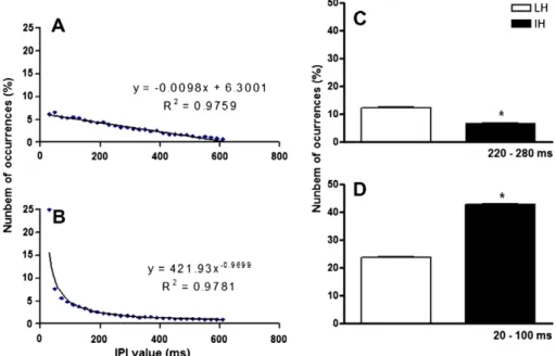
The difference in seizure threshold between LH and IH is not easily understood, and further manipulations of the temporal pat-tern of stimulation may help clarify the basis for this phenome-non. Although they have a distribution over a wide range of IPI values, the histograms of LH and IH have very distinct shapes. The LH histogram can be fit (R2= 0.98) by a linear equation of inclination close to 1/100, whereas IH is fit (R2= 0.98) by a power equation of exponent close to 1 (Figs. 3A and B, respec-tively). A first consequence of these different shapes is that each of them accumulates IPI occurrences in distinct ranges. Two dif-ferences are worth noting. First, LH has a higher count of IPI in the range 220–280 ms (Fig. 3C) than does IH. As mentioned be-fore, this is in the range of epileptiform activity frequency of dis-charge, and thus, IPIs in this range are probably resonating and convulsant, as suggested by the periodic group. Additionally, IH has a considerably higher count of IPIs within the range 20– 100 ms than does LH (Fig. 3D). One could assume that this means IH has a higher content of high-frequency stimulation, thus cor-roborating previous work, mainly on deep-brain stimulation, which would have anticonvulsant properties. However, this is not true, once high-frequency stimulation is delivered at frequen-cies over 100 Hz[7,21,35] to produce its inhibitory effects. Also, burst stimulation in this study, which is composed mainly of brief periods of high-frequency stimulation, was not effective in sup-pressing seizures. The authors propose an alternative hypothesis that relies on the recognition of different temporal codes of pulses or cortical motifs, also calledcortical songs[37], by special-ized neuronal circuits arranged, for example, as the synfire chains
[38]. The existence of such circuits is strongly suggested by experimental studies [39–41]; a series of biologically plausible proposals have been described [42–45] and their features have been modeled and studied in silico[38,46–48]. According to these studies, pattern recognition circuits would reverberate when neu-ronal inputs have a specific well-defined temporal structure[38], and two or more of these circuits may synchronously couple when sharing the same temporal pattern of activity[49]. Finally, cortical motifs have strict timing constraints, and their constitu-tive pulses must be grouped within a certain time limit. Although there is no consensus on a value, some authors suggest that the whisking frequency (10 Hz or 100-ms period) is a restrictive time window for somatosensory processing [39]. In this sense, our understanding is that IH randomly activates multiple distinct cir-cuits by ‘‘sending” randomized cortical motifs through efferents of the amygdala at a much faster rate than does LH, because it has a higher count of short IPIs in the range 20–100 ms. This would severely impair neural synchronization of local circuits, once they are activated by distinct patterns, preventing them from being coupled. It would also impair the transfer of epilepti-form activity to broader circuits once the amygdala would fire in a pattern that may be not synchronized.
Fig. 2.PTZ threshold for two convulsive behaviors—forelimb clonus (A) and generalized tonic–clonic seizures (B)—according to stimulus pattern. IH stimulation increased drug threshold for both forelimb clonus and generalized tonic–clonic seizures when compared with all groups (both P< 0.0001, one way ANOVA; *P< 0.05, **P< 0.01, ***P< 0.001, all post hoc Tukey). Moreover, periodic stimula-tion decreased drug threshold for forelimb clonus (P< 0.05, post hoc Tukey).
Table 1
Contingency table for occurrence of rearing behavior
With rearing Without rearing
Control 0 26
Periodica 6 13
Burst 0 6
LH 0 9
IHb 4 10
a,bPeriodic and IH stimulated groups manifested more rearing behavior compared with the control group (aP< 0.01 andbP< 0.05, respectively, Fischer’s exact test).
Table 2
Contingency table for survival ratioa
Survived Died
IH stimulus 3 11
Others 1 59
a All other groups (periodic, burst, LH, and control) have been pooled together under ‘‘Others.”
In short, a reasonable hypothesis for the difference between the two stimulation patterns is based on the proportions of reso-nating and antiresoreso-nating power present in each of them. The proportions of antiresonating and resonating power of LH stimu-lation patterns would not be high enough, probably close to one in a proper scale, canceling each other out. In contrast, the IH pat-tern would have a proportion such that the antiresonating power would overcome resonating power by far. This would make IH a seizure-suppressing stimulus, whereas LH would have no effect. A possible way to correlate antiresonating power with a random-ized pattern construction algorithm would be to apply different limits around fixed 4-Hz time stamps (0, 250, 500, and 750 ms) for randomization of pulses and analyzing their effects in seizure suppression.
Our results suggest that desynchronizing neural activity by neurostimulation is an alternative to be considered in the treat-ment of epileptic disorders in clinical practice. A next logical step would be to run clinical trials of human deep-brain stimulation activated in LH and IH patterns. Such a strategy may overcome some unwanted collateral effects of classic high-frequency stimu-lation for the treatment of human epilepsy, such as a higher energy transfer to brain tissue. However, much work must be done in ani-mal models to better assess the synchronizing/desynchronizing effects of ES. Moreover, behavior modulation through random pat-terns and other temporal codes of stimulation may provide fruitful insights into the mechanisms of seizure genesis, propagation, and termination.
Conflict of interest statement
The authors state that no other people or organization have inappropriately influenced this work. Therefore, there is no perti-nent claim of a conflict of interest.
Acknowledgments
We are grateful to Gioconda Alves de Assumpção for technical assistance and to CNPq, FAPEMIG, CAPES, and PRPq /UFMG for
financial support. Márcio Flávio Dutra Moraes and Maria Carolina Doretto are recipients of CNPq research fellowships.
References
[1] French JA. Refractory epilepsy: clinical overview. Epilepsia 2007;48(Suppl. 1):3–7.
[2] Löscher W, Schmidt D. New horizons in the development of antiepileptic drugs. Epilepsy Res 2002;50:3–16.
[3] Wuttke TV, Lerche H. Novel anticonvulsant drugs targeting voltage-dependent ion channels. Expert Opin Invest Drugs 2006;15:1167–77.
[4] Centeno R, Yacubian E, Sakamoto E, Ferraz A, Carrete Junior H, Cavalheiro S. Pre-surgical evaluation and surgical treatment in children with extratemporal epilepsy. Child’s Nerv Syst 2006;22:945–59.
[5] Wiebe S, Blume WT, Girvin JP, Eliasziw M. For the Effectiveness and Efficiency of Surgery for Temporal Lobe Epilepsy Study Group. A randomized, controlled trial of surgery for temporal-lobe epilepsy. N Engl J Med 2001;345:311–8.
[6] Spencer SS. When should temporal-lobe epilepsy be treated surgically? Lancet Neurol 2002;1:375–82.
[7] Theodore WH, Fisher RS. Brain stimulation for epilepsy. Lancet Neurol 2004;3:111–8.
[8] Ben Menachem E. Vagus-nerve stimulation for the treatment of epilepsy. Lancet Neurol 2002;1:477–82.
[9] Binnie CD. Vagus nerve stimulation for epilepsy: a review. Seizure 2000;9:161–9.
[10] Valencia I, Holder DL, Helmers SL, Madsen JR, Riviello J. Vagus nerve stimulation in pediatric epilepsy: a review. Pediatr Neurol 2001;25: 368–76.
[11] DeGiorgio CM, Shewmon DA, Whitehurst T. Trigeminal nerve stimulation for epilepsy. Neurology 2003;61:421–2.
[12] Fanselow EE, Reid AP, Nicolelis MAL. Reduction of pentylenetetrazole-induced seizure activity in awake rats by seizure-triggered trigeminal nerve stimulation. J Neurosci 2000;20:8160–8.
[13] Benabid AL, Minotti LMD, Koudsie AMD, Saint Martin AMD, Hirsch EMD. Antiepileptic effect of high-frequency stimulation of the subthalamic nucleus (corpus luysi) in a case of medically intractable epilepsy caused by focal dysplasia: a 30-month follow-up [technical case report]. Neurosurgery 2002;50:1385–92.
[14] Hodaie M, Wennberg RA, Dostrovsky JO, Lozano AM. Chronic anterior thalamus stimulation for intractable epilepsy. Epilepsia 2002;43:603–8. [15] Vonck K, Boon P, Claeys P, Dedeurwaerdere S, Achten R, Van Roost D.
Long-term deep brain stimulation for refractory temporal lobe epilepsy. Epilepsia 2005;46(Suppl. 5):98–9.
[16] Hamani C, Ewerton FIS, Bonilha SM, Ballester G, Mello LE, Lozano AM. Bilateral anterior thalamic nucleus lesions and high frequency stimulation are protective against pilocarpine-induced seizures and status epilepticus. Neurosurgery 2004;54:191–7.
Fig. 3.Curve fitting for the mean histogram (n= 8 simulated histograms) of the two randomized stimulus patterns. The LH mean histogram is fit by a linear equation of inclination close to 1/100 (A), and the IH mean histogram is fit by a power curve of exponential close to 1 (B), both with high correlation factors (R2= 0.9759 and
[17] Mirski MA, Rossell LA, Terry JB, Fisher RS. Anticonvulsant effect of anterior thalamic high frequency electrical stimulation in the rat. Epilepsy Res 1997;28:89–100.
[18] Chabardès S, Kahane P, Minotti L, Koudsie A, Hirsch E, Benabid A. Deep brain stimulation in epilepsy with particular reference to the subthalamic nucleus. Epileptic Disord 2002;4:83–93.
[19] Benabid A, Minotti L, Koudsie A, Saint Martin A, Hirsch E. Antiepileptic effect of high-frequency stimulation of the subthalamic nucleus (corpus luysi) in a case of medically intractable epilepsy caused by focal dysplasia: a 30-month follow-up [technical case report]. Neurosurgery 2002;50:1385–91. [20] Vonck K, Boon P, Achten E, De Reuck J, Caemaert J. Long-term
amygdalohippocampal stimulation for refractory temporal lobe epilepsy. Ann Neurol 2002;52:556–65.
[21] McIntyre CC, Savasta M, Walter BL, Vitek JL. How does deep brain stimulation work? Present understanding and future questions. J Clin Neurophysiol 2004;21:40–50.
[22] McIntyre CC, Savasta M, Kerkerian-Le Goff L, Vitek JL. Uncovering the mechanism(s) of action of deep brain stimulation: activation, inhibition, or both. Clin Neurophysiol 2004;115:1239–48.
[23] Rizzone M, Lanotte M, Bergamasco B, et al. Deep brain stimulation of the subthalamic nucleus in Parkinson’s disease: effects of variation in stimulation parameters. J Neurol Neurosurg Psychiatry 2001;71:215–9.
[24] Moraes MFD, Mishra PK, Jobe PC, Garcia-Cairasco N. An electrographic analysis of the synchronous discharge patterns of GEPR-9s generalized seizures. Brain Res 2005;1046:1–9.
[25] Moraes MFD, Chavali M, Mishra PK, Jobe PC, Garcia-Cairasco N. A comprehensive electrographic and behavioral analysis of generalized tonic– clonic seizures of GEPR-9s. Brain Res 2005;1033:1–12.
[26] Vouimba RM, Richter-Levin G. Physiological dissociation in hippocampal subregions in response to amygdala stimulation. Cereb Cortex 2005;15:1815–21.
[27] Akirav I, Richter-Levin G. Mechanisms of amygdala modulation of hippocampal plasticity. J Neurosci 2002;22:9912–21.
[28] Moraes MFD, Galvis-Alonso OY, Garcia-Cairasco N. Audiogenic kindling in the Wistar rat: a potential model for recruitment of limbic structures. Epilepsy Res 2000;39:251–9.
[29] Hirsch E, Danober L, Simler S, et al. The amygdala is critical for seizure propagation from brainstem to forebrain. Neuroscience 1997;77:975–84. [30] Racine RJ. Modification of seizure activity by electrical stimulation: I.
After-discharge threshold. Electroencephalogr Clin Neurophysiol 1972;32:269–79. [31] Knowles WD. Normal anatomy and neurophysiology of the hippocampal
formation. J Clin Neurophysiol 1992;9:252–63.
[32] Paxinos G, Watson C. The rat brain in stereotaxic coordinates. San Diego: Academic Press; 1998.
[33] Velisek L, Kubova H, Pohl M, Stankova L, Mares P, Schikerova R. Pentylenetetrazol-induced seizures in rats: an ontogenetic study. Naunyn-Schmiedeberg’s Arch Pharmacol 1992;346:588–91.
[34] MacDonald RL, Barker JL. Pentylenetetrazol and penicillin are selective antagonists of GABA-mediated post-synaptic inhibition in cultured mammalian neurones. Nature 1977;267:720–1.
[35] Eells JB, Clough RW, Browning RA, Jobe PC. Comparative fos immunoreactivity in the brain after forebrain, brainstem, or combined seizures induced by electroshock, pentylenetetrazol, focally induced and audiogenic seizures in rats. Neuroscience 2004;123:279–92.
[36] Uhlhaas PJ, Singer W. Neural synchrony in brain disorders: relevance for cognitive dysfunctions and pathophysiology. Neuron 2006;52: 155–68.
[37] Ikegaya Y, Aaron G, Cossart R, et al. Synfire chains and cortical songs: temporal modules of cortical activity. Science 2004;304:559–64.
[38] Jin DZ. Spiking neural network for recognizing spatiotemporal sequences of spikes. Phys Rev E 2004;69:21905–13.
[39] Ehud A, Sebastian H, Miriam Z. Decoding temporally encoded sensory input by cortical oscillations and thalamic phase comparators. Proc Natl Acad Sci USA 1997;94:11633–8.
[40] Hopfield JJ, Carlos DB. What is a moment? Transient synchrony as a collective mechanism for spatiotemporal integration. Proc Natl Acad Sci USA 2001;98:1282–7.
[41] Rauschecker JP, Tian B, Hauser M. Processing of complex sounds in the macaque nonprimary auditory cortex. Science 1995;268:111–4.
[42] Buonomano DV, Merzenich MM. Temporal information transformed into a spatial code by a neural network with realistic properties. Science 1995;267:1028–30.
[43] deCharms RC, Blake DT, Merzenich MM. Optimizing sound features for cortical neurons. Science 1998;280:1439–44.
[44] Hansel D, Mato G. Existence and stability of persistent states in large neuronal networks. Phys Rev Lett 2001;86:4175.
[45] Shimegi S, Akasaki T, Ichikawa T, Sato H. Physiological and anatomical organization of multiwhisker response interactions in the barrel cortex of rats. J Neurosci 2000;20:6241–8.
[46] Hayon G, Abeles M, Lehmann D. A model for representing the dynamics of a system of synfire chains. J Comput Neurosci 2005;18:41–53.
[47] Abeles M, Hayon G, Lehmann D. Modeling compositionality by dynamic binding of synfire chains. J Comput Neurosci 2004;17:179–201.
[48] Tank DW, Hopfield J. Neural computation by concentrating information in time. Proc Natl Acad Sci USA 1987;84:1896–900.
[49] Womelsdorf T, Schoffelen JM, Oostenveld R, et al. Modulation of neuronal interactions through neuronal synchronization. Science 2007;316:1609–12.