ABSTRACT
ORIGINAL AR
Maria Aparecida Tristão
2 or 3 in HIV-infected women
treated by large loop excision of
the transformation zone (LLETZ)
Instituto Fernandes Figueira, Fundação Oswaldo Cruz (IFF-Fiocruz),
Rio de Janeiro, Brazil
CONTEXT AND OBJECTIVE: Women infected by HIV are more likely to have cervical cancer and its precursors. Treatment of the precursor lesions can prevent this neoplasia. The aim of this study was to assess the likelihood of recurrent cervical intraepithelial neoplasia grades 2 or 3 (CIN 2-3) in HIV-infected women, compared with HIV-negative women, all treated by large loop excision of the transformation zone (LLETZ).
DESIGN AND SETTING: A cohort study in Instituto Fernandes Figueira, Fundação Oswaldo Cruz (IFF-Fiocruz), Rio de Janeiro.
METHOD: 55 HIV-positive and 212 HIV-negative women were followed up after LLETZ for CIN 2-3 (range: 6-133 months).
RESULTS: The incidence of recurrent CIN 2-3 was 30.06/10,000 woman-months in the HIV-posi-tive group and 4.88/10,000 woman-months in the HIV-negative group (relative risk, RR = 6.16; 95% confidence interval, CI: 2.07-18.34). The likelihood of recurrence reached 26% at the 62nd month of follow-up among the HIV-positive women, and remained stable at almost 0.6% at the 93rd month of follow-up among the HIV-nega-tive women. We were unable to demonstrate other prognostic factors relating to CIN recur-rence, but the use of highly active antiretroviral therapy (HAART) may decrease the risk of this occurrence among HIV patients.
CONCLUSION: After LLETZ there is a higher risk of recurrence of CIN 2-3 among HIV-positive women than among HIV-negative women. This higher risk was not influenced by margin status or grade of cervical disease treated. The use of HAART may decrease the risk of this occurrence in HIV patients.
KEY WORDS: Cervical intraepithelial neopla-sia. Prognosis. HIV seropositivity. Recurrence. Electrosurgery.
Introduction
The HIV epidemic is worldwide. The Joint United Nations Programme on HIV/ AIDS (UNAIDS) has indicated that more than 39 million people were living with HIV around the world at the end of 2006, of which 1.7 million were in Latin America and one third of these in Brazil.1
The improving survival rates observed in Brazil and other countries are a con-sequence of better clinical management, prophylaxis against common infections and the use of highly active antiretroviral therapy (HAART).1-3 These factors have turned
at-tention towards chronic and degenerative diseases that were, until now, irrelevant. For example, cervical precancer is more prevalent among HIV-positive patients,4-8
and is more likely to progress to a higher grade of the disease.9,10
Several studies have consistently shown that HIV-positive women present higher risk of cervical intraepithelial neoplasia (CIN) persistence or recurrence after standard therapy.9,11-16 These features have led some
clinicians to reevaluate the efficacy of tradi-tional therapy for CIN grades 2 or 3 (CIN 2-3) among HIV-infected women.17-22 However,
prognostic factors such as CD4 count, positive margins from excisional procedures and use of HAART have not been consistently correlated with this event.12,15,16,23-25
OBJECTIVE
The aim of this study was to report on the incidence of recurrent disease after large loop excision of the transformation zone (LLETZ)26 performed to treat CIN 2-3 in
HIV-infected women in Rio de Janeiro, Bra-zil; and on their relative risk compared with HIV-negative women and the likelihood of this occurrence over time.
Materials and methods
Fifty-five infected and 212 HIV-negative women were followed up after treatment for CIN 2-3 in Rio de Janeiro, Brazil, from 1994 to 2006. All these patients underwent LLETZ at Instituto Fernandes Figueira, Fundação Oswaldo Cruz (IFF-Fiocruz). All the cases presented satisfactory colposcopy examinations before treatment, and the transformation zones were fully visible in the ectocervical region or within the first centimeter of the endocervical canal.
LLETZ was performed under local anesthesia in an outpatient setting. The histological specimen was comprehensively examined in order to rule out invasion. Two tests, enzyme-linked immunosorbent assay (ELISA) and immunofluorescence, were performed on two different samples to detect any cases of HIV seropositivity. HIV absence was defined as a negative ELISA test at the time of patient inclusion and two and four years after treatment.
During the follow-up, all of the patients underwent a Pap smear examination and, on a subsequent visit, colposcopy was per-formed by one of the investigators (FR or MJC), every six months. Patients who failed to show up for any appointment received a letter or personal contact to schedule their next medical visit.
When an atypical area was observed, a new biopsy was taken for histological examina-tion. Recurrence was documented when CIN 2-3 or worse was reported.
we considered the CD4 cells count per mm3 and the use of HAART to be possible
prognostic factors.
Although all our HIV-infected patients were attended by infectious disease specialists within public hospital settings where CD4 count testing was available and antiretroviral therapy was available free of charge, we had no consistent information regarding CD4 count results, or about who was using this testing method, or whether all of our HIV patients had adhered to this. Because of this limitation, we took into consideration CD4 counts carried out less than 90 days before or after the last appointment (or the date of detecting recurrence) and the use of HAART at this time, only for those patients for whom this information was accurate.
In order to calculate the sample size, we used an estimate of seven times greater risk (which was our previously observed relative risk between these two groups), alpha error of 5%, power of 80%, the ratio of non-HIV-infected women to HIV-infected women in our setting (4:1) and an expected incidence of recurrent CIN 2-3 in unexposed subjects of 2.6%. This gave us a sample size of 245 women (196 HIV-negative and 49 HIV-infected women) (using Epi-Info version 6.04d).
The information on each visit was entered into a database and the analyses were performed using the Statistical Package for the Social Sci-ences (SPSS) software (version 8.0 – SPSS Inc, 1997) and Epi-Info (version 6.04d).
The characteristics of the study popula-tion are shown in Table 1.
In order to accommodate different fol-low-up periods, we used the person-length-of-observation concept to give us numbers for estimating absolute and relative risks of recurrence. To estimate the risk of recur-rence over the course of the follow-up, we performed survival analysis using the Ka-plan-Meyer method (SPSS, version 8.0).
The local Ethics Committee approved the study protocol and all patients signed an in-formed consent statement before inclusion.
Results
We found seven cases of recurrence in the HIV-positive group and six in the control group. This produced an incidence of recurrence of CIN 2-3 of 30.06/10,000 woman-months in the HIV-infected group and 4.88/10,000 woman-months in the HIV-negative group. This resulted in a relative risk (RR) of 6.16 (95% confidence interval, CI: 2.07-18.34) (Table 2). Table 1. Study population (Rio de Janeiro, Brazil, 2006)
Characteristics HIV-positive HIV-negative p-value
n (%) 55 (20.6) 212 (79.4)
-Age (years)
Mean (SD) 31.56 (7.19) 32.36 (7.61) 0.468*
Median (variance) 31.35 (51.642) 31.83 (57.922)
Minimum-maximum 19-51 17-54
Age at end of follow-up period (years)
Mean (SD) 35.09 (7.41) 37.20 (8.31) 0.070*
Median (variance) 34.23 (54.931) 37.01 (69.118)
Minimum-maximum 23-54 19-63
Length of follow-up after LLETZ (months)
Mean (SD) 42.35 (27.8) 58.04 (31.09) 0.000*
Median (variance) 33.40 (773.068) 60.53 (966.409)
Minimum-maximum 7-113 6-133
CIN grade treated
CIN-2 (% in row - % in column) 32 (28.6 - 58.2) 80 (71.4-37.7) 0.02†
CIN-3 (% in row - % in column) 20 (13.2-36.4) 131 (86.8-61.8)
CIN 2-3‡ (% in row - % in column) 3 (75-5.5) 1 (25-0.5)
Margin involvement in LLETZ specimen
Endocervical margin (% in row - % in column) 13 (28.9-23.6) 32 (71.1-15.1) 0.112† Endocervical margin not accessed
or damaged§ (% in row - % in column)
2 (40.0-3.6) 3 (60.0-1.4)
Ectocervical margin (% in row - % in column) 12 (35.3-21.8) 22 (64.7-10.4) 0.02† Ectocervical margin not accessed or damaged§
(% in row - % in column)
2 (33.3-3.6) 4 (66.7-1.9)
Stromal margin (% in row - % in column) 1 (100-1.8) 0 0.205||
Stromal margin not accessed or damaged§
(% in row - % in column) 1 (25.0-1.8) 3 (75.0-1.4)
At least one margin involved (% in row
- % in column)§ 20 (31.3-36.4) 44 (68.8-20.7) 0.016
†
Mean CD4 cell/mm3 count (SD)¶ 557.42 (295.59) -
% using HAART** 84.6 -
-SD = standard deviation; LLETZ = large loop excision of the transformation zone; CIN = cervical intraepithelial neoplasia; HAART = highly active antiretroviral therapy. *Two-tailed Student’s t test, without assuming equal variance; †Chi-squared test; ‡In these cases it was not possible to differentiate CIN-2 from CIN-3 (excluded from chi-squared statistics); §Including cases in which margin assessment was impossible due to thermal artifact or specimen segmentation (these cases were excluded from the hypothesis test of association with recurrence of disease); ||Fisher’s exact test; ¶For 19 HIV patients for whom this count was available 90 days before or after the last appointment; **For 26 HIV patients for whom this information was available at the last appointment.
Table 2. Incidence and recurrence of risk of cervical intraepithelial neoplasia (CIN 2-3) in study groups (Rio de Janeiro, Brazil, 2006)
HIV-positive HIV-negative
Number of patients 55 212
Number of recurrences 7 6
Total number of months of follow-up 2,329 12,305
Incidence (in woman-months) 30.06/10,000 4.88/10,000
Incidence (in woman-years) 3.61/100 0.58/100
Overall incidence (in woman-months) 8.88/10,000
Overall incidence (in woman-years) 1.07/100
Relative risk (95% CI) 6.16 (2.07-18.34)
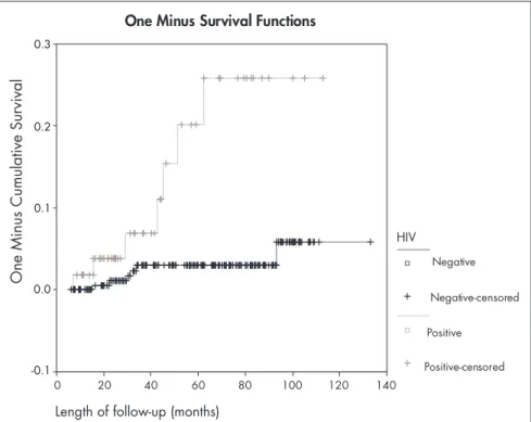
The risk of recurrence over time is shown in Figure 1, as obtained using the Kaplan-Meyer method (1-survival). The difference between the curves showed sta-tistical significance (log-rank test = 14.32; p = 0.0002).
Table 3 shows the relationships be-tween possible prognostic factors for recurrence of CIN, in order to highlight any confounding factors.27 CIN grade and
margin involvement failed to show any significant relationship with the outcome. Length of follow-up was not equally dis-tributed between the groups, in relation to the outcome.
Analysis of CD4 count and HAART use also failed to demonstrate any statistically sig-nificant relationship, which was probably due to the small number of patients from whom this information was available (Table 4). However, HIV patients using HAART seemed to have less risk of recurrence than did HIV patients who were not using it. This is shown in Figure 2, in which the Kaplan-Meyer method was used to compare the likelihood of recur-rence between these two groups (log-rank test = 4.32; p = 0.0377). This trend can also be seen in Figure 3, which shows that the recur-rence of CIN 2-3 was more frequent in HIV patients who had CD4 counts of less than 500 cells/mm3, but the difference between
these two curves was not significant (Log-rank test = 0.13; p = 0.7178).
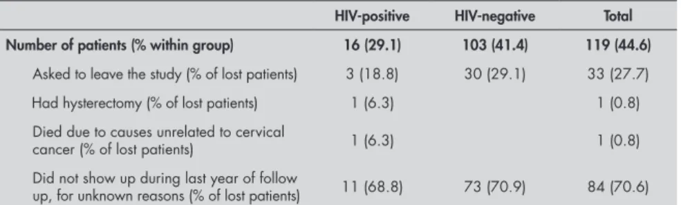
Patients who had not attended any visit over the last year of the study, or had asked to leave the study, or had left the cohort for other reasons, were considered to have been lost from the follow-up. Table 5 shows the numbers of losses in each group and the known reasons for this event.
Reanalyzing the censored cases, if it were considered that all of the lost HIV-positive patients had presented recurrence and none of the lost HIV-negative patients had had recurrence of CIN 2-3, the inci-dence of this outcome in each group would have been 987.5/10,000 patient-months and 48.0/10,000 patient-months, respec-tively, and the RR would have been as high as 20.56 (95% CI = 8.37-50.50). Similarly, if it were considered that none of the lost HIV-positive patients had presented this outcome and all of the lost HIV-negative patients had had recurrence, the incidence of this event in each group would have been 30.1/10,000 patient-months and 87.2/10,000 patient-months respectively, and the RR would have been 0.34 (95% CI = 0.16-0.74).
One Minus Survival Functions
Length of follow-up (months)
140 120 100 80 60 40 20 0
0.3
0.2
0.1
0.0
-0.1
HIV
Negative
Negative-censored
Positive
Positive-censored
One
Minus
Cumulative
Survival
Table 3. Possible prognostic factors for cervical intraepithelial neoplasia (CIN 2-3) recurrence (Rio de Janeiro, Brazil, 2006)
Recurrence
detected Recurrence not detected p-value
Cases (%) 13 (4.9) 254 (95.1)
Age at time of performing LLETZ – mean (SD) 32.5 (7.2) 32.2 (7.6) 0.880* Age at end of follow-up period – mean (SD) 35.6 (7.5) 36.9 (8.2) 0.563* Length of follow-up in months – mean (SD) 36.8 (22.9) 55.7 (31.2) 0.013* CIN grade treated – n (%)
CIN 2 (% in row; % in column) 7 (6.3-53.8) 105 (93.8-41.3) 0.40†
CIN 3 (% in row; % in column) 6 (4.0-46.2) 145 (96.0-57.1)
CIN 2-3‡ (% in row; % in column) 4 (100.0-1.6)
LLETZ specimen margin involvement
Any involvement (% in row; % in column) 2 (3.1-15.4) 62 (96.9-24.4)
No involvement§ (% in row; % in column) 11 (5.4-84.6) 192 (94.6-75.6) 0.74||
SD = standard deviation; LLETZ = large loop excision of the transformation zone; CIN = cervical intraepithelial neoplasia. *Student’s t test, without assuming equal variance; † Chi-squared test; ‡Not possible to differentiate between CIN-2 or 3 and therefore excluded from the statistical test of association of CIN grade with recurrence; §Including cases in which margin involve-ment could not be assessed due to thermal artifact or excessive fraginvolve-mentation; || Fisher’s exact test.
Table 4. Possible prognostic factors for cervical intraepithelial neoplasia (CIN 2-3) recurrence in HIV patients (Rio de Janeiro, Brazil, 2006)
Recurrence detected
Recurrence not
detected p-value
Cases (%) 7 (12.7) 48 (87.3)
Less than 500 CD4 cells/mm3 (% in row - % in column)* 2 (20.0-66.7) 8 (80.0-50.0)
500 CD4 cells/mm3 or more (% in row - % in column)* 1 (11.1-33.3) 8 (88.9-50.0) 1.00†
Not using HAART‡ 2 (50.0-40.0) 2 (50.0-9.5)
Using HAART‡ 3 (13.6-60.0) 19 (86.4-90.5) 0.155†
One Minus Survival Functions
Length of follow-up (in months)
120 100
80 60
40 20
0
One
Minus
Cumulative
Survival
0.7
0.6
0.5
0.4
0.3
0.2
0.1
0.0
-0.1
Using HAART?
Yes
Yes-censored
No
No-censored
Figure 2. Likelihood of recurrence during follow-up period for 26 HIV patients, accord-ing to whether they were usaccord-ing highly active antiretroviral therapy (HAART), usaccord-ing the Kaplan-Meyer method (1-survival function). Log-rank test = 4.32; p = 0.0377 (Rio de Janeiro, Brazil, 2006).
One Minus Survival Functions
Length of follow-up (in months)
120 100
80 60
40 20
0
One
Minus
Cumulative
Survival
0.3
0.2
0.1
0.0
-0.1
CD4 count
<500
<500-censored
>=500
>=500-censored
Figure 3. Likelihood of recurrence during follow-up period for 19 HIV patients, accord-ing to CD4 count (less than 500 cells/mm3 versus 500 cells/mm3 or more), usaccord-ing the Kaplan-Meyer method (1-survival function). Log-rank test = 0.13; p = 0.7178 (Rio de Janeiro, Brazil, 2006).
The distribution of the possible prog-nostic factors relating to recurrence in the group of lost patients, compared with those who remained in the study, is shown in Table 6.
Discussion
We found a higher risk of recurrence of CIN 2-3 in HIV-infected Brazilian women than in non-HIV-infected women. Even tak-ing into account the wide range of the confi-dence interval, recurrence was at least twice as frequent as in non-HIV-infected women.
The risk of CIN 2-3 recurrence after LLETZ in our study was lower than what was observed in the cohort studied by Heard et al.25 They found an absolute risk
of 8.6 per 100 patient-years, which is twice as high as our finding of 30.06/10,000 woman-months in HIV-infected patients (which can be converted to 3.61 per 100 patient-years). This can be partially explained by the lower median level of CD4 count.
We observed an increasing risk of recur-rence over time among the HIV-positive women, which reached 26% at the 62nd
month. Among the HIV-negative women, this likelihood stabilized after 93 months at almost 0.6%of the women, which there-fore suggests that HIV-positive patients need longer follow-ups than HIV-negative women do, and that LLETZ is an effective method for treating HIV-negative women. For HIV-positive women, the same manage-ment protocol may apply in the event of recurrence, for their retreatment.
Since CIN grade and margin involve-ment failed to show any significant relation-ship with the outcome, we did not test for confounding. Length of follow-up, however, was not equally distributed between the groups in relation to the outcome. None-theless, if this were a confounder, it would bias the result such that the HIV-positive group would be favored (greater length of follow-up in the HIV-negative group would show more recurrences in this group, if this factor were a confounder). CD4 count and HAART use were not statistically related to recurrence in HIV patients, but those who were using HAART seemed to have a better prognosis.
In our study groups, the only significant factor relating to recurrence of CIN 2-3 treated by LLETZ was HIV status.
Table 5. Losses in each study group and the known reasons for this event (Rio de Janeiro, Brazil, 2006)
HIV-positive HIV-negative Total Number of patients (% within group) 16 (29.1) 103 (41.4) 119 (44.6) Asked to leave the study (% of lost patients) 3 (18.8) 30 (29.1) 33 (27.7)
Had hysterectomy (% of lost patients) 1 (6.3) 1 (0.8)
Died due to causes unrelated to cervical
cancer (% of lost patients) 1 (6.3) 1 (0.8)
Did not show up during last year of follow
up, for unknown reasons (% of lost patients) 11 (68.8) 73 (70.9) 84 (70.6)
Table 6. Distribution of possible prognostic factors for cervical intraepithelial neoplasia (CIN 2-3) recurrence in the group lost from the follow-up, in comparison with patients who remained in the cohort (Rio de Janeiro, Brazil, 2006)
Lost from follow-up
Not lost from
follow-up p-value Age at time of performing LLETZ – mean (SD) 31.87 (7.2) 32.5 (7.7) 0.400* Age at end of follow-up period – mean (SD) 34.7 (7.4) 38.5 (8.4) <0.0001* Length of follow-up in months – mean (SD) 35.2 (20.9) 71.9 (27.5) <0.0001† CIN grade treated – n (%)
CIN 2 (% in row; % in column) 57 (50.9-47.9) 55 (49.1-37.2) 0.072‡
CIN 3 (% in row ;% in column) 60 (39.7-50.4) 91 (60.3-61.5)
CIN 2-3§ (% in row; % in column) 2 (50.0-1.7) 2 (50.0-1.4) LLETZ specimen margin involvement
Any involvement (% in row; % in column) 25 (39.1-21.0) 39 (60.9-26.4)
No involvement|| (% in row; % in column) 94 (46.3-79.0) 109 (53.7-73.6)) 0.309‡
SD = standard deviation; LLETZ = large loop excision of the transformation zone; CIN = cervical intraepithelial neoplasia. *Student’s t test, without assuming equal variance; †Student’s t test, assuming equal variance; ‡Chi-squared test; §Not possible to differentiate between CIN-2 or 3 and therefore excluded from the statistical test of association of CIN grade with being lost to follow-up ; || Including cases for which margin involvement could not be assessed due to thermal artifact or excessive fragmentation.
up more closely because of their disease. Re-analysis of the censored cases showed that if all HIV-negative patients who were lost had had recurrence, our results would have been negative (RR < 1.0). However, this would not be a reasonable assumption.
The losses from the follow-up seem not to have biased our results. As shown in Table 5, the known causes of losses were not related to the outcome. The losses for which the reasons for leaving the cohort were unknown were similarly distributed between the study groups. Furthermore, the groups of lost patients and continuing pa-tients were similar with regard to age at the time of treatment, CIN grade and margin involvement, thus showing that the losses were not related to the prognostic factors studied (Table 6).
Conclusion
AUTHOR INFORMATION Fábio Russomano, MD, PhD. Gynecologist in charge of
Colposcopy Clinic, Instituto Fernandes Figueira, Fundação Oswaldo Cruz (IFF-Fiocruz), Rio de Janeiro, Brazil
Aldo Reis, MD, PhD. Professor, Faculdade de Medicina de Cam-pos (FMC), CamCam-pos dos Goytacazes, Rio de Janeiro, Brazil
Maria José Camargo, MD, PhD. Gynecologist, Instituto Fer-nandes Figueira, Fundação Oswaldo Cruz (IFF-Fiocruz), Rio de Janeiro, Brazil
Beatriz Grinsztejn, MD, PhD. Infectologist, Instituto de Pesquisa Clínica Evandro Chagas, Fundação Oswaldo Cruz (IPEC-Fiocruz), Rio de Janeiro, Brazil
Maria Aparecida Tristão, MD, MSc. Pathologist, Instituto Fernandes Figueira, Fundação Oswaldo Cruz (IFF-Fiocruz), Rio de Janeiro, Brazil
Address for correspondence: Fábio Russomano
Instituto Fernandes Figueira — Fundação Oswaldo Cruz (IFF-Fiocruz)
Avenida Rui Barbosa, 716 — 3o andar — Flamengo Rio de Janeiro (RJ) — Brasil — CEP 22250-020 Tel./Fax. (+ 55 21) 2554-1738
E-mail: [email protected]
Copyright © 2008, Associação Paulista de Medicina
RESUMO Recorrência de neoplasia intra-epitelial cervical graus 2 ou 3 em mulheres infectadas pelo HIV tratadas pela exérese da zona de transformação por alça diatérmica (EZTAD)
CONTEXTO E OBJETIVO: Mulheres infectadas pelo HIV têm maior probabilidade de apresentar câncer cervical e seus precursores. O tratamento dessas lesões pode prevenir a neoplasia. O objetivo deste estudo foi verificar a probabilidade de recorrência de neoplasia intra-epitelial cervical graus 2 ou 3 (NIC 2-3) em mulheres infectadas pelo HIV (HIV+), comparando-a com a de mulheres soronegativas (HIV-) tratadas pela exérese da zona de transformação por alça diatérmica (EZTAD).
TIPO DE ESTUDO E LOCAL: Estudo de coorte conduzido no Instituto Fernandes Figueira — Fundação Oswaldo Cruz (IFF-Fiocruz), Rio de Janeiro, Brazil.
MÉTODO: 55 HIV+ e 212 HIV- foram acompanhadas após tratamento de NIC 2-3 pela EZTAD (faixa: 6-133 meses).
RESULTADOS:A incidência de NIC 2-3 recorrente foi de 30,06/10.000 mulheres-mês no grupo HIV+ e 4,88/10.000 mulheres-mês no grupo HIV- (risco relativo, RR = 6,16; intervalo de confiança, IC 95%: 2,07-18,34). A probabilidade de recorrência alcançou 26% aos 62 meses de acompanhamento em mulheres HIV+, e manteve-se estável em cerca de 0,6% no 93o mês de acompanhamento em mulheres HIV-. Não pudemos demon-strar outros fatores prognósticos relacionados à recorrência de NIC, mas o uso de terapia antiretroviral potente (highly active antiretroviral therapy - HAART) pode reduzir o risco dessa ocorrência em pacientes HIV+. CONCLUSÕES: Mulheres HIV+ têm maior risco de recorrência de NIC 2-3 após EZTAD comparadas a mulheres HIV-. Esse maior risco não foi influenciado pelo status da margem ou grau de doença tratada. O uso de HAART pode reduzir o risco desta ocorrência em mulheres HIV+.
PALAVRAS-CHAVE: Neoplasia intra-epitelial cervical. Prognóstico. Soropositividade para HIV. Recidiva. Eletrocirurgia. 1. UNAIDS. AIDS epidemic update: special report on HIV/AIDS.
Geneva: Joint United Nations Programme on HIV/AIDS (UN-AIDS) and World Health Organization (WHO); 2006. p. 1-96. 2. Palella FJ Jr, Delaney KM, Moorman AC, et al. Declining
morbidity and mortality among patients with advanced hu-man immunodeficiency virus infection. HIV Outpatient Study Investigators. N Engl J Med. 1998;338(13):853-60. 3. Brito AM, Castilho EA, Szwarcwald CL. Regional patterns of the
tempo-ral evolution of the AIDS epidemic in Brazil following the introduction of antiretroviral therapy. Braz J Infec Dist. 2005;9(1):9-19.Braz J Infec Dist. 2005;9(1):9-19. 4. Massad LS, Ahdieh L, Benning L, et al. Evolution of cervicalEvolution of cervical
abnormalities among women with HIV-1: evidence from surveillance cytology in the women’s interagency HIV study. J Acquir Immune Defic Syndr. 2001;27(5):432-42. 5. Grinsztejn B. Prevalência de infecções sexualmente transmissíveis
e estudo dos fatores de risco para infecção pelo HPV numa coorte de mulheres infectadas pelo HIV no Rio de Janeiro. [thesis]. Rio de Janeiro: Faculdade de Medicina da Universidade Federal do Rio de Janeiro; 2001.
6. Russomano F, Bastos FI, Camargo MJ, et al. High prevalence of genital intra-epithelial neoplasia in a cohort of HIV infected women in Rio de Janeiro (RJ), Brazil. In: XIV International AIDS Conference. July 7-12, 2002 (abstract no ThPeC7549).
Available from: http://gateway.nlm.nih.gov/MeetingAb-stracts/102256183.html. Accessed in 2007 (Nov 9). 7. Suárez Rincón AE, Vázquez Valls E, Ramírez Rodríguez M,
Mon-toya Fuentes H, Covarrubias Rodríguez M de L, Sánchez Corona. J. Lesiones escamosas intraepiteliaes en pacientes VIH seropositivas. Su frecuencia y asociación con factores de riesgo para neoplasia cervical. [Squamous intra-epithelial lesions in HIV seropositive females. Their frequency and association with cervical neoplasia risk factors]. Ginecol Obstet Mex. 2003;71:32-43.
8. Hluangdansakul W, Phinchantra P, Bowonwatanuwong C. Prevalence of SIL and SCCA in human immunodeficiency virus-seropositive women at anonymous clinic in Chonburi Hospital. J Med Assoc Thai. 2006;89(3):289-93. 9. Petry KU, Scheffel D, Bode U, et al. Cellular immunodeficiency
enhances the progression of human papillomavirus-associated cervical lesions. Int J Cancer. 1994;57(6):836-40. 10. Olaitan A, Mocroft A, McCarthy K, Phillips A, Reid W,
John-son M. Cervical abnormality and sexually transmitted disease screening in human immunodeficiency virus-positive women. Obstet Gynecol. 1997;89(1):71-5.
11. Wright TC Jr, Koulos J, Schnoll F, et al. Cervical intraepithelial neoplasia in women infected with the human immunodeficiency virus: outcome after loop electrosurgical excision. Gynecol Oncol. 1994;55(2):253-8.
12. Fruchter RG, Maiman M, Sedlis A, Bartley L, Camilien L, Arrastia CD. Multiple recurrences of cervical intraepithelial neoplasia in women with the human immunodeficiency virus. Obstet Gynecol. 1996;87(3):338-44.
13. Robinson WR, Hamilton CA, Michaels SH, Kissinger P. Effect of excisional therapy and highly active antiretroviral therapy on cervical intraepithelial neoplasia in women infected with human immuno-deficiency virus. Am J Obstet Gynecol. 2001;184(4):538-43. 14. Chirenje ZM, Rusakaniko S, Akino V, Munjoma M, Mlingo
M. Effect of HIV Disease in Treatment Outcome of Cervical Squamous Intraepithelial Lesions Among Zimbabwean Women. J Low Genit Tract Dis. 2003;7(1):16-21.
15. Tebeu PM, Major AL, Mhawech P, Rapiti E. The recur-rence of cervical intraepithelial neoplasia in HIV-positive women: a review of the literature. Int J STD AIDS. 2006;17(8):507-11.
16. Massad LS, Fazzari MJ, Anastos K, et al. Outcomes after treat-Outcomes after treat-ment of cervical intraepithelial neoplasia among women with HIV. J Low Genit Tract Dis. 2007;11(2):90-7.
17. Schwartz LB, Carcangiu ML, Bradham L, Schwartz PE. Rapidly progressive squamous cell carcinoma of the cervix coexisting with human immunodeficiency virus infection: clinical opinion. Gynecol Oncol. 1991;41(3):255-8.
18. Del Priore G, Lee MJ, Barnes M, Garcia P, Till M. The inad-equacy of standard treatment of dysplasias in a woman with acquired immune deficiency syndrome. Int J Gynaecol Obstet. 1994;47(3):273-4.
19. Stratton P, Ciacco KH. Cervical neoplasia in the patient with HIV infection. Curr Opin Obstet Gynecol. 1994;6(1): 86-91. 20. Holcomb K, Matthews RP, Chapman JE, et al. The efficacyThe efficacy
of cervical conization in the treatment of cervical intraepi-thelial neoplasia in HIV-positive women. Gynecol Oncol. 1999;74(3):428-31.
21. Kuhn L, Sun XW, Wright TC Jr. Human immunodeficiency virus infection and female lower genital tract malignancy. Curr Opin Obstet Gynecol. 1999;11(1):35-9.
22. Robinson W 3rd. Invasive and preinvasive cervical neoplasia in human immunodeficiency virus-infected women. Semin Oncol. 2000;27(4):463-70.
23. Tate DR, Anderson RJ. Recrudescence of cervical dysplasia among women who are infected with the human immunodeficiency virus: a case-control analysis. Am J Obstet Gynecol. 2002;186(5):880-2. 24. Gilles C, Manigart Y, Konopnicki D, Barlow P, Rozenberg S.
Management and outcome of cervical intraepithelial neoplasia lesions: a study of matched cases according to HIV status. Gynecol Oncol. 2005;96(1):112-8.
25. Heard I, Potard V, Foulot H, Chapron C, Costagliola D, Kazatchkine MD. High rate of recurrence of cervical intraepi-thelial neoplasia after surgery in HIV-positive women. J Acquir Immune Defic Syndr. 2005;39(4):412-8.
23. Heard I, Tassie JM, Kazatchkine MD, Orth G. Highly ac-tive antiretroviral therapy enhances regression of cervical intraepithelial neoplasia in HIV-seropositive women. AIDS. 2002;16(13):1799-802.
24. Maiman M, Fruchter RG, Serur E, Levine PA, Arrastia CD, Sedlis A. Recurrent cervical intraepithelial neoplasia in human immunodeficiency virus-seropositive women. Obstet Gynecol. 1993;82(2):170-4.
25. Frega A, Biamonti A, Maranghi L, et al. Follow-up of high-grade squamous intra-epithelial lesions (H-SIls) in human immu-nodeficiency virus (HIV)-positive and human papillomavirus (HPV)-positive women. analysis of risk factors. Anticancer Res. 2006;26(4B):3167-70.
26. Prendiville W. Large loop excision of the transformation zone. In: Prendiville W, editor. Large loop excision of the transformation zone. A practical guide to LLETZ. London: Chapman & Hall Medical; 1993. p. 36-58.
27. Kleinbaum DG, Kupper LL, Morgerstern H. Confounding. In: Kleinbaum DG, Kupper LL, Morgerstern H, editors. Epidemiologic research: Principles and quantitative methods. New York: John Wiley & Sons; 1982. p. 242-65.
Acknowledgment: We are very grateful to Walter Prendiville (of the Royal College of Surgeons in Ireland) and Fiona Lyons (GUM Specialist Registrar, Department of Gynae-cology, AMNCH, Tallaght Hospital, Dublin, Ireland) who kindly reviewed the initial manuscript and added important contributions to the text.
Sources of funding: None
Conflicts of interest: None
Date of first submission: February 22, 2007
Last received: January 9, 2008
Accepted: January 9, 2008