In vitro corrosion of metallic orthodontic brackets: Influence of artificial saliva with and without fluorides
original article
Mônica Pereira Saporeti1, Enio Tonani Mazzieiro2, Wisley Falcco Sales3
Objective: This in vitrostudy verified the resistance to corrosion of metallic brackets, evaluating the superficial aspects in
scanning electron microscopy (SEM) and the residual components. Methods: The sample consisted of 17 sets of brackets of
four different metallic alloys: Titanium, Cobalt-Chromium, Stainless steel with low nickel concentration and with titanium nitride coating (NiTi). Twelve sets were submitted to corrosion by immersion in 50 ml of artificial saliva (pH 6.5) and four in saliva (pH 6.5) containing fluoride (2 g/l), all at a temperature of 37 ºC and analyzed after 7, 9 and 11 weeks. One was kept as control set. The analysis consisted in qualitative evaluation of the corrosion by the images obtained on the SEM, in semi-quantitative evaluation of chemical composition of the surface residue by SEM-EDS and the amount of ions released in saliva
on evaluation of atomic absorption spectrophotometry. Results: The results showed that the pure titanium brackets and the
ones with low nickel concentration were superior regarding resistance to corrosion. The cobalt-chromium alloy showed the greatest corrosion. In the presence of fluoride, it was observed greater variation in all alloys, especially in the ones of NiTi
coat-ed steel and the ones of cobalt-chromium. Conclusion: Although observed corrosion on the SEM, the spectrophotometry
showed low ions release in the artificial saliva, however, the presence of fluoride negatively affected the corrosion resistance.
Keywords: Corrosion. Fluoride. Orthodontic brackets. Stainless steel. Titanium. Chromium alloys.
How to cite this article: Saporeti MP, Mazzieiro ET, Sales WF. In vitro corrosion of metallic orthodontic brackets: Influence of artificial saliva with and without fluo-rides. Dental Press J Orthod. 2012 Nov-Dec;17(6):24.e1-7.
Submitted: October 31, 2008 - Revised and accepted: August 16, 2012
» The author reports no commercial, proprietary or financial interest in the products or companies described in this article.
» Patients displayed in this article previously approved the use of their facial and in-traoral photographs.
1 MSc in Orthodontics, PUC-MG. 2 MSc and PhD in Orthodontics, USP, Bauru. 3 MSc and PhD in Mechanical Engineering, UFU.
Contact address: Enio Tonani Mazzieiro
Av. Contorno 8000 sala 1004, Santo Agostinho, Belo Horizonte, MG – Brazil CEP 30110-932 – Brazil
E-mail: [email protected]
Objetivo: este estudo in vitro verificou a resistência à corrosão de braquetes metálicos, avaliando-se os aspectos
superficiais em microscopia eletrônica de varredura (MEV) e os componentes residuais formados. Métodos: a
amostra consistiu de 17 conjuntos de braquetes de quatro diferentes ligas metálicas: titânio, cobalto-cromo, aço inoxidável com baixa concentração de níquel e com cobertura de nitreto de titânio (TiN). Doze conjuntos foram submetidos à corrosão por imersão em 50ml de saliva artificial (pH 6,5) e 4 em saliva (pH 6,5) contendo flúor (2g/l), todos sob temperatura de 37ºC, e analisados após 7, 9 e 11 semanas. Uma montagem foi mantida como con-trole. As análises consistiram na avaliação qualitativa da corrosão por meio das imagens obtidas no MEV, na ava-liação semiquantitativa da composição química dos resíduos superficiais por meio de MEV-EDS e da quantidade
de íons liberados nas salivas na avaliação da espectrofotometria de absorção atômica. Resultados: os resultados
demonstraram que os braquetes com liga de titânio puro e os de aço com baixa concentração de níquel foram supe-riores em relação à resistência à corrosão. A liga de cobalto-cromo foi a que apresentou maior corrosão. Na presença do flúor, observaram-se maiores alterações em todas as ligas, com destaque para as de aço cobertos com TiN e as de
cobalto-cromo. Conclusão: apesar de se observar a corrosão no MEV, a espectrofotometria mostrou baixo
despren-dimento de íons nas salivas artificiais, porém a presença do flúor interferiu negativamente na resistência à corrosão.
INTRODUCTION
The release of metallic ions and the associated biological effects of the nickel alloys have received attention in literature of biomedical materials. Some researches6,17,25 have focused the in vitro phenomenon
that occurs with it, including toxicity and allergenic-ity. The biocompatibility of the use of nickel alloys in the oral cavity for long periods of time has promoted the study of alternative materials for orthodontic patients. The oral environment is particularly ideal for biodegradation of metals,1 facilitating the
corro-sion process on orthodontic appliances. For this rea-son, nickel-free alloys or alloys of stainless steel with reduced amount of nickel have been tested.17
Stud-ies1,4,8,11,12,18,27 confirmed the possibility of in vivo and
in vitro corrosion of steel brackets and associated a lower or greater potential of corrosion not only to the metal used, but to the manufacturing process and to the final structure of these appliances. The pure tita-nium was recently introduced as alternative material for metallic brackets for its low allergenicity, greater corrosion resistance10 (inferior to gold and platinum
only), biocompatibility and good mechanical resis-tance.6,13 Some authors, however, have questioned the
behavior of titanium brackets in fluoridated environ-ment.3,14,22,24 The titanium alloys (Ti
6Al4V) and the
co-balt-chromium alloys have also been used. Filmore,9
evaluating the soft temper cobalt-chromium alloy and comparing it to two titanium alloys, observed that the
chemical composition and the thermodynamic treat-ment of the cobalt-chromium alloy present impact on its resistance to corrosion. Considering the peculiari-ties inherent to the oral cavity and the mechanical re-quirements during the orthodontic treatment, it is es-sential the development of more researches related to biocompatibility, mechanical resistance and anticor-rosion properties of these alternative alloys, so they can settle as a possibility on the bracket manufactur-ing process. This study evaluates the corrosion of me-tallic alloys in artificial saliva with and without fluo-rides, evaluating the superficial aspects in scanning electron microscopy and the residual components, so that they can be used alternatively to stainless steel in patients hypersensitive to nickel.
MATERIAL AND METHODS
The sample consisted of 64 straight-wire premo-lars brackets, distributed in groups according to man-ufacturer and type of alloy (Table 1). Twelve groups were immersed in artificial saliva, pH 6,5 (Table 2), for periods of 7, 9 and 11 weeks and four in artificial saliva (pH 6,5) with 2g/l of NaF for 11 weeks. One sample was not immersed (Table 3).
The brackets were set with the aid of tweezers (Univer, Duflex, Rio de Janeiro, Brazil) in sterile Pe-tri dishes, 90 x 15 mm, containing 50 ml of artificial saliva, sealed and kept in kiln (EES, Olidef, Ribeirão Preto, Brazil) at 37 °C constant, in static conditions.
Table 1 - Distribution of sample according to experimental groups.
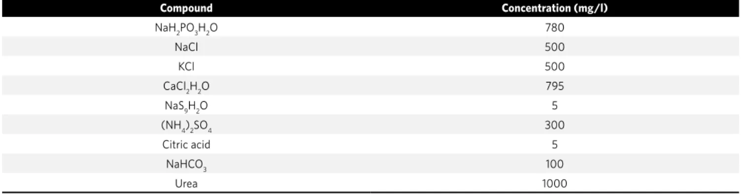
Table 2 - Artificial saliva composition (mg/l).
Brand Lot Alloy Control group Group 1 Group 2 Group 3 Group 4 Total
Morelli Nickel- Free - Sorocaba, Brasil 791448 Ni-free steel 1 4 4 4 4 17
Morelli Golden Line - Sorocaba, Brasil 632079 TiN coating 1 4 4 4 4 17
TP Orthodontics - La Porte, USA 1395LJR Co-Cr 1 4 4 4 4 17
Dentaurum- Inspringen, Germany 345360 Ti 1 4 4 4 4 17
Total 4 16 16 16 16 68
Compound Concentration (mg/l)
NaH2PO3H2O 780
NaCl 500
KCl 500
CaCl2H2O 795
NaS9H2O 5
(NH4)2SO4 300
Citric acid 5
NaHCO3 100
Saporeti MP, Mazzieiro ET, Sales WF
At the end of each week, about 3 ml of saliva was re-placed, preventing the solutions saturation by the cor-rosion byproducts. The corcor-rosion process was qualita-tively analyzed on the brackets surface through scan-ning electron microscopy (SEM) (JSM-5310, JEOL, Japan). The panoramic images were obtained with angulation of 60° and magnification of 15x. The tie-wing area and region of transition between body and base of the bracket were selected to be evaluated with magnification of 200 and 500x. The chemical compo-sition of surface residue resultant from the corrosive process was semi-quantitatively evaluated through SEM linked to EDS system (XL30, Philips, Eindhoven, Netherlands). Table 4 presents the alloys composition before the corrosive process.
The quantitative evaluation of released products concentration in solutions of artificial saliva by corro-sion was performed by atomic absorption spectropho-tometry (Varian 220 FS, New York, USA).
RESULTS
Results for steel samples with low nickel concentration
The visual inspection of brackets from the control group revealed a unibody architecture, having little porosity on the surface. The tie-wing area, under-neath it and on the area of transition between body and base presented a quite irregular and rough sur-face, probably due to the difficult access to polishing in that region. On experimental groups immersed in
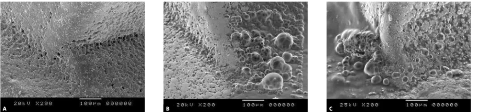
artificial saliva it was observed color changes in iso-lated areas of the brackets that were proved time-de-pendent. It was not noticed superficial deposits sug-gesting corrosion. In the group of 11 weeks of immer-sion in solution of saliva with fluoride showed stain-ing, granulation in all surfaces and some superficial deposit (Fig 1). The EDS analysis did not show for-mation of superficial oxides in measurable amount. The atomic absorption spectrophotometry did not identify ions release after 11 weeks.
Results for steel samples with titanium nitride coating
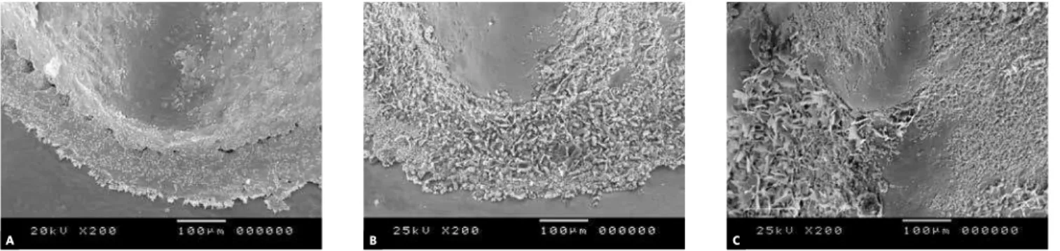
The visual inspection revealed that these brack-ets also present a unibody architecture. According to manufacturer, the bracket has the same chemical composition as the steel with low concentration of nickel, but a thin coating of titanium nitride (TiN) is applied. The control group presented a rough and ir-regular surface. On experimental groups immersed in artificial saliva, small whitish deposits were seen on all brackets that are more susceptible to surface defects for the TiN coating. Extending the immersion period, isolated granules were seen on the bracket surface and some cracks also could be noticed. A more in-tense corrosive process was noticed on these brackets when compared to the group with low concentration of nickel. On the group of 11 weeks in artificial saliva with fluoride it was observed greater granulation and formation of vesicles (Fig 2). In the semi-quantitative
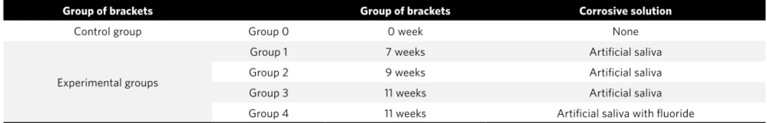
Table 3 - Distribution of brackets groups according to period of immersion and solution utilized.
Table 4 - Initial chemical composition of the brackets alloys.
Group of brackets Group of brackets Corrosive solution
Control group Group 0 0 week None
Experimental groups
Group 1 7 weeks Artificial saliva
Group 2 9 weeks Artificial saliva
Group 3 11 weeks Artificial saliva
Group 4 11 weeks Artificial saliva with fluoride
Chemical composition(% by weight)
Brand Fe Cr Ni Si Mo Mn Ti W Co Cu H O N C
stainless
steel
TiN
Morelli Nikel-free 64.69 17.5 0.01 1.0 3.5 12.0 - - - 0.8 0.2 -
-Morelli Golden-line 64.69 17.5 0.01 1.0 3.5 12.0 - - - 0.8 0.2 - Superficial coating
Dentaurum - - - 99.0 - - - 0.06 0.35 0.35 0.05 0.3
-evaluation of chemical composition of surface resi-dues (SEM-EDS), it was identified the presence of titanium (95,88%) and oxygen (4,12%) which shows formation of oxides on the surface of the material, re-sultant from the corrosion (Fig 3). Despite the pres-ence of this corrosion after 11 weeks, there was no significant amount of ions on the atomic absorption spectrophotometry, showing that despite the corro-sion, the ions release in saliva was low.
Results for titanium samples
The titanium bracket from the control group present-ed the most irregular surface, full of fenestrations and in-creased rugosity in relation to the other brackets, despite also being unibody. This bracket presented little altera-tion and superficial deposits during the experimental pe-riod. Brackets from groups of 9 and 11 weeks of immersion in artificial saliva presented areas of staining and it was noticed the appearance of isolated granulation.
Figure 3 - Result of the SEM-EDS evaluation of chemical composition
of surface residues from the titanium nitride alloy after 9 weeks of im-mersion.
Figure 1 - Superficial aspects of the transition area between body and base of steel alloy with low nickel concentration brackets (magnification of 200x).
A) control; B) 11 weeks without fluoride; C) 11 weeks with fluoride.
Figure 2 - Superficial aspects of the transition area between body and base of steel alloy with titanium nitride coating brackets (magnification of 200x).
A) control; B) 11 weeks without fluoride; C) 11 weeks with fluoride.
A B C
A B C
O Ka
1.00
Element O K TiK Total
4.12 11.40 0.0041 0.9519 0.9920 88.60
100.00 95.88 100.00
1.1613 0.0848 1.0007
1.0000 1.0000
Wt% At% K-Ratio Z A F
2.00 3.00 4.00 TiKa
TiKb
Saporeti MP, Mazzieiro ET, Sales WF
In the presence of fluoride, it was noticed an in-crease of these color variations and of granulations, which suggests a lower resistance of the titanium alloy to the fluoride (Fig 4). The EDS analysis did not show clear signs of oxidation and after 11 weeks, the solu-tions of saliva did not present measurable amount of ions on the atomic absorption spectrophotometry.
Results for cobalt-chromium samples
The assessment of the cobalt-chromium bracket proved to be the only one in the sample formed by two parts connected by a weld interface. The con-trol group already presented staining on the surface and some porosity. The weld bead was irregular and with spongy aspect, which may facilitate the be-ginning of the corrosion process. Galvanic, inter-granular and crevice corrosions are more frequent in brackets with soldering area. The experimental group showed the greatest alterations during exper-iment, when compared to the other tested alloys.
Figure 4 - Superficial aspects of the transition area between body and base of titanium alloy brackets (magnification of 200x). A) control; B) 11 weeks
without fluoride; C) 11 weeks with fluoride.
Figure 5 - Superficial aspects of the transition area between body and base of cobalt-chromium alloy brackets (magnification of 200x). A) control;
B) 11 weeks without fluoride; C) 11 weeks with fluoride.
After 9 weeks of immersion in artificial saliva it was observed a great deposition of material in the solder-ing area. The sample of 11 weeks showed significant alterations, with formation of crystals on the body/ base interface, suggesting a deposition of corrosion byproducts in these areas. The group of 11 weeks in solution of saliva with fluoride experienced greater corrosion in relation to the others. The bracket pre-sented a superficial granulated aspect, with forma-tion of crystals and with release of metal chips from the surface of the bracket (Fig 5). The semi-quanti-tative analysis (SEM-EDS) of chemical composition of surface residues after 11 weeks of immersion in artificial saliva present a predominance of cobalt (50,86%), chromium (19,32%) and silver (16,83%). The presence of oxygen (5,98%) shows formation of oxides on the surface of the material (Fig 6). The analysis of the solutions by atomic absorption spec-trophotometry did not show clear signs of ions re-lease on the solution of artificial saliva.
A B C
Figure 6 - Result of the SEM-EDS evaluation of chemical composition of surface residues from the cobalt-chromium alloy after 11 weeks of immersion.
DISCUSSION
The corrosion of metallic alloys used on the orth-odontic bracket manufacturing process has been re-searched. The corrosion release of nickel and chro-mium ions has received special attention due to its potential carcinogenic, mutagenic and allergenic effects. It is estimated that about 10% of the popula-tion present allergic reacpopula-tions to nickel.6,18,26 In the
oral cavity this reaction can manifest through edema and gingival bleeding, often of difficult differential diagnosis in relation to periodontal inflammatory re-actions of patients in orthodontic treatment and that present bad sanitation. The orthodontic industry have sought alternatives to minimize these allergic reactions, altering the composition of stainless steel alloy, reducing the concentration of nickel or using alternative alloys as titanium-aluminium-vanadium alloy, pure titanium, cobalt-chromium or superficial coating of titanium nitride. The present study evalu-ated the corrosion of some of these alloys, keeping the brackets immersed in solution of artificial saliva, pH 6,5, at constant temperature of 37 °C. Similar methodology was used by some researchers.1,6,10,16,20
Other works10,19 evaluated the bracket corrosion
us-ing solution of sodium chloride at 0,9%. Although the solution of NaCl is potentially more corrosive than artificial saliva, no laboratory corrosive envi-ronment is capable to simulate every event that oc-curs in the oral cavity. The interaction between the characteristics of the human saliva, alterations of pH due to food variety, bacterial colonization and its by-products make the oral cavity an extremely favorable
environment to corrosive process and of difficult re-production in vitro.5,6 The addition of fluoride to
ar-tificial saliva diminished the corrosion resistance in all tested alloys. Similar result was found in several works,3,10,14,15,17,23,25 indicating a greater susceptibility
to corrosion in the presence of fluorides. Clinically, this fact represents a concern regarding cavity pre-vention in orthodontic patients, once that astringent solutions and fluoridated toothpaste, normally rec-ommended as a way to prevent cavity, may increase the appliance corrosion, releasing greater amount of ions in the oral cavity. The evaluations of the mi-croscopy images showed a different behavior of the alloys. The steel alloy with low concentration of nick-el presented better performance when compared to the others. After 11 weeks submitted to the corrosive process it was not observed the presence of surface residues in the groups with and without fluoride. It was not identified the presence of oxygen on the EDS evaluation and the atomic absorption spectro-photometry did not reveal ions release. Similar re-sults were found in a previous study,19 although the
saline solution was used as corrosive environment. The titanium nitride bracket presented greater gal-vanic and crevice corrosion process than the steel brackets with low concentration of nickel. Defects on this coating during the manufacturing process may facilitate it.2,6,20,21 Titanium brackets presented good
resistance to corrosion. The titanium is considered a stable metal, biocompatible and resistant to corro-sion.3,4,7,8,10,14,15,17,23,25 The analysis of the images showed
little alteration, mostly, only chromatic, and even in presence of fluoride, the increase of alterations was not sufficient to cause ions release in the solution. The cobalt-chromium alloy was the one that pre-sented lowest resistance to corrosion. The presence of fluoride increased the corrosion, indicating a low resistance to fluorides. Associated to this fact, these brackets were the only ones that presented a solder-ing area between the body and the bracket bases. The presence of this area and thermal treatment increase the susceptibility of this alloy to corrosion.7,8,9,16,20,21
The solutions saturated by corrosion byproducts were analyzed in order to verify the types and concen-trations of released metals after the corrosion tests of the different samples in all three experimental in-tervals. The atomic absorption spectrophotometer
1.00 Element O K ClK AlK AgL O Ka AlKa CIKa AgLa AgLb CrKa CrKb CoKa CoKb CoLa S Ka S K CrK CoK Total 5.98 18.73 2.83 0.0209 0.0142 0.1991 0.0157 0.1479 0.0179 0.4865 1.0902 1.1304 1.0778 1.9061 1.0040 0.9877 6.21 7.81 43.22 2.59 18.61 100.00 3.35 1.66 16.83 50.86 2.00 19.32 100.00 1.1731 0.2972 0.7495 0.9645 1.4300 0.8152 0.9627 0.9685 1.0020 1.0113 1.0056 1.0018 0.0170 1.0660 1.0000
Wt% At% K-Ratio Z A F
Saporeti MP, Mazzieiro ET, Sales WF
is capable to quantify chemical elements in surfaces, solid materials and or solutions, through detecting its atoms.12 Several works about corrosion of
brac-es12,13,19,20,25 used the atomic absorption
spectropho-tometer to quantify chemical elements in these solu-tions. In the present study, this method proved that the corrosion of alloys, observed on the SEM, was of low intensity, with low ions release in the solution of saliva. This result suggests that the alloys used on the bracket manufacturing process presented fine biocompatibility and may be used on patients with nickel hypersensitivity.
CONCLUSION
Through interpretation of the results obtained from this study, we concluded that the cobalt-chromi-um alloy showed the greatest corrosion, quantitative-ly. The pure titanium alloys and the steel alloy with low concentration of nickel presented the greatest corrosion resistance. The presence of fluoride caused greater alterations in all tested alloys. The quantita-tive evaluations, through atomic absorption spectro-photometry, did not identify measurable amount of ions in the solution of artificial saliva, suggesting fine biocompatibility of the alloys.
1. Barrett RD, Bishara SE, Quinn JK. Biodegradation of orthodontic appliances. Part I. Biodegradation of nickel and chromium in vitro. Am J. Orthod Dentofacial Orthop. 1993;103(1):8-14.
2. Callister WD. Ciência e engenharia de materiais. 5ª ed. Rio de Janeiro: LTC;2002. 3. Conz MB, Soares GA, Ponciano JAC. Efeito da aplicação de fluoretos sobre a
superfície de uma liga de Ti-Al-V. Rev Bras Implant. 2002;8(1):10-3. 4. Deguchi T, Ito M, Obata A, Koh Y, Yamagishi T, Oshida Y. Trial production of
titanium orthodontic brackets fabricated by metal injection molding (MIM) with sintering. J Dent Res. 1996;75(7):1491-6.
5. Eliades T, Eliades G, Brantley WA. Orthodontic brackets. In: Brantley WA, Eliades T. Orthodontic materials: scientific and clinical aspects. New York: T Thieme; 2001. p. 143-71.
6. Eliades T, Athanasiou AE. In vivo aging of orthodontic alloys: implications for corrosion potential, nickel release, and biocompatibility. Angle Orthod. 2002;72(3):222-37.
7. Es-Souni M, Fischer-Brandies H, Es-Souni M. On the in vitro biocompatibility of Elgiloy, a co-based alloy, compared to two titanium alloys. J Orofacial Orthop. 2003;64(1):16-26.
8. Ferreira TL. Avaliação da resistência à corrosão de materiais metálicos utilizados em aparelhos ortodônticos fixos [tese]. Rio de Janeiro (RJ): Universidade Federal do Rio de Janeiro; 2005.
9. Filmore GM, Tomlinson JL. Heat treatment of cobalt-chromium alloy wire. Angle Orthod. 1976;46(2):187-95.
10. Gioka C, Bourauel C, Zinelis S, Eliades T, Silikas N, Eliades G. Titanium orthodontic brackets: structure, composition, hardness and ionic release. Dent Mater. 2004;20(7):693-700.
11. Gontijo LP, Mazzieiro ET, Landre J Jr. Composição química e resistência mecânica da base de bráquetes straight-wire. Rev Dental Press Ortod Ortop Facial. 2004;9(4):52-9.
12. Goldstein JI, Newbury DE, Joy DC, Lyman CE, Echlin P, Lifshin E, et al. Scannig Electron Microscopy end X-ray microanalysis. 2nd ed. New York: Plenum Press; 1992. 13. Grimsdottir MR, Gjerdet NR, Hensten-Pettersen A. Composition and in vitro
corrosion of orthodontic appliances. Am J Orthod Dentofacial Orthop. 1992 Jun;101(6):525-32.
REFERENCES
14. Hamula DW, Hamula W, Sernetz F. Pure titanium orthodontic brackets. J Clin Orthod. 1996 ;30(3):140-4.
15. Harzer W, Schröter A, Gedrange T, Muschter F. Sensivity of titanium brackets to the corrosive influence of fluoride-containing toothpaste and tea. Angle Orthod. 2001;71(4):318-23.
16. Huang TH, Yen CC, Kao CT. Comparison of ion release from new and recycled orthodontic brackets. Am J Orthod Dentofacial Orthop. 2001;120(1):68-75. 17. Huang HH, Chiu YH, Lee TH, Wu SH, Yang HW, Su KH, et al. Ion release from
NITI orthodontic wires in artificial saliva with various acidities. Biomaterials. 2003;24(20):3585-92.
18. Kusy RP. Clinical response to allergies in patients. Am J Orthod Dentofacial Orthop. 2004;125(5):544-7.
19. Leite THM. Estudo comparativo do comportamento in vitro de diferentes bráquetes ortodônticos [dissertação]. Belo Horizonte (MG): Pontifícia Universidade Católica de Minas Gerais; 2003.
20. Maijer R, Smith DC. Corrosion of orthodontic bracket bases. Am J Orthod. 1982;81(1):43-8.
21. Matasa CG. Flaws in bracket manufacturing. J Clin Orthod. 1990;24(3):149-52. 22. Mondelli J. Ligas alternativas para restaurações fundidas. 1ª ed. São Paulo:
Panamericana; 1995.
23. Pröbster L, Lin W, Hutteman H. Effect of fluoride prophylactic agents on titanium surface. Int J Oral Maxillofacial Implants. 1992;7(3):390-4.
24. Profit WR. Aparelhos fixos contemporâneos. In: Proffit WR. Ortodontia contemporânea. 2ª ed. Rio de Janeiro: Guanabara Koogan; 1995. p. 312-40. 25. Schiff N, et al. Corrosion of titanium and its alloys in fluoridated and acidic
environment. J Dental Res. 2002;81 Spec. Issue B, p. 273..
26. Staffolani N, Damiani F, Lilli C, Guerra M, Staffolani NJ, Belcastro S, et al. Ion release from orthodontic appliances. J Dent. 1999;27(6):449-54.