Evaluation of the stability of open bite treatment using a removable appliance with
palatal crib combined with high-pull chincup
Fernando Pedrin Carvalho Ferreira1, Renato Rodrigues de Almeida2, Fernando César Torres3, Renata Rodrigues de Almeida-Pedrin4, Marcio Rodrigues de Almeida5, Roberto Santana Filho6
Objective: The aim of this prospective study was to cephalometrically analyze the stability of dentoalveolar and skeletal changes produced by a removable appliance with palatal crib associated to high-pull chincup in individuals with anterior open bite treated for 12 months, and compare them to individuals with similar malocclusion and age, not submitted to orthodontic treatment, also followed for the same period. Methods: Nineteen children with a mean age of 9.78 years old treated for 12 months with a removable appliance with palatal crib associated with chincup therapy were evaluated after 15 months (post-treatment period) and compared with a control group of 19 subjects with mean age of 9.10 years with the same malocclusion that was followed-up for the same period. Seventy-six lateral cephalograms were evaluated at T1 (after correction) and T2 (follow-up) and cephalometric variables were analyzed by statistical tests. Results: The results did not show significant skeletal, soft tissue or maxillary dentoalveolar changes. Overall, treatment effects on the experimental group were maintained at T2 evaluation with an increase of 0.56 mm in overbite. Overjet and maxillary incisors/molars position (vertical and sagittal) remained es-sentially unchanged during the study period. Only mandibular incisors showed significant changes (labial inclination and protrusion) com-pared to control group. Conclusions: Thus, it can be concluded that the early open bite treatment with a removable appliance and palatal crib associated with high-pull chincup therapy provided stability of 95%.
Keywords: Open bite. Angle Class I malocclusion. Orthodontics.
How to cite this article: Ferreira FPC, Almeida RR, Torres FC, Almeida-Pedrin RR, Almeida MR, Santana Filho R. Evaluation of the stability of open bite treatment us-ing a removable appliance with palatal crib combined with high-pull chincup. Dental Press J Orthod. 2012 Nov-Dec;17(6):52-60.
Submitted: November 24, 2008 - Revised and accepted: June 26, 2009
» Patients displayed in this article previously approved the use of their facial and in-traoral photographs.
Contact address: Fernando Pedrin Carvalho Ferreira
Rua Vereador Leandro dos Santos Martins, 2-20 – Jardim Estoril – Bauru/SP – Brazil CEP: 17017-900 – E-mail: [email protected]
1 MSc and PhD in Orthodontics, FOB/USP. Professor and Coordinator of the
Specialization Courses, CORA – Vilhena/RO.
2 PhD in Orthodontics, USP. Professor of Orthodontics, FOB/USP.
3 MSc and PhD in Orthodontics, FOB/USP. Associate Professor of the Department of
Orthodontics, UNICID.
4 Post-Doc in Orthodontics and Specialist in Radiology, FOB/USP. Professor of
Graduation and Post-Graduation Courses, Sagrado Coração University.
5 Post-Doc in Orthodontics, FOB/USP.
6 Specialist in Orthodontics, CORA – Bauru. Professor of Orthodontics, CORA –
Vilhena/RO.
» The author reports no commercial, proprietary or financial interest in the products or companies described in this article.
INTRODUCTION AND LITERATURE REVIEW
Stability in orthodontic treatments has always been a challenge for the orthodontists. Correc-tion of vertical dysplasia, such as anterior open bite, has presented high indexes of relapse. Several treatment protocols have been indicated, in dif-ferent ages, with the same objectives, for reaching an occlusal and facial harmony,1,8,11-13,19,25 and, long-term stability.
Systematically evaluating the orthodontic lit-erature, focusing on anterior open bite treatments in the mixed dentition,24 only two randomly con-trolled studies reached the requirements for this selection. The study of Erbay et al13 in which used Frankel functional regulator (FR-4), and other study by Ferreira-Pedrin et al14 in which a remov-able appliance with a palatal crib combined to a chincup was used. Other systematic reviews8,18 evi-denced the lack of studies investigating the stabil-ity in the mixed dentition, using a treatment pro-tocol and comparing to a matched control group.
Huang et al19 studied stability, after a minimum period of 1 year, in patients with (n = 26) and with-out (n = 7) growth treated with palatal crib. In the group without growth, no patients showed relapse; in the group with growth, 17.4% showed open bite relapse, suggesting that palatal crib treatment pro-vides good results besides stability, probably due to the normalization of tongue posture. Almeida et al3 reported that early treatment promoted ante-rior open bite correction, and outcome stability.
Another protocol employed, which also veri-fied long-term stability, was performed by Lopes-Gavitto et al.25 In the study, the sample comprised of 41 patients who had undergone a conventional treatment with a fixed and extraoral appliance and were compared to a normal occlusion sample. They concluded that more than 35% of patients ex-perimented relapse of 3 mm or more.
Using the same sample of Lopez-Gavito et al,25 Zuroff,30 in 1990, revaluated the stability of the cases using other cephalometric variables for mea-suring overbite. Overbite was measured by the in-cisal edges of the upper and lower incisors regard-ing menton-nasion line. As a result, it was verified that after 10 years, 60% of patients showed lack of incisal contact.
According to Katsaros and Berg22 evaluating pa-tients with anterior open bite (mean of -1.9 mm) treat-ed with treat-edgewise fixtreat-ed appliance (n = 20) and func-tional appliance (n = 1), stability could reach indexes up to 75%. Kim et al,23 also evaluated stability after 2 years of anterior open bite correction with fixed ap-pliances, in patients with (n = 29) and without (n = 26) growth. They observed minimum alterations on over-bite and concluded that their results were stable.
Believing that instability of the results obtained after anterior open bite closure would be due to tongue positioning, Justus,21 used spurs in order to avoid anterior tongue posture. Treatment protocol showed effectivness on correcting malocclusion and obtaining post-treatment stability.
Huang18 performed a literature review focusing on orthodontic and/or surgical treatment of ante-rior open bite. The evaluated literature suggested that about 80% of patients who showed anterior open bite, presented after the retention period a positive overbite, regardless of the treatment type (orthodontic or surgical). However, the author highlighted that those results should be cautiously analyzed, since several of these articles had showed methodological failures (small sample and bias dur-ing sample selection) and suggested further studies on anterior open bite stability.
Janson et al20 evaluated anterior open bite stabil-ity after a mean period of 5 years in patients (n = 21) treated with fixed appliance. Results showed sig-nificant open bite relapse. The main factor that contributed for the relapse was the deficient verti-cal development of upper and lower incisors, in the post-treatment period. However, 61.9% of the cases treated showed a “clinical” stability. Nor the initial magnitude of anterior open bite, neither the total amount of malocclusion correction was correlated to treatment relapse. Freitas et al15 showed long-term stability data of treatment with extractions in the permanent dentition after 8.35 years and con-cluded that the sample showed clinical stability of 74.2% of open bite correction.
Emphasizing the need of new researches, Ren27
Due to the lack of studies with appropriate meth-odology for evaluating treatment stability of ante-rior open bite in the mixed dentition, it was aimed to cephalometrically analyze the stability of dento-skeletal and profile changes in the treatment with removable appliance with palatal crib associated to high-pull chincup therapy in young patients with an-terior open bite who had been treated for 12 months, and to compare them to similar individuals who had not undergone orthodontic treatment.
MATERIAL AND METHODS
The present study was derived from the research of Ferreira Pedrin in 2006,14 which consisted of a prospective randomized study with the treatment of 30 individuals (mean age of 8.33 years) with anterior open bite, who had been compared to a similar con-trol group composed of 30 individuals (mean age of 8.61 years). These authors verified after a 12-month treatment the correction of the malocclusion in 24 individuals who had undergone treatment with a mean overbite closure of 5.01 mm, while control group showed 1.38 mm, allowing the spontaneous correction in only 4 subjects.
It was also aimed in the present study to verify the stability of the effects obtained with the use of the removable appliance with palatal crib com-bined to high-pull chincup in individuals with mixed dentition.
The sample was composed of 76 lateral cephalo-grams of 38 young Brazilian subjects of both genders (19 from the treated group and 19 from the control group). Data collected for this study comprised two time pe-riods: T1 (treatment completion) and T2 (15 months post-treatment).
Sample homogeneity
The criteria for sample selection were based on the following characteristics:
» Young individuals who had undergone treat-ment of Angle’s Class I malocclusion with an-terior open bite, for 12 months, with remov-able appliance with palatal crib associated to high-pull chincup.
» Young individuals with ages varying from 7 to 12 years and presenting the upper permanent first molars in occlusion.
» Caucasian individuals, descending from Ital-ian, Portuguese, and Spanish.
» No dental agenesis or permanent teeth loss. » This study did not aim to evaluate oral habits
(pacifier, thumb suction or oral breathing), or other etiologic factors.
» No young individuals had undergone dental extractions.
Group 1 (control)
This group comprised 19 young subjects, 17 female and 2 male, with Class I malocclusion with anterior open bite, who had not been submitted to any type of orthodontic treatment, with initial mean age of 9.10 years-old (ranging from 7.31 to 11.51 years). These individuals showed the following initial cephalomet-ric characteristics: ANB = 4.86° (ranging from -0.50° to 8.50°), SN.GoGn = 35.04° (ranging from 28.20° to 47.8°), and negative overbite of 2.66 mm (ranging from -0.10 mm to -9.00 mm). This group was selected from the files of the Department of Orthodontics of Bauru Dental School – University of São Paulo. The mean interval between the two radiographic exami-nations used in this group was 15.15 months.
Group 2 (treated)
Group 2 was composed of 19 young individuals, 13 female and 6 male, who had undergone treat-ment with removable appliance with palatal crib combined to high-pull chincup, for 12 months, and showed, previously to treatment, a Class I malocclu-sion with anterior open bite. Mean age at T1 (treat-ment completion) was 9.78 years (raging from 8.43 to 11.96 years). Patients presented the following ceph-alometric characteristics at T1: ANB = 5.73° ing from 2.00° to 12.20°), SN.GoGn = 35.43° (rang-ing from 27.8° to 46.10°) and overbite = 0.94 mm (ranging from -3.00 mm to 3.80 mm). From the 30 patients of the previous study sample,14 11 could not participate in this present research because 6 moved out of town and 5 patients were using fixed orthodontic appliance. Mean Interval of radio-graphic exams between T1 and T2 phases used in this group was 15.19 months.
The cephalometric measurements were grouped in sectors regarding to:
1) Maxillary component: SNA and Co-A. 2) Mandibular component: SNB, Ar.GoMe,
Ar-Go, Co-Gn.
3) Maxillomandibular relationship: ANB. 4) Vertical relationship: SN.GoGn, SN.PP,
NS.Gn, AFH, PFH, AIFH.
5) Dental component: overbite, overjet, 1.NA, 1-NA, 1-PP, 6-FHp, 6-PP, 1.NB, 1-NB, 1-GoMe, 6-FHp, 6-GoMe.
6) Soft tissues component: ANL, AML, facial convexity, Ls-P’Sn, and Li-P’Sn.
Aiming to determine the results reliability, 25 ra-diographs from the two groups studied were random-ly selected. All radiographs were again traced and digitized by the same researcher after a 1-month pe-riod from the initial tracing, according to the guide-lines of Midtgard et al.26 The difference between the first and second measurement of each radiograph was determined and the Dalberg’s formula10 was ap-plied for visualizing the casual error. The systematic error, was obtained by Student’s t test, at the signifi-cance level of 5% (p < 0.05).
Statistic analysis was executed employing the non-paired t test, and the results were considered statistically significant for p < 0.05. Therefore, the following factors were verified: The similarity de-gree between Group 1 and Group 2 regarding to the chronological age and evaluation period, the simi-larity between the cephalometric measurements of the two groups, and, specially, the cephalometric changes occurred in the experimental period.
RESULTS
Patients age-group
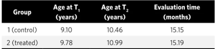
The patients studied showed initial and final mean age compatibility and were evaluated by a same follow-up period, shown in Tables 1 and 2.
Among the 29 measurements evaluated, it was observed at the significance level of 5% that no mea-surement showed systematic error. Regarding casual error, only 3 measurements were greater than 1 mm for the linear measurements: 1-GoMe (1.29 mm), 6-FHp (1.27 mm) and 6-FHp (1.26 mm). However, errors from dental measurements are expected ac-cording to Baumrind et al.7 These values showed
that the landmark delimitation or location did not interfere on the obtainment of the cephalometric measurements and did not impair the results, which has been frequently observed on researches, as re-ported by Baumrind et al7 and Midtgard et al.26
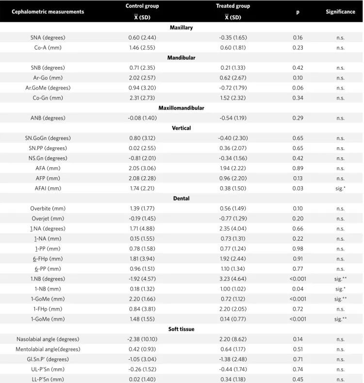
Table 3 shows the result of the non-paired t test in the inter-groups comparison, the mean changes for both the control group and the treated group in the final phase of the stability assessment.
Figure 1 shows the difference between groups evidencing the changes due to both craniofacial growth and development and treatment, in the final phase of stability assessment.
In Figure 2, it is possible to observe 3 intraoral anterior photos of a patient during the phases of pre-treatment, 12-month post-treatment, and 15 months after the appliances removal (stability).
In Figure 3, it is observed two intraoral anterior photos of a patient representing control group in the initial and 15-month follow-up phase.
DISCUSSION
A research published in 2006,14 performed the comparison of the initial cephalometric measure-ments between groups, using Student’s non-paired
t test and the same group of the present study. The purpose of the present paper, however, is to verify stability. The groups showed total similarity in the pre-treatment phase, none of the evaluated vari-ables showed statistical significant difference, re-inforcing the reliability between the groups’ parity.
Table 1 - Mean age of the individuals in the two groups and the mean time of evaluation.
Group Age at T1
(years)
Age at T2 (years)
Evaluation time (months)
1 (control) 9.10 10.46 15.15
2 (treated) 9.78 10.99 15.19
Sig. = significant for p < 0.05; n.s. = non significant.
Table 2 - Statistic comparison between the mean age and evaluation pe-riods.
Group p SIG.
1 (control) x 2 (treated) age T2 0.06 n.s.
Table 3 - Difference of the mean changes (X) T2-T1, standard deviation (SD), p value and statistical significance level.
Cephalometric measurements
Control group
X (SD)
Treated group
X (SD)
p Significance
Maxillary
SNA (degrees) 0.60 (2.44) -0.35 (1.65) 0.16 n.s.
Co-A (mm) 1.46 (2.55) 0.60 (1.81) 0.23 n.s.
Mandibular
SNB (degrees) 0.71 (2.35) 0.21 (1.33) 0.42 n.s.
Ar-Go (mm) 2.02 (2.57) 0.62 (2.67) 0.10 n.s.
Ar.GoMe (degrees) 0.94 (3.20) -0.72 (1.79) 0.06 n.s.
Co-Gn (mm) 2.31 (2.73) 1.52 (2.32) 0.34 n.s.
Maxillomandibular
ANB (degrees) -0.08 (1.40) -0.54 (1.19) 0.29 n.s.
Vertical
SN.GoGn (degrees) 0.80 (3.12) -0.40 (2.30) 0.65 n.s.
SN.PP (degrees) 0.02 (2.55) 0.36 (2.07) 0.65 n.s.
NS.Gn (degrees) -0.81 (2.01) -0.34 (1.56) 0.42 n.s.
AFA (mm) 2.05 (3.06) 1.94 (2.22) 0.89 n.s.
AFP (mm) 2.08 (2.28) 0.96 (2.20) 0.13 n.s.
AFAI (mm) 1.74 (2.21) 0.38 (1.50) 0.03 sig.*
Dental
Overbite (mm) 1.39 (1.77) 0.56 (1.49) 0.10 n.s.
Overjet (mm) -0.19 (1.45) -0.77 (1.29) 0.20 n.s.
1.NA (degrees) 1.71 (4.88) 2.35 (4.04) 0.66 n.s.
1-NA (mm) 0.15 (1.55) 0.73 (1.31) 0.22 n.s.
1-PP (mm) 0.78 (1.58) 0.77 (1.24) 0.98 n.s.
6-FHp (mm) 1.81 (3.94) 1.92 (2.44) 0.91 n.s.
6-PP (mm) 0.96 (1.51) 1.10 (1.34) 0.77 n.s.
1.NB (degrees) -1.92 (4.57) 3.23 (4.64) <0.001 sig.**
1-NB (mm) 0.18 (1.32) 1.00 (1.02) 0.04 sig.*
1-GoMe (mm) 2.20 (1.66) 0.72 (1.12) <0.001 sig.**
1-FHp (mm) 0.84 (3.81) 2.20 (2.05) 0.72 n.s.
1-GoMe (mm) 1.48 (1.55) 0.14 (0.77) <0.001 sig.**
Soft tissue
Nasolabial angle (degrees) -2.38 (10.10) 2.20 (8.62) 0.14 n.s.
Mentolabial angle(degrees) 0.42 (0.93) 0.64 (1.17) 0.51 n.s.
Gl.Sn.P' (degrees) -1.05 (3.04) -1.38 (2.48) 0.71 n.s.
UL-P'Sn (mm) -0.26 (1.52) -0.44 (1.74) 0.74 n.s.
LL-P'Sn (mm) 0.02 (1.40) 0.34 (1.18) 0.45 n.s.
n.s. = non significant. Sig.* = significant (p ≤0.05) Sig.** = significant (p ≤0.01).
It was observed that there was a greater number of young females than males. This fact did not im-pair the comparison of changes between groups, be-cause an appropriate proportion of gender between treated and control groups could also be observed.
re-garding stability during a period of about 15 months was assessed. The obtained values were compared to a control group with similar characteristics.
Several studies describe anterior open bite treatment, however, the literature lacks for studies
evaluating outcome stability. Others15,20,23 have eval-uated stability, however, using distinct protocols in patients with more advanced ages.
No study with proper methodology has evaluated this issue.8,18,24,27
Figure 1 - Total superimposition (S-N) of the means of the control group (white) and the treated group (red) in the fi nal phase of the stability assessment.
Figure 3 - Intraoral photos of control group:
A) initial (T1); B) 15 months of follow-up (T2). A B
Figure 2 - Intraoral photos of treated group: A) Initial (T0); B) after 12 months (T1); C) 15 months after appliances removal (T2).
Changes comparisons between groups
In the inter-groups comparison, it was possible to identify small dentoskeletal and soft tissue changes, showing that the early treatment of anterior open bite seemed to be very stable.
From all skeletal measurements, only AIFH was statistically different between groups, which was in-fluenced by the dental factor. This occurred due to a greater extrusion of lower molars in the control group (2.20 mm) compared to treated group (0.72 mm).
Aiming to verify the stability of the effects obtained on treatment regarding anterior overbite, the method-ology employed in this research assessed the distance between the incisal edges of the upper and lower inci-sors perpendicular to the occlusal plane.14,20
A previous study published by Ferreira-Pedrin et al, in 2006,14 verified a mean decrease of overbite of 5.01 mm for the group treated with removable appliance with palatal crib combined with high-pull chincup, while control group showed 1.38 mm. Due to the ini-tial anterior open bite of -3.95 mm in the treated group, overbite correction was possible to be achieved in 24 out of 30 patients. In the control group, anterior open bite was initially -4.01 mm, which was sufficient for the overbite correction in only 4 out of 30 individuals.
When evaluating the stability of both control and treated groups, these presented initial mean values of -2.66 mm and 0.94 mm, respectively, which were sta-tistically different. This was already expected since group 2 (treated) had been treated for 12 months, while group 1 (control) had not undergone any treatment, maintaining the negative overbite.
After a mean radiographic follow-up period of 15 months, it was verified that the control group showed a mean increase of overbite of 1.39 mm. Out of 19 young individuals of the control group, 7 showed a positive overbite, 5 had an overbite reduction worsening the malocclusion, and 7 increased the values of this vari-able. However, these values were not sufficient for cor-rection, maintaining the anterior open bite.
In the treated group, a mean increase of overbite of 0.56 mm was verified, resulting in an overbite improve-ment in 12 of the 19 young individuals evaluated. This value was reached due to the influence of the intrinsic growth of the treated patients besides the results ob-tained from the treatment protocol used. Seven of the 19 individuals of the treated group showed overbite
reduction, and only one patient showed anterior open bite relapse, with initial overbite of 0.7 mm and final of -0.7 mm. Another young individual with initial over-bite of -3 mm presented a spontaneous improvement of overbite and finished with -1 mm, without malocclu-sion correction.
The differences between the two groups were not statistically significant. However, it was estimated that the lowest increase in the treated group occurred due to the loss of the mechanism that instructed the tongue posture regarding the interference and contact between teeth, which could have generated new inter-ferences,16 while the control group only suffered the influence of growth and development, with aging.
When verifying the mean changes the lower in-cisors variables showed statistical significant dif-ferences between groups. In the control group, the means of the incisor changes represented retrocli-nation (1.NB = -1.92°), protrusion (1-NB = 0.18 mm) and extrusion (1-GoMe = 2.20 mm). In the treated group, the mean change represented proclination (1.NB = 3.23°), protrusion (1-GoMe = 1.00 mm), and extrusion (1-GoMe = 0.72 mm).
The probable factor responsible for the proclina-tion of the lower incisors in the treated group is tongue action on the incisors due to the removal of the palatal crib. Ferreira-Pedrin et al14 and Torres et al29 evidenced the retroclination of these same teeth, due to the dif-ficulty of tongue contact promoted by the palatal crib, therefore making the lower lip act more strongly on these teeth. This action promoted a statistical signifi-cant retroclination of these teeth during the use of the appliance, therefore, treatment provided an improve-ment in lip sealing. On the other hand, in average, the control group showed lingual tipping probably because they would still present some oral habit, since no treat-ment had been employed at that motreat-ment.
Corroborating with these results, other authors20 also observed proclination and protrusion of lower in-cisors in the post-treatment period.
When verifying stability, Freitas et al15 also observed significant extrusion of incisors.
In relation to vertical displacement, lower molars showed an extrusion of 1.4 mm in the control group and 0.14 mm in the treated group, with statistically sig-nificant difference. This could be explained by the oc-clusal improvement and probable muscular balance in the treated group, while in the control group this fact did not occur (occlusal balance), contributing to the stability of anterior open bite correction.
Some studies that assessed stability23 found molar extrusion in the post-treatment phase, which could have contributed for the anterior open bite relapse.
The effects promoted by 12 months of treatment with the use of removable appliance with palatal crib combined with high-pull chincup evidenced about 95% of stability. Other studies9,15,19,22,23,25,30 which veri-fied treatment stability of anterior open bite obtained distinct values and did not make a comparison with a matched control group regarding malocclusion.
Table 4 lists the main studies that evaluated treat-ment stability of anterior open bite, exposing the per-centages of maintenance results.
CLINICAL CONSIDERATIONS
Due to the results obtained in this prospective research and evaluating the stability of the clinical effects after using removable appliance with palatal
crib combined with high-pull chincup in the treat-ment of the anterior open bite, it was demonstrated that during a follow-up period of 15 months after ap-pliance removal it was possible to maintain the good occlusal relationship obtained by the treatment, sat-isfying the functional (occlusal), esthetical and social necessities of the treated young individuals.
The effects of craniofacial growth and develop-ment on patients with anterior open bite who did not undergo treatment (control group) were maintained constant, perpetuating the malocclusion, verifying the need of intervention in 12 of the 19 patients. In pri-vate clinic routine, aiming to treat anterior open bite, appliances have been used until reaching a positive overbite. It is worth using retainers after early correc-tion of this malocclusion, besides the interaccorrec-tion with other specialties (otorhinolaryngology, speech ther-apy) and periodical follow-up, which would probably provide a better stability.
CONCLUSIONS
The results of this study indicated that the ante-rior open bite treatment in the mixed dentition, us-ing removable appliance with palatal crib combined with high-pull chincup promoted 95% of dental and skeletal stability because only lower incisors pre-sented significant changes when compared to the control group.
Table 4 - Studies on anterior open bite stability.
Authors Appliance used Sample OB Control group % Stability
Huang, Justus,
Kennedy19 (1990) Palatal crib
Group with growth
Group with no growth No
82.6% 100%
Lopez-Gavito et al25 (1985) Fixed appliances with elastics Adults No 65%
Zuroff30 (1990) Fixed appliances with elastics Adults No 40%
Katsaros, Berg22 (1993) Fixed and functional appliances Adults No 75%
Kim et al23 (2000) MEAW fixed appliance
Group with growth (mean age 18 yrs) Group with no growth
(mean age 18 yrs)
No 97%
95%
Janson et al20 (2006) Non-extraction fixed appliance Young adults No 61.9%
Freitas et al15 (2004) Extraction fixed appliance Young adults No 72.4%
Crepaldi9 (2008) Occlusal adjustment Young adults No 66.7%
Ferreira-Pedrin14 (2006) Removable crib appliance with
1. Alexander CD. Open bite, dental alveolar protrusion, Class I malocclusion: A successful treatment result. Am J Orthod Dentofacial Orthop. 1999;116(5):494-500. 2. Almeida RR, Almeida-Pedrin RR, Almeida MR, Ferreira FPC, Pinzan A, Insabralde
CMB. Displasias verticais: mordida aberta anterior - tratamento e estabilidade. Rev Dental Press Ortod Ortop Facial. 2003;8(4):91-119.
3. Almeida RR, Henriques JFC, Almeida MR, Vasconcelos MHF. Early treatment of anterior open bite - prevention of orthognatic surgery. In: Davidovitch Z, Mah J, editors. Biological mechanisms of tooth eruption, resorption and replacement by implants. Boston: Harvard Society for the Advancement of Orthodontics; 1998. p. 585-8. 4. Almeida RR, Ursi WJS. Anterior open bite. Etiology and treatment. Oral Health.
1990;80:27-31.
5. Almeida RR, Garib DG, Henriques JFC, Almeida MR, Almeida RR. Ortodontia preventiva e interceptadora: mito ou realidade. Rev Dental Press Ortod Ortop Maxilar. 1999:(6):87-108.
6. Baccetti T, Franchi L, McNamara JA Jr. An improved version of the cervical vertebral maturation (CVM) method for the assessment of mandibular growth. Angle Orthod. 2002;72(4):316-23.
7. Baumrind S, Miller D, Molthen R. The reliability of head film measurements: 3. Tracing superimposition. Am J Orthod. 1976;70(6):617-44.
8. Cozza P, Baccetti T, Franchi L, Mucedero M, Polimeni A. Sucking habits and facial hyperdivergency as risk factors for anterior open bite in the mixed dentition. Am J Orthod Dentofacial Orthop. 2005;128(4):517-9.
9. Crepaldi MV. Estabilidade do tratamento da mordida aberta com ajuste oclusal [dissertação]. Bauru (SP): Universidade de São Paulo; 2008.
10. Dahlberg G. Statistical methods for medical and biological students. New York: Interscience; 1940.
11. Defraia E, Marinelli A, Baroni G, Franchi L, Baccetti T. Early orthodontic treatment of skeletal open-bite malocclusion with the open-bite bionator: A cephalometric study. Am J Orthod Dentofacial Orthop. 2007;132(5):595-8.
12. English JD. Early treatment of skeletal open bite malocclusions. Am J Orthod Dentofacial Orthop. 2002;121:563-5.
13. Erbay E, Uğur T, Ulgen M. The effects of Frankel function regulator (FR-4) therapy on the treatment of Angle Class I skeletal anterior open bite malocclusion. Am J Orthod Dentofacial Orthop. 1995;108(1):9-21.
14. Pedrin F, Almeida MR, Almeida RR, Almeida-Pedrin RR, Torres F. A prospective study of the treatment effects of a removable appliance with palatal crib combined with high-pull chincup therapy in anterior open-bite patients. Am J Orthod Dentofacial Orthop. 2006;129(3):418-23.
15. Freitas MR, Beltrão RT, Janson G, Henriques JF, Cançado RH. Long-term stability of anterior open bite extraction treatment in the permanent dentition. Am J Orthod Dentofacial Orthop. 2004;125(1):78-87.
REFERENCES
16. Haryett RD, Hansen FC, Davidson PO. Chronic thumb-sucking. A second report on treatment and its psychological effects. Am J Orthod. 1970;57(2):164-78. 17. Houston WJB. The analysis of errors in orthodontic measurements. Am J Orthod.
1983;83(5):382-90.
18. Huang GJ. Long-term stability of anterior open-bite therapy: a review. Semin Orthod. 2002;8(3):162-72.
19. Huang GJ, Justus R, Kennedy DB, Kokich VG. Stability of anterior open bite treated with crib therapy. Angle Orthod. 1990;60(1):17-24; discussion 25-6.
20. Janson G, Valarelli FP, Beltrão RT, de Freitas MR, Henriques JF. Stability of anterior open-bite extraction and nonextraction treatment in the permanent dentition. Am J Orthod Dentofacial Orthop. 2006;129(6):768-74.
21. Justus R. Correction of anterior open bite with spurs: long-term stability. World J Orthod. 2001;2(3):219-31.
22. Katsaros C, Berg R. Anterior open bite malocclusion: a follow-up study of orthodontic treatment effects. Eur J Orthod. 1993;15(4):273-80.
23. Kim YH, Han UK, Lim DD, Serraon ML. Stability of anterior openbite correction with multiloop edgewise archwire therapy: a cephalometric follow-up study. Am J Orthod Dentofacial Orthop. 2000;118(1):43-54.
24. Lentini-Oliveira D, Carvalho FR, Qingsong Y, Junjie L, Saconato H, Machado MA, et al. Orthodontic and orthopaedic treatment for anterior open bite in children (Cochrane Review). Cochrane Database Syst Rev. 2007;18(2):CD005515. 25. Lopez-Gavito G, Wallen TR, Little RM, Joondeph R. Anterior open-bite
malocclusion: a longitudinal 10 years post retention evaluation of orthodontically treated patients. Am J Orthod. 1985;87:175-86.
26. Midtgård J, Björk G, Linder-Aronson S. Reproducibility of cephalometric landmarks and errors of measurements of cephalometric cranial distances. Angle Orthod. 1974;44(1):56-61.
27. Ren Y. Is early treatment of skeletal open-bite malocclusion effective? Evidence-Based Dent. 2006;7:81-2.
28. Shapiro P. Stability of open bite treatment. Am J Orthod Dentofacial Orthop. 2002;121(6):566-8.
29. Torres FC, Almeida RR, Almeida MR, Almeida-Pedrin RR, Pedrin F,Henriques JFC. Anterior open bite treated with a palatal crib and high-pull chin cup therapy. A prospective randomized study. Eur J Orthod. 2006;28(6):610-7.