Evaluation of occlusal factors in patients with
temporomandibular joint disorder
Max Dória Costa1, Gontran da Rocha Torres Froes Junior2, Carlos Neanes Santos3
Objective: The aim of this study was to determine the prevalence and the relation between the main occlusal factors and the tem-poromandibular disorder (TMD). Methods: We analyzed 100 patients (50 diagnosed with TMD and 50 asymptomatic volunteers, control group) through a questionnaire that classified TMD as absent, mild, moderate and severe. Then, an evaluation was made of intraoral occlusal factors: Absence of posterior teeth, wear facets, overjet, overbite, open bite, posterior crossbite, sagittal rela-tionship (Class I, II and III), centric relation discrepancy for maximum intercuspation, anterior guidance and balancing occlusal interference. The χ2 examined the association between TMD and considered occlusal variables. Results: The prevalence of studied
occlusal factors was higher in patients with moderate and severe TMD. Statistically significant results were found on: Absence of five or more posterior teeth, overbite and overjet greater than 5 mm, edge-to-edge bite, posterior crossbite, Class II and III, the absence of effective anterior guide and balancing side interferences. Conclusions: Indeed, it is concluded that there is a relationship between TMD and occlusal factors, however it can not be told to what extent these factors are predisposing, precipitating or perpetuating the disease. Therefore, despite its multifactorial etiology, one can not neglect the occlusal analysis of these patients.
Keywords: Dental occlusion. Malocclusion. Temporomandibular joint dysfunction syndrome.
How to cite this article: Costa MD, Froes Junior GRT, Santos CN. Evaluation of occlusal factors in patients with temporomandibular joint disorder. Dental Press J Orthod. 2012 Nov-Dec;17(6):61-8.
Submitted: January 12, 2009 - Revised and accepted: November 13, 2009
Contact address: Departamento de Odontologia da Universidade Federal de Sergipe – Av. Mal. Rondon, s/n – Cidade universitária – São Cristóvão/SE – Brazil CEP: 49100-000 – E-mail: [email protected]
1 MSc and PhD Student of Rehabilitation, FOB – USP. 2 Specialist in Radiology, UFBA.
3 Associate Professor of Odontology, Occlusion and Integrated Clinic Discipline,
UFS.
» The author reports no commercial, proprietary or financial interest in the products or companies described in this article.
Objetivo: o presente estudo teve como objetivo verificar a prevalência e relação dos principais fatores oclusais com a disfunção tem-poromandibular. Métodos: foram analisados 100 pacientes (50 com diagnóstico de DTM e 50 voluntários assintomáticos, grupo con-trole) através de um questionário para classificação do grau de DTM, em ausente, leve, moderada e severa. Em seguida, foi realizada uma avaliação intrabucal dos fatores oclusais ausência de dentes posteriores, facetas de desgaste, overjet, overbite, mordida aberta anterior, mordida cruzada posterior, relação sagital (Classe I, II e III), discrepância da relação cêntrica para máxima intercuspidação habitual, guia anterior e interferência oclusal em balanceio. O teste qui-quadrado analisou a associação entre DTM e as variáveis oclusais consideradas. Resultados: a prevalência dos fatores oclusais analisados foi maior nos pacientes que apresentaram DTM moderada e severa. Resultados estatisticamente significativos foram encontrados para ausência de cinco ou mais dentes posteriores, overjet e overbite maiores que 5mm, relação dos incisivos topo a topo, mordida cruzada posterior, Classe II e III, ausência de guia anterior efetiva e interferências no lado de balanceio. Conclusão: conclui-se que realmente existe uma relação entre DTM e os fato-res oclusais; entretanto, não se pode afirmar até que ponto esses fatofato-res são predisponentes, desencadeantes ou perpetuantes dessa disfunção. Portanto, apesar de sua etiologia multifatorial, não se pode negligenciar a análise oclusal desses pacientes.
INTRODUCTION
The temporomandibular disorder (TMD) is de-fined as a set of functional and pathological con-ditions affecting the temporomandibular joint (TMJ), masticatory muscles and tissue adjacent
components.9 It is characterized by several signs
and symptoms that include facial muscle and joint pain, limitation and / or mandibular deviation in the trajectory, joint noises, headaches, earaches and
pain of cervical origin.20
Because of the etiological complexity and variety of signs and symptoms that may represent other pa-thologies, recognition and differentiation of TMDs is not very clear to the professional. Therefore, routine screening should be combined with the anamnesis and selective clinical examination, for the profes-sional to perform an accurate diagnosis and
there-fore develop the proper treatment plan.2 Several
di-agnostic systems have been used in literature, play-ing an important role in characterizplay-ing and
classify-ing patients with TMD.4,11
Although there is not a defined etiology for TMD, functional, structural and psychological factors characterize the multifactorial origin of this dys-function. Some conditions, such as malocclusion, parafunctional habits, emotional stress, trauma, sleep disorders, postural abnormalities, systemic factors, are present with particular frequency in pa-tients with TMD signs. However, it cannot be stated that these factors are predisposing TMD or are only
coincidental.25
The occlusion is now treated not only as the ra-tio of contact between teeth, but as a dynamic, mor-phological and functional relation between all com-ponents of the stomatognathic system, presenting a
great influence on chewing, swallowing and speech.19
There is a huge controversy in literature regarding the association between occlusion and TMD. Some authors have reported high turnout of occlusal
fac-tors in signs and symptoms of TMD.1,19 Others are
skeptical about it6,10,16 and others believe that
occlu-sion plays a limited role, but it cannot be
underesti-mated.3.8,12,14,17,18,22,23,26,27
Taking all together, many studies have found relationship between TMD and occlusal
chang-es: Premature occlusal contacts,19,20 no anterior
guide,19,27 balancing side interference,12,19 sagittal
relation Class II,17,19,27 and Class III,7,13,19 anterior
open bite,14,19,25,26 crossbite,3,13,14,19,26 overjet / deep
overbite greater than 5 mm,14,18,26 great centric
relation discrepancy (CR) to usual maximum
intercuspation(MI) greater than 2 to 4 mm3,12,14,19,26
and 5 or more missing posterior teeth.14,19,26
The aim of this present study was to verify the prevalence and relationship between TMD and sev-eral occlusal factors.
MATERIAL AND METHODS
This research was submitted and approved by the Ethics Committee in Research of Federal Univer-sity of Sergipe - CEP / UFS UniverUniver-sity Hospital (No. CAAE - 0053.0.107.000-08).
One hundred patients from the Department of Operative Dentistry, Federal University of Ser-gipe, were evaluated. Fifty patients had TMD and the control group was composed by 50 volunteers with no TMD symptoms. In both groups, it was ad-opted the following exclusion criteria: Previous orthodontic treatment and/or orthopedic and pros-thetic rehabilitation treatment. All patients signed a consent form.
A single investigator was used to collect the data from the clinical records. Initially, the patient un-derwent an interview (anamnesis) using the
ques-tionnaire proposed by Fonseca et al4 composed
of 10 questions related to the signs and symptoms of TMD, such as the presence of TMJ sounds, im-paired mouth movement, pain and muscle fatigue during chewing, headaches, neck pain, earaches or pain in the region of the joints. For each question, the possible responses were”yes”, “sometimes” and “no”, which are assigned the values “10”, “5” and “0”, respectively. For the questionnaire analysis, “yes”, “sometimes” and “no” were added up togeth-er. Subjects were classified according to the value found in TMD: “Absent,” “Mild,” “Moderate” and “Severe” (ranging from “0-15”, “20-40”, “45-65” and “70-100”, respectively). For inclusion in the control group they were asked to present the classification of TMD “absent” in range “0-15”.
Next, it was performed an intraoral clinical exami-nation for evaluation of occlusal characteristics using a second form:
B) Number of posterior missing teeth (excluding third molars).
C) Overjet and overbite measured with the use of a millimeter ruler and using a dry point com-pass. Edge-to-edge bite were considered in cases of no overjet and overbite. The negative overjet was obtained by the distance between the end of the incisal edge of lower incisors and the anterior surface of the maxillary incisor, measured horizontally.
D) Presence of anterior open bite (negative over-bite).
E) Presence of posterior crossbite (unilateral or bilateral).
F) Centric relation (CR) discrepancy to maximum intercuspation (MHI), obtaining the position of CR with the technique of manipulating the tip of the chin ( unforced guide) of the patient and comparing the position of MHI, measuring the discrepancy between these positions: G) Classes I, II or III malocclusion.
H) The type of anterior guide. The patient was asked to do the laterality move, right and left, (absent, canine, total or partial group func-tion) and protusive movement (absent or pres-ent) to the top position.
I) Presence of non-working side interference during laterality and disarticulation of the pos-terior teeth in protrusion, verified with cello-phane strip on the posterior teeth.
The frequencies of the different variables were ex-pressed as percentage and to obtain the results it was
applied the χ2 test examining the association between
TMD and the occlusal variables. The implementation
of graphics and statistical testing were performed using the softwares Microsoft Office Excel 2007 and SPSS (Statistical Package for Social Sciences) version 13.0 for Windows.
RESULTS
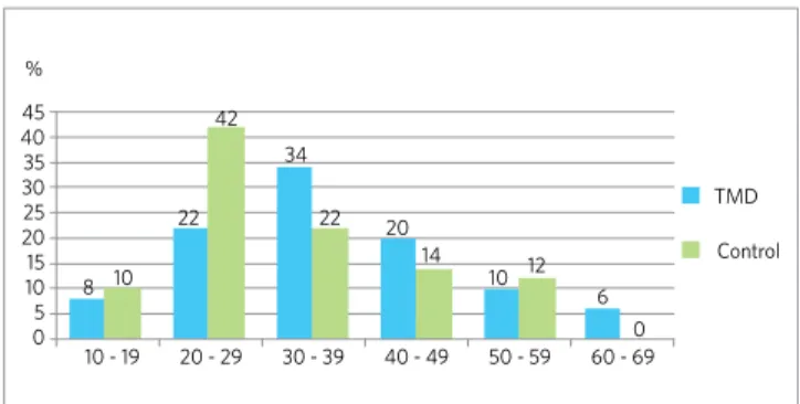
The distribution of the sample regarding gender and age can be observed in Figures 1 and 2, respec-tively. The positive responses of the anamnestic questionnaire for the diagnosis of TMD can be found
in Figure 3. Regarding Fonseca et al4 index, adding
up the scores of answers (10 answer “yes”, 5 “some-times” and 0 “no”) to the ten questions in the an-amnestic questionnaire came to the classification in symptomatic group that 36% of the sample had mod-erate TMD (“45-65” scores) and 64% severe (“70-100”). In the control group all patients showed the classification of absent TMD (“0-15”).
In the TMD group, 32% had posterior crossbite (20% unilateral and 12% bilateral), 8% open bite, 18% overbite and 10% overjet greater than 5 mm, about 38% edge-to-edge bite, 22% did not have over-jet, overbite or edge-to-edge due to the absence of incisors and 62% absence of 5 or more teeth. The number of teeth with dental wear found was 20% for 1 to 4 teeth, 12% for 5 to 10 teeth and 18% with more than 10 worn teeth. Considering the sagittal rela-tion, 42% were Class I, 26% Class II and 32% Class III. The discrepancy between centric relation (CR) and maximum intercuspation (MHI) was 68% for 0 to 2 mm, 30% for 2 to 4 mm and 2% for greater than 4 mm. Figure 4 shows the distribution of main occlusal factors studied, both in the TMD group as well as in the control group.
Figure 1 - Distribution of the sample according to gender. Figure 2 - Distribution of the sample according to age.
100 %
12 88
36 64 90
80 70
60 30
35 40 45 %
10 - 19 20 - 29 30 - 39 40 - 49 50 - 59
TMD
Control
60 - 69
50 25
40 20
30 15
TMD
Male
Female
Control
20 10
10 5
0 0
8 10 22
42
34
22 20
14 10 12
Figure 3 - Distribution of positive responses to the questionnaire.
Figure 4 - Distribution of occlusal factors.
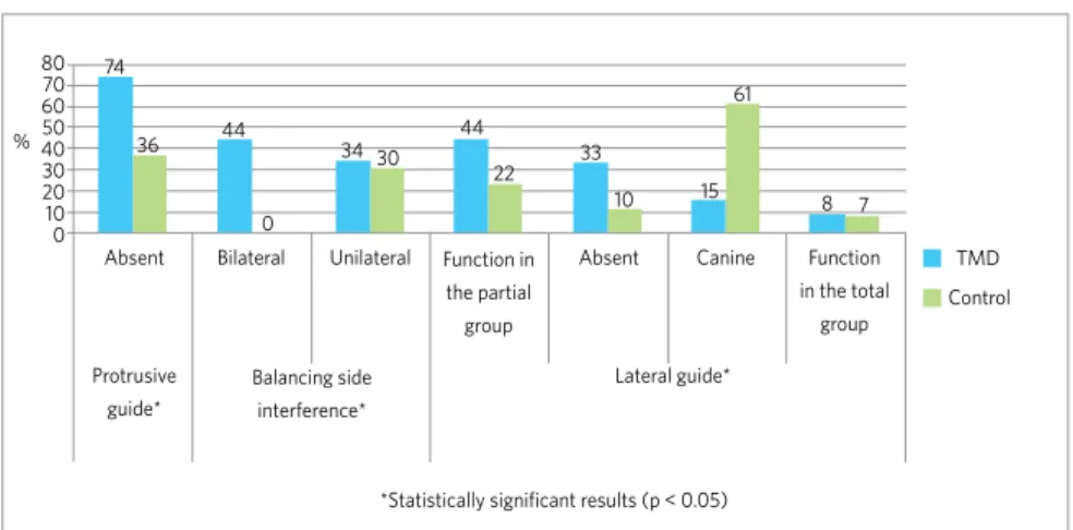
During the lateral mandibular movement in TMD patients, it was observed high percentage of group function, partial (46% right and 42% left) and total (10% right and 6% left), with a low frequency of ca-nine guide (14% right and 16% left). The lateral guide was absent due to tooth loss, being 30% on the right and 36% on the left side. Balancing side interferences were found in 78% of TMD patients (34% unilateral and 44% bilateral). During the protusive movement 74% had malocclusion of the posterior teeth guided by anterior teeth. The summarized data related to the functional aspect of occlusal anterior guidance in both groups can be observed in Figure 5.
The χ2 test detected a statistically significant
asso-ciation (p < 0.05) for absence of five or more posterior teeth, overbite and overjet greater than 5 mm, edge-to-edge bite, posterior crossbite (uni and bilateral),
Class II and III, different types of anterior guide and balancing side interference.
DISCUSSION
This study evaluated the prevalence and rela-tion of several occlusal factors in TMD patients di-agnosed. More than a half of patients (54%) were 30 to 49 years old with a strong female predominance
(88%), which corroborates with other studies.9,11,13,20
According Luther,13 the high incidence in women
may be related to hormonal changes that occur at this stage of life. A greater concentration of women (64%) was also observed in the control group.
Regarding the questionnaire, it is noticed that about 70 to 80% of TMD patients showed positive responses to TMJ sounds, muscle discomfort or pain while chewing, headaches and pain in the cervical
1. Is it difficult to open your mouth?
Anterior open bite
0 10 20 %
*Statistically significant results (p < 0.05)
30 40 50 60 70
Control
Control TMD
TMD 26
4
26
10
4
6
10
0
0
4
48 44
76 70
72 44
84 68
82 82
2. Do you feel it difficult to move your jaw sideways?
Overjet > 5 mm* 3. Do you feel tired or muscular pain when chewing?
Overbite > 5 mm* 4. Do you have frequent headaches?
Class II* 5. Do you have neck pain or stiff neck?
5 or more teeth with wear facets 6. Do you have earache or pain in the surrounding areas of the TMJ?
Class III* 7. Have you noticed noise in the TMJ when chewing or opening your mouth?
Discrepancies of CR/MHI > 2 mm 8. Have you noticed if you have any habits such as clenching or grinding your teeth?
Posterior crossbite* 9. Do you feel that your teeth do not fit well together?
Edge-to-edge bite* 10. Do you consider yourself a stressed (nervous) person?
Absence of 5 or more posterior teeth*
0 10 20 30 40 50 60 70 80 90 %
34
8
4
38
32
32
30
26
18
10
8
62
4
32 18
16
4
2
0
region, agreeing with work related to the main
sig-nals and symptoms of patients with TMD.7,11,20 Signs
and symptoms of TMD can occur in healthy people2,14
as found in the control group in this study, that
de-spite the classification as “absent” by Fonseca et al4
index, it is observed the presence mainly of headache (10%) and joint noise (10%).
The TMJ joint noises are frequent, even in an asymptomatic population, while the absence of joint noise cannot be used as a rule for articulation
normality.2,7,14 One must perform a careful clinical
examination of signs and symptoms of TMD, be-fore starting the orthodontic treatment, for the pa-tient to be alerted in case of presence of these signs and / or symptoms.
The emotional factor was also directly related to TMD in this study, since 82% of patients reported
some type of stress.12,25,28 Stress in the control group
also showed a significant participation (26%), show-ing its influence on society today. Both stress and occlusion have different involvement in the occur-rence of TMD, depending on the adaptive capacity of the patient, explained by different degrees of physi-ological tolerance. Emotional stress can cause mus-cle hyperactivity, characterizing the so-called brux-ism or clenching, so when an emotional component is associated with a physical factor, such as occlusal changes, stress relief by the stomatognathic system
produces symptoms of pain and dysfunction.12,28
The habit of grinding and / or clenching was pres-ent in 26% of the control group, and 68% in the TMD sample, showing the important relationship be-tween these habits and TMD as observed in previous
studies.5,28,29 The high incidence of these habits
clini-cally reflected in the presence of 5 or more teeth with wear facets, which were observed in 16% from con-trol and 30% in patients with dysfunction,
corrobo-rating with other studies21,29 that also observed an
as-sociation between TMD, dental wear and the habit of grinding or clenching.
The most prevalent occlusion factor in patients with TMD was the absence of 5 or more posterior teeth observed in 62% of the sample. Due to the adaptive process of the TMJ before the functional
changes related to tooth loss, several studies14,26,30
report that the loss of posterior teeth is associated with joint changes, particularly increasing the risk of cracking, and disc displacement. This was also the most prevalent occlusal factor in asymptomatic pa-tients, although with a lower percentage (34%).
The occlusal instability caused by the lost of posterior teeth may cause TMD since the occlusal changes, muscle changes and joint changes exceed the adaptive threshold of the stomatognathic
sys-tem.14.26 Making prostheses to replace missing teeth
in symptomatic patients is not the ideal treatment, because in these cases joint changes have already occurred, thus requiring a complex multidisci-plinary rehabilitation treatment to reestablish the physiological function. The early prosthetic reha-bilitation treatment in asymptomatic patients may be indicated in order to prevent the occlusal col-lapse and, consequently, to reduce the risk of future joint problems.
Previous studies3,13,14,15,26 reported an association
between TMD and crossbite. In this study, 32% of Figure 5 - Distribution of functional occlusal factors of the anterior guide.
Control TMD 80
Absent Bilateral Unilateral Function in
the partial group
Protrusive guide*
Balancing side interference*
Lateral guide*
*Statistically significant results (p < 0.05)
Absent Canine Function in the total
group 70 74
36 44
0
34 30 44
22 33
10 15 8 7
61 60
the patients had TMD and only 4% were from the control group. The crossbite generates an interfer-ence with the contact of posterior teeth, displacing the mandible to a more effective contact. Thus, this abnormal movement of the mandible may have long-term effects on the growth and development of teeth and jaws, creating greater pressure on the
mandibu-lar muscles and joints.13
For some authors,14,26 patients with crossbite have
increased risk to develop TMD and, in these cases, early orthodontic treatment would help to reduce the adaptive demands on the masticatory system, preventing the occurrence of TMD signs and symp-toms in the future. On the other hand, the orthodon-tic treatment in adults to prevent the development of TMD is probably not guaranteed because the bone adaptation has already occurred. The literature is very controversial and now it can not be scientifi-cally proved that the orthodontic treatment alone, prevents, cure, or causes TMD, because its etiology is multifactorial and complex, i.e., limiting several functional, structural and psychological factors such as: emotional stress, trauma, sleep disorders, pos-tural abnormalities, systemic factors, muscle hyper-activity and / or TMJ overload, among others, may trigger this disorder.
In the study by John et al10 there was no
relation-ship between TMD and high values of overbite / over-jet (greater than 5 mm), which are compatible with the normal function of the masticatory muscles and TMJ. In the healthy individuals of this study, there was no occurrence of high values of overbite and/or overjet (2% and 0%), respectively. However many
studies14,18,26 corroborate that overjet and/or overbite
greater than 5 mm are related to the increasing risk of developing TMD, being found in 10% and 18% re-spectively, of patients with dysfunction in this study.
Despite few reports in literature3,6 regarding the
association between TMD and edge-to-edge bite, it is observed a relatively high frequency of 38% of the sample with TMD and only 8% of asymptomatic
pa-tients. For some authors15,26 minimal overjet /
over-bite are associated with TMJ problems.
Some studies14,26 reported an association between
the anterior open bite and TMD, but a low frequency was found in 8% of the TMD group and 2% of the con-trol group. This occlusal feature causes the absence
of the anterior guide and the presence of posterior
interference.14,26 According to some authors,14 the aim
of early orthodontic treatment in these patients is to improve the function of the stomatognathic system and to avoid adaptive changes of the TMJ, preventing the occurrence of TMD symptoms.
It is noteworthy that 22% of the sample with dys-function showed no relation between upper and low-er incisors due to the absence of these teeth. When it was added this percentage (22%) with anterior open bite (8%), overjet and/or overbite greater than 5 mm (20%), edge-to-edge bite (38%), it was observed that 88% of patients with TMD present changes in the re-lation between the incisors.
This high prevalence of problems in the upper and lower incisors relationship reflected directly on the anterior guide of patients, which is consid-ered essential for the health of the stomatognathic system. In the effective anterior guide it occurs the disocclusion of posterior teeth guided by harmonic contact of the lingual surfaces of maxillary anterior teeth and maxillary anterior incisors during lateral
movements and protrusion.19
However, during protusive movement, 74% of surveyed patients with TMD had no efficient guide, different from that observed in the control group, in which showed 64% efficiency of this guide. In the lateral movement of the patients with TMD, there is a predominance of group function (44% partial and 8% total), besides that 30% had no guide due to lack of teeth. The canine guide considered ideal was found in 61% of the control group and in only 15% of the sample with TMD, and the absence of canine guide is considered a risk factor for the
develop-ment of TMD.20,24,27
The effect of disarticulation of the posterior teeth in the treatment of TMD symptoms was found in
other studies,20,24,27 reporting a decrease of symptoms
after restoring the anterior guide. During the lateral movement it was observed that there is interference on the non-working or balancing side in 78% of the sample with TMD (34% unilateral and 44% bilateral).
According to Landi et al,12 non-working side
About 58% of TMD patients had problems regard-ing the sagittal relationship (26% Class II and 32%
Class III ), as reported by other authors.8,13
Individu-als with Class II malocclusion have great freedom of mandible movement, unlike Class III mandibular
movement that is limited.17 Some studies17,26 indicate
a greater participation of Class II in temporoman-dibular disorders considered an important risk fac-tor for these patients. The Class I relation was the most frequent in 42% of the sample with TMD and in control groups with 92%.
Regarding the discrepancy between the positions of CR and MHI, deviations greater than 2 mm were found in 32% of patients with TMD. Discrepancies
from 0 to 2 mm are considered normal22 and in this
study they were found in 82% of asymptomatic pa-tients and in 68% of TMD papa-tients showing that this discrepancy was common both in the control group as in patients with TMD, so there is direct relation between this occlusal factor with TMD.
CONCLUSION
The relation between occlusal factors, orthodontics and TMD remains controversial, and there is not a con-sensus in literature yet. In this study, from the observed variables, it was found a statistically significant associa-tion for five or more missing posterior teeth, overbite and overjet greater than 5 mm, edge-to-edge bite, pos-terior crossbite, Class II and III, different types of ante-rior guide and balancing side interference.
1. Alarcón MA, Paredes DA, Balarezo JA. Evidencias de asociación entre los factores oclusales y los desordenes temporo-mandibulres mediante um análisis de regresión logística. Rev Estomatol Herediana. 2002;13(1-2):12-8.
2. Delboni MEG, Abrão J. Estudo dos sinais de DTM em pacientes ortodônticos assintomáticos. Rev Dental Press Ortod Ortop Facial. 2005;10(4):88-96. 3. Egermark I, Magnusson T, Carlsson GE. A 20-year follow-up of signs and
symptoms of temporomandibular disorders and malocclusions in subjects with and without orthodontic treatment in childhood. Angle Orthod. 2003;73(2):109-15. 4. Fonseca DM, Bonfante G, Valle AL, Freitas SFT. Diagnóstico pela anamnese da
disfunção craniomandibular. RGO: Rev Gaúch Odontol. 1994;42(1):23-8. 5. Fujita Y, Motegi E, Nomura M, Kawamura S, Yamaguchi D, Yamaguchi H. Oral
habits of temporomandibular disorder patients with malocclusion. Bull Tokyo Dent Coll. 2003;44(4):201-7.
6. Gesch D, Bernhardt O, Kocher T, John U, Hensel E, Alte D. Association of malocclusion and functional occlusion with signs of temporomandibular disorders in adults: results of the population-based study of health in Pomerania. Angle Orthod. 2004;74(4):512-20.
7. Henrikson T, Nilner M, Kurol J. Signs of temporomandibular disorders in girls receiving orthodontic treatment. A prospective and longitudinal comparison with untreated Class II malocclusions and normal occlusion subjects. Eur J Orthod. 2000;22(3):271-81.
8. Henrikson T, Nilner M. Temporomandibular disorders, occlusion and orthodontic treatment. J Orthod. 2003;30(2):129-37.
9. Huang GJ, Leresche L, Critchlow CW, Martin MD, Drangsholt MT. Risk factors for diagnostic subgroups of painful temporomandibular disorders (TMD). J Dent Res. 2002;81(4):284-8.
10. John MT, Hirsch C, Drangsholt MT, Mancl LA, Setz JM. Overbite and overjet are not related to self-report of temporomandibular disorder symptoms. J Dent Res. 2002;81(3):164-9.
11. John MT, Dworkin SF, Mancl LA. Reliability of clinical temporomandibular disorder diagnoses. Pain. 2005;118(1-2):61-9.
12. Landi N, Manfredini D, Tognini F, Romagnoli M, Bosco M. Quantification of the relative risk of multiple occlusal variables for muscle disorders of the stomatognathic system. J Prosthet Dent. 2004;92(2):190-5.
13. Luther F. TMD and occlusion part II. Damned if we don’t? Functional occlusal problems: TMD epidemiology in a wider context. Br Dent J. 2007;202(1):38-9. 14. McNamara JA, Seligman DA, Okeson JP. Occlusion, orthodontic treatment, and
temporomandibular disorders: a review. J Orofac Pain. 1995;1(9):73-90. 15. Mohlin BO, Derweduwen K, Pilley R, Kingdon A, Shaw WC, Kenealy P.
Malocclusion and temporomandibular disorder: a comparison of adolescents with moderate to severe dysfunction with those without signs and symptoms of temporomandibular disorder and their further development to 30 years of age. Angle Orthod. 2004;74(3):319-27.
REFERENCES
16. Mohlin B, Axelsson S, Paulin G, Pietilä T, Bondemark L, Brattstr MV, et al. TMD in relation to malocclusion and orthodontic treatment. Angle Orthod. 2007;77(3):542-8.
17. Pahkala RH, Laine-Alava MT. Do early signs of orofacial dysfunctions and occlusal variables predict development of TMD in adolescence? J Oral Rehabil. 2002;29(8):737-43.
18. Pahkala R, Qvarnstr MM. Can temporomandibular dysfunction signs be predicted by early morphological or functional variables? Eur J Orthod. 2004;26(4):367-73. 19. Palácios CGF, Casado AC, Trigo AF, Pérez-Varela JC. La oclusión como factor
etiopatológico em los trasntornos temporomandibulres. RCOE. 2007;12(1):37-47. 20. Pereira JF, Conti PCR. Alterações oclusais e a sua relação com a disfunção
temporomandibular. Rev Fac Odontol Bauru. 2001;9(3-4):139-44.
21. Pergamalian A, Rudy TE, Zaki HS, Greco CM. The association between wear facets, bruxism, and severity of facial pain in patients with temporomandibular disorders. J Prosthet Dent. 2003;90(2):194-200.
22. Pullinger AG, Seligman DA, Gornbein JA. A multiple logistic regression analysis of the risk and relative odds of temporomandibular disorders as a function of common occlusal features. J Dent Res. 1993;72(6):968-79.
23. Pullinger AG, Seligman DA. Quantification and validation of predictive values of occlusal variables in temporomandibular disorders using a multifactorial analysis. J Prosthet Dent. 2000;83(1):66-75.
24. Rinchuse DJ, Kandasamy S, Sciote J. A contemporary and evidence-based view of canine protected occlusion. Am J Orthod Dentofacial Orthop. 2007;132(1):90-102. 25. Santos ECA, Bertoz FA, Pignatta LMB, Arantes FM. Avaliação clínica de sinais e
sintomas da disfunção temporomandibular em crianças. Rev Dental Press Ortod Ortop Facial. 2006;11(2):29-34.
26. Seligman DA, Pullinger AG. The role of functional occlusal relationships in temporomandibular disorders: a review. J Craniomandib Disord. 1991;5(4):265-79. 27. Selaimen CM, Jeronymo JC, Brilhante DP, Lima EM, Grossi PK, Grossi ML. Occlusal
risk factors for temporomandibular disorders. Angle Orthod. 2007;77(3):471-7. 28. Van Selms MK, Lobbezoo F, Visscher CM, Naeije M. Myofascial
temporomandibular disorder pain, parafunctions and psychological stress. J Oral Rehabil. 2008;35(1):45-52.
29. Van ‘t Spijker A, Kreulen CM, Creugers NH. Attrition, occlusion, (dys)function, and intervention: a systematic review. Clin Oral Implants Res. 2007;18 Suppl 3:117-26. 30. Wang MQ, Cao HT, Liu FR, Chen C, Li G. Association of tightly locked occlusion