www.saber.ula.ve/avancesenquimica
Avances en Química, 5(2), 95-98 (2010)
Artículo científico
95
Flavonoids from
Urena sinuata
L.
Adakarleny Sosa & Carmelo Rosquete
*
Laboratorio de Productos Naturales, Departamento de Química, Facultad de Ciencias. Universidad de los Andes, Mérida 5101, Venezuela.
(*)
Recibido: 12/02/2010 Revisado: 28/06/2010 Aceptado: 07/07/2010
---
Resumen
En este manuscrito se reportan los resultados obtenidos del estudio fitoquímico de las hojas frescas de Urena sinuata L. (cadillo de perro). Los componentes mayoritarios identificados fueron los flavonoides: 3'-β -D-glucopiranosil-6,7-O-dimetilquercetagetina (I), 4'-β-D-glucopiranosil-6,7-O-dimetilquercetagetina (II) y 3-β -D-glucopiranosil-6,7-O-dimetil-quercetagetina (III). Los compuestos aislados fueron caracterizados mediante sus constantes físicas y el análisis de sus espectros ultravioleta, de masas y de resonancia magnética nuclear mono- y bi-dimensionales. También se muestran los resultados obtenidos para estos compuestos en el ensayo de citotoxicidad sobre Artemia salina.
Palabras claves:Urena sinuata; flavonoides; glicósidos de flavonoles; derivados de quercetagetina Abstract
In this work it is exposed the obtained results of the phytochemical study of the fresh leaves of Urena sinuata L. (dog wart). The major components are the flavonoids: quercetagetin-6,7-O-dimethylether-3'-β-D-gluco-pyranoside (I), quercetagetin-6,7-O-dimethylether-4'-β-D-glucopyranoside (II), and quercetagetin-6,7-O-dimethylether-3-β -D-glucopyranoside (III). These products were characterized through their physical constants, UV, MS, and one- and two-dimensional NMR studies. By other hand, the obtained results of the Artemia salina cytotoxicity bioassay carried out to the isolated products are exposed.
Keywords:Urena sinuata; Flavonoids; Flavonol glycosides; Quercetagetin derivatives
Introduction
Urena L. (Malvaceae) is a genus composed by two species:
Urena lobata L. and Urena sinuata L. although some Botanist suggest that U. sinuata is subspecie of U. lobata. This plant (U. lobata) has been phytochemically studied by some authors, and steroids (stigmasterol and β-sitosterol)1, xanthones (mangiferin)2, flavonoids (quercetina2-4, kaempferol, hypolaetin, gossypetin, luteolin, apigenin and chrysoeriol3), sugars (glucose, mannose, xylose and fructose)5, and vitamins (ascorbic acid)5 has been reported. For U. sinuata only has been reported fatty acids: sterculic and malvalic acids6.
In Venezuela, the infusion of foliage from Urena sinuata L. is used as anti-inflammatory, analgesic, and against kidney pain and gall stone7. For U. lobata antiparasitic8,9, antibacterial10-12, antidiarrheal13, and immunomodulatory activity14 have been mentioned.
Experimental
General Experimental Procedures
IR spectra were recorded as KBr disc on a Perkin Elmer FT-IR Spectrometer 1725X. UV spectra were recorded on
an UV Varian Scan 3 using methanol as solvent. NMR spectra were run on Bruker Avance DRX 400 using CDCl3
as solvent and TMS as internal standard. Mass spectra were recorded on a Hewlett-Packard Mass Spectrometer model 5930a (70 eV). Si gel 60 (Merck, 70-230 mesh) with dry assembly was used in CC and Si gel Merck HF 254 (10 – 40 µ) on glass sheet (0.25 and 0.5 mm thickness, respectively) was used as adsorbent for TLC and PTLC.
Plant Material
Urena sinuata L. (Malvaceae) was collected at San Cristóbal suburbs (Táchira State-Venezuela). Voucher specimens were stored at MERC Herbarium, Sciences Faculty, Universidad de los Andes-Venezuela.
Extraction and Isolation
4-A Sosa & C Rosquete /4-Avances en Química, 5(2), 95-98 (2010)
96
monoitaconate (500 mg) as mainly product. Acetone extract (8.9 g) was also percolated on Sephadex LH-20® column for eliminate chlorophylls using firstly a mixture of n-hexane/chloroform/methanol (1:1:1) and then methanol, as solvents. From methanol eluate (525 mg) were obtained, before PTLC using n-hexane/acetone (1:8) x 8 as eluent, quercetagetin-6,7-O-dimethylether-3'-β -D-glucopyranoside, I (63 mg), quercetagetin-6,7-O-dimethyl-ether-4'-β-D-glucopyrano-side, II (72 mg), and querceta-getin-6,7-O-dimethylether-3-β-D-glucopyranoside, III (45 mg).
Biological Essay
Artemia salina cytotoxicity test was performed following the procedure described by Meyer et al.16
Quercetagetin-6,7-O-dimethylether-3'-β -D-glucopyrano-side, I: dark yellow prisms, mp > 300 ºC. IR (KBr) υmax:
3377, 2923, 1654, 1554, 1130, 812 cm-1. UV (MeOH): see Table 2. 1H- and 13C-NMR (CDCl3): see table 1. MS m/z:
[M]+. 508 (< 1), [M – Glu + H]+. 346 (100), [M – Glu – H2O + H]
+.
328 (31), [M – Glu – H2C=C=O + H] +.
303 (66), [M – Glu – H2C=C=O – H2O + H]
+.
285 (16), [M – Glu – H2C=C=O – H2O – CH3 + H]
+
260 (9), [A1 – OCH3
– CO]+ 137 (19).
Quercetagetin-6,7-O-dimethylether-4'-β -D-glucopyrano-side, II: dark yellow prisms, mp > 300 ºC. IR (KBr) υmax:
3378, 2929, 1654, 1547, 1131, 811 cm-1. UV (MeOH): see Table 2. 1H- and 13C-NMR (CDCl3): see table 1. MS m/z:
[M]+. 508 (< 1), [M – Glu + H]+. 346 (100), [M – Glu – H2O + H]
+.
328 (34), [M – Glu – H2C=C=O + H] +.
303 (70), [M – Glu – H2C=C=O – H2O + H]
+.
285 (18), [M – Glu – H2C=C=O – H2O – CH3 + H]
+
260 (9), [A1 – OCH3
– CO]+ 137 (19).
Quercetagetin-6,7-O-dimethylether-3-β -D-glucopyrano-side, III: pale yellow prisms, mp > 300 ºC. IR (KBr) υmax:
3413, 2945, 1658, 1556, 1134, 806 cm-1. UV (MeOH): see Table 2. 1H- and 13C-NMR (CDCl3): see table 1. MS m/z:
[M]+. 508 (< 1), [M – Glu + H]+. 346 (100), [M – Glu – H2O
+ H]+. 328 (31), [M – Glu – H2C=C=O + H] +.
303 (68), [M – Glu – H2C=C=O – H2O + H]
+.
285 (16), [M – Glu – H2C=C=O – H2O – CH3 + H]+ 260 (9), [A1 – OCH3 – CO]+
137 (19).
Results and Discussion
All compounds, I to III, showed very similar IR, mass and
13
C-NMR spectra and only slight differences at 1H-NMR spectra (see table 1). The whole analysis of 1D spectroscopic data allowed us to determine that I to III correspond to flavone-type compounds, O-substituted at 3, 5, 6, 7, 3', and 4' positions, with one β-D-glucopyranosyl (mainly identified by 13C-NMR chemical shift), two
methoxyl, and three hydroxyl groups as substituents. HMBC experiments confirmed us that methoxyl groups were placed at 6 and 7 positions for all three compounds, remain therefore to insert the β-D-glucopyranosyl group between 3, 5, 3' and 4' positions, which was made using shift reagent for UV spectra.
During UV spectra analysis of the compounds (see table 2) were observed: For compound I, the bathochromic shift for band I (+ 42 nm) showing a low intensity decrease with in time (hypochromic effect) observed when UV spectrum was recorded in methanol + sodium methoxyde can let us to place hydroxyl group on positions 3 and 4'. When UV spectrum was recorded in methanol + AlCl3, a + 53 nm
bathochromic shift was observed; that shift remain unchanged after hydrochloric acid addition, which place hydroxyl group on positions 3 and 5. In consequence, the
β-D-glucopyranosyl moiety should be located on 3' carbon, and compound I should be quercetagetin-6,7-O -dimethylether-3'-β-D-glucopyranoside.
Similar to I, compound II showed for band I, a bathochromic shift (+ 54 nm) in presence of AlCl3 and
AlCl3 + HCl, which place hydroxyl groups on positions 3
and 5 too. The bathochromic displacement (+ 42 nm) of band I observed after sodium methoxyde addition without in time intensity decrease, together with the existence of C3 hydroxyl group previously established, place in this case,
the β-D-glucopyranosyl moiety on C4', and compound II should be quercetagetin-6,7-O-dimethylether-4'-β -D-glucopyranoside.
Different to previously described compounds, band I of UV spectrum in methanol + AlCl3 of compound III was
affected after HCl addition, underwent a hypsochromic shift of -37 nm (respect to methanol + AlCl3) which
indicated the existence of an orto-dihydroxyl moiety on B-ring (3' and 4' positions), which was corroborated by band I hypsochromic displacement (-32 nm) observed on methanol + sodium acetate UV spectrum after boric acid addition. The stability in time of the methanol + sodium methoxyde, together with the 12 nm bathochromic shift remanent respect to methanol UV spectrum observed on band I of methanol + AlCl3 + HCl, place the third hydroxyl
group on C5 carbon, and therefore compound III should be quercetagetin-6,7-O-dimethylether-3-β-D-glucopyranoside. Although this compound has been reported from Brickellia dentata15, none spectroscopic data has been published in reference 15 and references cited therein.
A Sosa & C Rosquete /Avances en Química, 5(2), 95-98 (2010)
97
OR3
OH
OR1
O O
O
OR2
I II III
R1 Glu H H
R2 H Glu H
R3 H H Glu
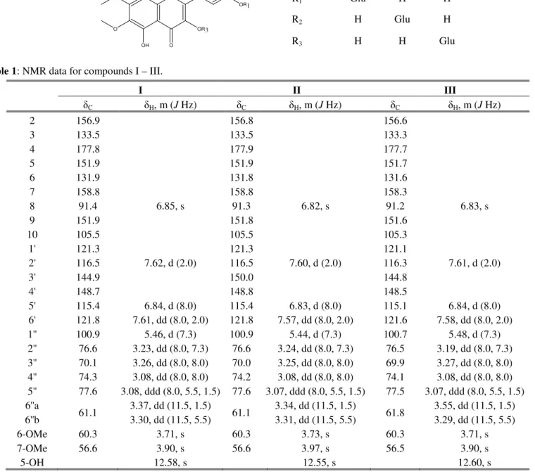
Table 1: NMR data for compounds I – III.
I II III
δC δH, m (J Hz) δC δH, m (J Hz) δC δH, m (J Hz)
2 156.9 156.8 156.6
3 133.5 133.5 133.3
4 177.8 177.9 177.7
5 151.9 151.9 151.7
6 131.9 131.8 131.6
7 158.8 158.8 158.3
8 91.4 6.85, s 91.3 6.82, s 91.2 6.83, s
9 151.9 151.8 151.6
10 105.5 105.5 105.3
1' 121.3 121.3 121.1
2' 116.5 7.62, d (2.0) 116.5 7.60, d (2.0) 116.3 7.61, d (2.0)
3' 144.9 150.0 144.8
4' 148.7 148.8 148.5
5' 115.4 6.84, d (8.0) 115.4 6.83, d (8.0) 115.1 6.84, d (8.0) 6' 121.8 7.61, dd (8.0, 2.0) 121.8 7.57, dd (8.0, 2.0) 121.6 7.58, dd (8.0, 2.0) 1'' 100.9 5.46, d (7.3) 100.9 5.44, d (7.3) 100.7 5.48, d (7.3) 2'' 76.6 3.23, dd (8.0, 7.3) 76.6 3.24, dd (8.0, 7.3) 76.5 3.19, dd (8.0, 7.3) 3'' 70.1 3.26, dd (8.0, 8.0) 70.0 3.25, dd (8.0, 8.0) 69.9 3.27, dd (8.0, 8.0) 4'' 74.3 3.08, dd (8.0, 8.0) 74.2 3.08, dd (8.0, 8.0) 74.1 3.08, dd (8.0, 8.0) 5'' 77.6 3.08, ddd (8.0, 5.5, 1.5) 77.6 3.07, ddd (8.0, 5.5, 1.5) 77.5 3.07, ddd (8.0, 5.5, 1.5) 6''a
61.1 3.37, dd (11.5, 1.5) 61.1 3.34, dd (11.5, 1.5) 61.8 3.55, dd (11.5, 1.5) 6''b 3.30, dd (11.5, 5.5) 3.31, dd (11.5, 5.5) 3.29, dd (11.5, 5.5)
6-OMe 60.3 3.71, s 60.3 3.73, s 60.3 3.71, s
7-OMe 56.6 3.90, s 56.6 3.97, s 56.5 3.90, s
5-OH 12.58, s 12.55, s 12.60, s
Table 2: UV data for compounds I – III.
UV λmax (nm)
I II III
Band I Band II Band I Band II Band I Band II
MeOH 352 256 352 259 353 258
MeOH+NaOMe 394↓ 272↓ 405 270 393 270
MeOH+NaOAc 404 258 376 266 407 256
NaOAc+H3BO3 404 258 376 266 375 258
MeOH+AlCl3 405 271 406 276 402 273
MeOH+AlCl3+HCl 408 272 406 274 365 266
A Sosa & C Rosquete /Avances en Química, 5(2), 95-98 (2010)
98
Due to the frequently ingestion of U. sinuata leaves infusion by Andean peoples and, by other hand, by the possibility of use of the three flavonoids in pharmacological essays, the cytotoxicity of these compounds was tested. Compounds I to III showed similar values for DL50≈ 1000 ppm, which point out the
low cytotoxicity showed for the three compounds to
Artemia salina16.
Hexadecyl 4-monoitaconate isolated from dichloro-methane extract probably arises from fungus of the
Aspergillus genus which colonized the plant material during the storage.
Conclusions
From Urena sinuata leaves, three quercetagetin glucosides were isolated and identified; two of them are new natural products. The presence of I-III in U. sinuata leaves difference chemically to this plant from U. lobata, from the which only flavonoid aglycones were isolated; this sentence support the location of these taxa in different species.
References
1. S Lin, T Pan, C Horg. Chemical constituents of Urena lobata
L. var. tomentosa (Blume) Walp (Malvaceae). Hua Hsueh,
41, 72-73 (1983).
2. K Srinivasan, S Subramanian. Isolation of mangiferin from
Urena lobata. Arogya, 7, 140-141 (1981).
3. I Matlawska. Investigation of flavonoid compounds of select species from Malvaceae family. Herba Polonica, 36, 65-69 (1990).
4. I Matlawska, M Sikorska. Flavonoid compounds in the flowers of Urena lobata L (Malvaceae). Acta Pol. Pharm., 56, 69-71 (1999).
5. D Bhatt, G Rawat. Chemical analysis of medicinal plants
Urena lobata L and Abutilon indicum L. Orient. J. Chem., 17, 341-342 (2001).
6. M Kittur, C Mahajanshetti, G Lakshminaroyana. Charac-teristics and composition Trichosanthes bracteata, Urena sinuata and Capparis divaricata seeds and oils. J. Oil Techn., 25, 34-41 (1993).
7. Banco de Datos TRAMIL. Investigaciones pendientes. http://funredes.org/endacaribe/investigPendientespag11.html. Visited on February, 9th, 2010. 8:55 h.
8. J Satalaya, J Rojas, B Ríos, M Grandez, E Rengifo, G Ruiz, D Gutiérrez, A Giménez, N Flores. Antiparasitic activity of medicinal plants from Peruvian Amazon. BIOFARBO, 17, 23-31 (2009).
9. J Nguyen-Pouplin, H Tran, H Tran, T Phan, C Dolecek, J Farrar, T Tran, P Caron, B Bodo, P Grellier. Antimalarial
and cytotoxic activities of ethnopharmacologically selected medicinal plants from South Vietnam. J.Ethno-pharmacol., 109, 417-427(2007).
10. U Antibacterial activity of Urena lobata root., 927-929 (2001).
11. O Adeloye, A Akinpelu, O Ogundaini, A Obafemi. Studies on antimicrobial, antioxidant and phytochemical analysis of
Urena lobata Leave extract. J. Phys. Nat. Sc.,1, 1-9 (2007). 12. R Mathappan, V Prasanth, C Jolly. M Somanath.
Comparative study on the antibacterial activity of the methanolic extract of Urena lobata root and a standard marketed herbal formulation. J. Pharm. Res., 3, 953-955 (2010).
13. A Yadav, V Tangpu. Antidiarrheal Activity of Lithocarpus
dealbata and Urena lobata. Extracts: Therapeutic
Implications. Pharm. Biol., 45, 223-229 (2007).
14. R Mathappan, V Prasanth, G Parthasarathy. Immuno-modulatory activity of the methanolic extract of Urena lobata Linn. Internet J. Pharmacol., 7, (2009).
15. A Ulubelen, B Timmermann, T Mabry. Flavonoids from
Brickellia chlorolepis and B. dentata. Phytochemistry, 19, 905-908 (1980).