J. Braz. Chem. Soc., Vol. 13, No. 5, 704-707, 2002. Printed in Brazil - ©2002 Sociedade Brasileira de Química 0103 - 5053 $6.00+0.00
Short Report
* e-mail: [email protected]
Additional Coumarins from Kielmeyera reticulata
Frederico G. Cruz*,a, Luciana de M. Moreiraa, Nelson A. S. Santosa and Maria L. S. Guedesb
a
Instituto de Química, Universidade Federal da Bahia, bInstituto de Biologia, Universidade Federal da Bahia, 40170-290, Salvador-BA, Brazil
Do extrato hexânico e da fase extraída com diclorometano do extrato metanólico de Kielmeyera reticulata foram isoladas três novas 4-fenilcumarinas, 5,7-diidroxi-6-(2’-hidroxi-3’-metil-3’-butenil)-8-(4”-cinamoil-3”-metil-1”-oxobutil)-4-fenilcumarina, 7-hidroxi-8-(4”-cinamoil-3”-metil-1”-oxobutil)-2’-(2-hidroxiisopropil)diidrofurano (5’,4’:5,6)-4-fenilcumarina e 5,7-diidroxi-8-(4”-cinamoil-3”-metil-1”-oxobutil)-4-fenilcumarina, além das substâncias já conhecidas, 7-hidroxi-8-(4”-cinamoil-3”-metil-1”-oxobutil)-2’,2’-dimetilpirano(6’,5’:5,6)-4-n-propilcumarina, 5-hidroxi-6-(4”-cinamoil-3”-metil-1”-oxobutil)-2’,2’-dimetilpirano(6’,5’:7,8)-4-n-propilcumarina, δ-tocotrienol e 2,3-metilenodioxixantona.
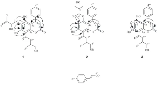
Three new 4-phenylcoumarins, 5,7-dihydroxy-6-(2’-hydroxy-3’-methyl-3’-butenyl)-8-(4”-cinnamoyl-3”-methyl-1”-oxobutyl)-4-phenylcoumarin, 7-hydroxy-8-(4”-cinnamoyl-3”-methyl-1”-oxobutyl)-2’-(2-hydroxyisopropyl)-dihydrofurano(5’,4’:5,6)-4-phenylcoumarin, and 5,7-dihydroxy-8-(4”-cinnamoyl-3”-methyl-1”-oxobutyl)-phenylcoumarin, along with the known
4-n-propylcoumarins, 7-hydroxy-8-(4”-cinnamoyl-3”-methyl-1”-oxobutyl)-2’,2’-dimethylpyrano (6’,5’:5,6)-4-n-propylcoumarin, 5-hydroxy-6-(4”-cinnamoyl-3”-methyl-1”-oxobutyl)-2’,2’-dimethylpyrano(6’,5’:7,8)-4-n-propylcoumarin, the xanthone, 2,3-methylenedioxyxanthone and δ -tocotrienol were isolated from the organic extracts of Kielmeyera reticulata stems .
Keywords: Kielmeyera reticulata, Guttiferae, Clusiacea, 4-phenylcoumarins, 4-propylcoumarins, neoflavonoids
Introduction
Kielmeyera reticulata is a wild shrub belonging to Guttiferae (Clusiaceae). Its stems were collected in sand dunes (restinga) on the Bahia state coast, Brazil. A previous work reported on the isolation of five prenylated 4-phenylcoumarins from this species.1 Studies of this genus
showed that species from the “cerrado” (savanna) of the Brazilian Central plateau are rich in xanthones,2-7 while
species from the “restinga” (sand dunes) are rich in prenylated 4-phenyl and 4-alkylcoumarins.1,8 Only two
species showed xanthones such as prenylated 4-phenyl and 4-alkylcoumarins.9,10
Experimental
General procedures
UV spectra were obtained on Cary 1-E, Varian in MeOH
and MeOH/NaOH. EIMS were taken with direct probe insert at 70 eV on HP 5973. NMR spectra were obtained from Gemini 300-Varian in CDCl3 solution with TMS as internal reference standard. Rotations were determined with 241 Perkin Elmer polarimeter.
Plant material
Stems of K. reticulata Saad were collected in the sand dunes of Parque Metropolitano do Abaeté, Salvador, Bahia, Brazil, in January 1992. A voucher specimen, 027415, is deposited in the “Alexandre Leal Costa” Herbarium, Instituto de Biologia, Universidade Federal da Bahia, Salvador, Bahia, Brazil.
Extraction and isolation
The dried stems (3.4 kg) were first extracted with hexane and after with methanol. The hexane extract (37.1 g) was
submitted to silica gel (40-63 µm, Merck) column
705 Additional Coumarins from Kielmeyera reticulata
Vol. 13, No. 5, 2002
fractions were rechromatographed on silica gel CC using hexane/EtOAc gradient to give 1 (276.9 mg), 2 (123.0 mg), 7-hydroxy-8-(4”-cinnamoyl-3”-methyl-1”-oxobutyl)-2’,2’-dimethylpyrano(6’,5’:5,6)-4-n-propylcoumarin (17.8 mg), 5-hydroxy-6-(4”-cinnamoyl-3”-methyl-1”-oxobutyl)-2’,2’-dimethylpyrano(6’,5’:7,8)-4-n-propylcoumarin (26.9 mg) and δ-tocotrienol (288.3mg). The methanol extract
was submitted to partition with MeOH/H2O/CH2Cl2
yielding a CH2Cl2 phase (32.1 g) that was submitted to silica gel column chromatography using hexane/acetone gradient. Several fractions were rechromatographed on silica gel CC using hexane/acetone gradient to give 3 (30.4 mg) and 2,3-methylenedioxyxanthone (6.7 mg).
5,7-dihydroxy-6-(2’-hydroxy-3’-methyl-3’-butenyl)-8-(4”-cinnamoyl-3”-methyl-1”-oxobutyl)-4-phenylcoumarin, (1)
C34H32O8.Amorphous yellow-greenish solid. NMR: see Tables 1 and 2. EIMS m/z (rel. int.): 568 [M]+ (1), 349 (99),
293 (59), 131 (100), 103 (54). [α]24 D –61.0
o (c0.410 CHCl 3).
7-hydroxy-8-(4”-cinnamoyl-3”-methyl-1”-oxobutyl)-2’-(2-hydroxyisopropyl) dihydrofuran-(5’,4’:5,6)-4-phenylcoumarin,(2)
C34H32O8. Amorphous yellow-greenish solid. NMR: see
Tables 1 and 2. EIMS m/z (rel. int.): 420
[M-C6H5C2H2CO2H]+ (7), 365 (9), 361 (8), 347 (8), 293 (9),
131 (100), 59 (30). [α]24 D +92.3
o (c 0.370 CHCl 3).
5,7-dihydroxy-8-(4”-cinnamoyl-3”-methyl-1”-oxobutyl)-4-phenylcoumarin,(3)
C29H24O7. Amorphous yellow-greenish solid. NMR: see
Tables 1 and 2. EIMS m/z (rel. int.): 484 [M]+ (2), 336 (39),
321 (100), 281 (44), 131 (63). [α]24
D +21.74
o (c 0.460
CHCl3).
Results and Discussion
From the hexane extract of K. reticulata two new 4-phenylcoumarins, 1 and 2, two 4-n-propylcoumarins, 7-
hydroxy-8-(4”-cinnamoyl-3”-methyl-1”-oxobutyl)-2’,2’-dimethylpyrano(6’,5’:5,6)-4-n-propylcoumarin,
5-
hydroxy-6-(4”-cinnamoyl-3”-methyl-1”-oxobutyl)-2’,2’-dimethylpyrano(6’,5’:7,8)-4-n-propylcoumarin,
previously isolated from K. argentea,8 and δ-tocotrienol11
were isolated. The CH2Cl2 phase of the methanol extract
yielded a new 4-phenylcoumarin, 3, and
2,3-methyl-enedioxyxanthone.10
Compounds 1, 2, and 3 are yellow-greenish amorphous solids and their molecular formulae and structures were established by UV, EI mass spectrometry, 1H and 13C NMR.
Complete structural assignments were made by analogy with literature data12-14 and by a combination of DEPT, 1H-13C COSY
one bond and multiple bonds. 1H NMR spectra of 1, 2,and 3
(Table 1) showed characteristic signals of a H-3 singlet, a 4-phenyl group, and one hydroxyl hydrogen (exchangeable with D2O) bonded to an acyl side chain, and a cinnamoyl group. The spectrum of 1 showed the additional presence of two broad singlets at δ 4.81 and δ 4.92 assigned to terminal double bond hydrogens, a broad doublet at δ 4.27 assigned to the oxymethine hydrogen H-2’, a doublet at δ 4.18 assigned to oxymethylene hydrogens H-4”, and at δ 3.39 and δ 3.26, two pairs of doublets of doublets assigned to 2”a and H-2”b respectively. The hydrogen H-1’a at δ 3.04 was a doublets of doublets (14.6 and 1.4 Hz) while H-1’b at δ 2.73, was partially overlapped with the multiplet of H-3”.
706 Cruz et al. J. Braz. Chem. Soc.
The 13C NMR spectrum (Table 2) confirmed the
presence of the groups above and their positions on the 4-phenylcoumarin nucleus were unequivocally assigned by
1H-13C long range correlations (COLOC). Thus, the
correlations between the signal at δ 5.97 (H-3) with the carbon signals at δ 159.4, 139.7 and 102.3 permitted to assign these resonances to C-2, C-1’”, and C-4a, respectively. The correlations of the signal at δ 14.62 (OH bonded) with the carbon signals at δ 166.7 (C-7), 110.1 (C-6) and 103.9 (C-8) made possible to place this hydroxyl group at C-7. The group 2’-hydroxy-3’-methyl-3’-butenyl was located at C-6 due to the observed correlations between the signal at δ 3.04 (H-1’) with the carbons at δ
166.7 (C-7), 161.1 (C-5), and 110.1 (C-6). Therefore, the 4”-cinnamoyl-3”-methyl-1”-oxobutyl group was placed at C-8. This assumption was corroborated by the observation of a low bathochromic shift ( 387 to 392 nm) of the longer wavelength band in the UV spectrum after alkali addition.13
The signals in the 1H NMR spectrum of 2 (Table 1) at δ
4.45, 2.87 and 2.99, in addition to the signals at δ 93.3 (C-2’), 72.1 (C-4’), 25.3 (C-5’) and 23.9 (C-6’) in the 13C NMR
spectrum (Table 2), suggested the presence of the 2’-(2-hydroxyisopropyl)dihydrofuran system. This group was confirmed by 1H-13C long range correlations and by the
presence of an abundant ion at m/z 59 (C3H7O+) in the mass
spectrum, which is characteristic of that system.14 The
positions of the groups in aromatic coumarin ring were deduced by long range correlations (COLOC). The
Table 2.13C NMR data for compounds 1, 2, and 3 (75 MHz, CDCl 3)
C 1 2 3
2 159.4 159.2 158.8
3 111.9 111.7 111.8
4 156.8* 155.4* 157.7*
4 a 102.3 99.3 101.2
5 161.1 162.9 160.4
6 110.1 110.6 101.0
7 166.7 164.0 168.4
8 103.9 105.7 104.8
8 a 156.4* 157.7* 155.0*
1 ’ 28.8 -
-2 ’ 76.9 93.3
-3 ’ 146.2 27.3
-4 ’ 110.6 72.1
-5 ’ 18.4 25.3
-6 ’ - 23.9
-1 ” 204.6 205.4 204.4
2 ” 48.5 49.1 48.5
3 ” 30.0 30.8 30.9
4 ” 68.9 69.4 68.9
5 ” 17.3 17.9 17.3
6 ” 167.1 167.4 167.2
7 ” 117.9 118.7 117.9
8 ” 144.8 145.2 144.9
9 ” 134.3 135.1 134.4
10”-14” 128.1 128.7 128.8
11”-13” 128.8 129.4 128.9
1 2 ” 130.3 130.7 130.2
1’” 139.7 138.6 137.4
2’”-6’” 127.1 128.7 127.3
3’”-5’” 127.6 129.4 128.1
4’” 128.2 128.00 129.6
* This signals may be interchangeable
Table1.1H NMR data for compounds 1, 2, and 3 (300 MHz, CDCl 3)
H 1 2 3
3 5.97, 1H, s 6.08, 1H, s 6.00, 1H,
6 6.22, 1H, s
1’a 3.04, 1H, dd (14.6, 1.4)
1’b 2.73, 1H, overlap
2 ’ 4.27, 1H, bd(8.4) 4.45, 1H, dd (9.7, 8.9)
3’a 2.87, 1H, dd (15.4; 8.9)
3’b 2.99, 1H, dd (15.4; 9.7)
4’a 4.81, 1H, s
4’b 4.92, 1H, s
5 ’ 1.75, 3H, s 0.91, 3H, s
6 ’ 0.98, 3H, s
2 ” a 3.26, 1H, dd ( 16.2; 6.2) 3.29, 1H, dd(16.3; 6.2) 3.28, 1H, dd (16.5; 6.3)
2”b 3.39, 1H, dd(16.2; 7.1) 3.46, 1H, dd (16.3; 7.2) 3.41, 1H, dd(16.5; 6.8)
3 ” 2.68, 1H, overlap 2.71, 1H, m 2.69, 1H, m
4 ” 4.18, 2H, d(5.9) 4.24, 2H, m 4.21, 2H, m
5 ” 1.13, 3H, d (6.7) 1.18, 3H, d(7.0) 1.16, 3H, d(6.8)
7 ” 6.38, 1H, d(16.2) 6.42, 1H, d(16.1) 6.41, 1H, d(16.0)
8 ” 7.61, 1H, d(16.2) 7.64, 1H, d(16.1) 7.65, 1H, d(16.0)
Phenyl (Cinnamoyl) 7.36, 3H, m 7.49, 2H, m 7.44, 3H, m 7.53, 2H, m 7.36,3H, m 7.52, 2H, m
4-phenyl 7.28, 2H, m 7.36, 3H, m 7.30, 2H, m 7.38, 3H, m 7.36, 2H, m 7.46, 3H, m
7-OH 14.62, 1H, s 14.10, 1H, s 14.00, 1H, s
707 Additional Coumarins from Kielmeyera reticulata
Vol. 13, No. 5, 2002
correlations observed between the signal at δ 6.08 (H-3) with the signals at δ 159.2 and δ 99.3 permitted the assignment of these resonances to carbons C-2 and C-4a, respectively. The correlations of the resonance at δ 14.10 (OH bonded) with the signals at δ 105.7 (C-8), 110.6 (C-6) and 164.0 (C-7) and those at δ 2.87 3’a) and 2.99 (H-3’b) with δ 110.6 (C-6) made evident the location of the bonded hydroxyl at C-7, the 2’-(2-hydroxyisopropyl)-dihydrofuran ring at C-5 and C-6 and the 4”-cinnamoyl-3”-methyl-1”-oxobutyl group at C-8. The UV spectrum of
2 after alkali addition showed a bathochromic shift (348 to 379 nm) of the longer wavelength band. The observed shift was lower than the obtained with a similar compound with the acyl side chain at C-6 (348 to 428 nm).15 In
conclusion, the UV data corroborated with the location of the acyl side chain at C-8.
The long-range correlation observed in the 1H-13C
COSYspectrum of 3,between the signal at δ 6.00 (H-3) and the carbon signal at δ 101.2 allowed the assignment of this signal to C-4a. The correlations of the hydrogen signal at δ 6.22 with the carbon signals at δ 160.4 (C-5), 168.4 (C-7) and 104.8 (C-8) allowed the placement of this hydrogen at C-6 and consequently the 4”-cinnamoyl-3”-methyl-1”-oxobutyl side chain was placed at C-8. The acyl side chain at C-8 was confirmed by the low bathochromic shift (382 to 388 nm) of the longer wavelength band in the UV spectrum after alkali addition.
Acknowledgements
Authors are grateful to Cecília Veronica Nuñes (IQ-USP) for recording the αD values, as well as to CNPq for the MSci. fellowships awarded to L. M. M. and N. A. S. S. This work was supported by grants from CNPq and FINEP.
References
1. Cruz, F. G.; Moreira, L. de M.; David, J. M.; Guedes, M. L. S.; Chávez, J. P.; Phytochemistry1998, 47, 1363.
2. Gottlieb, O. R.; Magalhães, M. T.; Stefani, G. M.; Tetrahedron
1966, 22, 1785.
3. Gottlieb, O. R.; Mesquita, A. A. L.; Nagem, T. J.; Phytochemis-try, 1971, 10, 2253.
4. Gottlieb, O. R.; Mesquita, A. A. L.; Oliveira, G. G.; Melo, M.
T.; Phytochemistry1970, 9, 2537.
5. Gottlieb, O. R.; Mesquita, A. A. L.; Silva, E. M.; Melo, M. T.;
Phytochemistry1969, 8, 665.
6. Gottlieb, O. R.; Stefani,G. M.; Phytochemistry1970, 9, 453. 7. Cortez, D. A.; Young, M. C. M.; Marston, A.; Wolfender, J-L.;
Hostettmann, K.; Phytochemistry1998, 47, 1367.
8. Cruz, F. G.; Santos, N.A. S.; David, J. M.; Guedes, M. L. S.; Chávez, J. P.; Phytochemistry 1998, 48, 703.
9. Nagem, T. J.; Silva, M. A.; Phytochemistry 1988, 27, 2961. 10. Cruz, F.G.; da Silva-Neto, J. T.; Guedes, M. L. S.; J. Braz.
Chem. Soc. 2001, 12, 117.
11. Monache, D. F.; Marta, M.; Mac-Qhae, M. M.; Nicoletti, M.;
Gazz.Chim. Ital.1984, 114, 135.
12. Crichton, E. G.; Waterman, P. G.; Phytochemistry1978, 17, 1783.
13. Crombie, L.; Jones, R. C. F.; Palmer, C. J.; J. Chem. Soc.,
Perkin Trans. I1987, 317.
14. Carpenter, I.; McGarry, E. J.; Scheinmann, F.; J. Chem. Soc.
(C), 1971, 3783.
15. Bandaranayake, W. M.; Selliah, S. S.; Sultanbawa, M. U. S.; Games, D. E.; Phytochemistry1975, 14, 265.