O R I G I N A L A R T I C L E UDC: 616-055.1/.3-071.3::611.96 DOI: 10.2298/VSP1111935J
Sex differences in anatomical parameters of acetabulum among
asymptomatic Serbian population
Polne razlike u anatomskim parametrima acetabuluma kod
asimptomatske srpske populacije
Dejan Jeremić, Ivana Živanović Mačužić, Maja Vulović
Medical Faculty, University of Kragujevac, Department of Anatomy and Forensic
Medicine, Kragujevac, Srbija
Abstract
Background/Aim. Anatomical parameters of the bony components of the hip joint are essential for better under-standing of etiopathogenesis of diseases like primary osteo-arthrosis of the hip joint. The aim of this reserch was to ex-amine the normal acetabular morphometry in Serbian popu-lation and to determine whether there are sex differences in anatomical parameters of the acetabulum among asympto-matic subjects. Methods. Pelvic radiographics of 320 adult asymptomatic patients (640 hips) were analyzed in 170 men and 150 women to determine the morphology of the ace-tabulum in Serbian population. For each hip the center edge angle of Wiberg (CEA), the acetabular angle of Sharp (AA), acetabular depth (AD), acetabular roof obliquity (ARO) and roof angle (RA) were measured. Results. The following aver-age measurements for acetabulum geometry were obtained (ґ± SD): CEA – 33.5 ± 6.5° (33.6 ± 5.8° in male, 33.3 ± 6.9° in female), AA – 38.0 ± 3.8° (37.5 ± 3.6° in male, 38.5 ± 3.9° in female), AD – 11.9 ± 2.8 mm (12.5 ± 2.7 mm in male, 11.2 ± 2.7 mm in female), ARO – 7.6 ± 5.7° (6.2 ± 4.9° in male, 9.0 ± 6.0° in female) and RA – 18.4 ± 10.0° (19.6 ± 8.5° in male, 17.1 ± 9.5° in female). There were significant differences in the CEA, AA, AD, ARO and RA related to gender (p < 0.01, t-test). Conclu-sion. There are significant gender differences in Serbian population for all the examined anatomical parameters of acetabulum. We found sex-related differences in acetabular morphology, female acetabulum being marginally more dys-plastic than male acetabulum. There is also a clear tendency of female hips to be more dysplastic than male ones.
Key words:
hip joint; acetabulum; anatomy; gender identity; serbia.
Apstrakt
Uvod/Cilj. Anatomski parametri koštanih struktura zgloba kuka najvažniji su za bolje razumevanje etiopatogeneze bole-sti kao što je primarna osteoartroza zgloba kuka. Cilj istraži-vanja bio je ispitivanje normalne acetabularne morfometrije i postojanje polnih razlika u anatomskim parametrima aceta-buluma, kod asimptomatskih osoba srpske populacije. Meto-de. Analizirani su radiografski snimci karlice 320 odraslih asimptomatskih ispitanika (640 zglobova kuka) srpske popu-lacije radi utvrđivanja normalne morfologije acetabuluma. Među ispitanicima bilo je 170 muškaraca i 150 žena. Za svaki zglob kuka mereni su Wiberg ugao (CEA), Šarp ugao (AA), acetabularna dubina (AD), kosina krova acetabuluma (ARO) i ugao krova acetabuluma (RA). Rezultati. Dobijene su
slede-će vrednosti acetabularne morfometrije (ґ± SD): CEA je 33,5 ± 6,5° (33,6 ± 5,8° kod muškaraca, 33,3 ± 6,9° kod že-na), AA – 38,0 ± 3,8° (37,5 ± 3,6° kod muškaraca, 38,5 ± 3,9° kod žena), AD – 11,9 ± 2,8 mm (12,5 ± 2,7 mm kod muškaraca, 11,2 ± 2,7 mm kod žena), ARO – 7,6 ± 5,7° (6,2 ± 4,9° kod muškaraca, 9,0 ± 6,0° kod žena) i RA – 18,4 ± 10,0° (19,6 ± 8,5° kod muškaraca, 17,1 ± 9,5° kod že-na). Uočene su statistički značajne razlike u odnosu na pol za sve ispitivane parametre: CEA, AA, AD, ARO i RA (p < 0,01, t test). Zaključak. Za sve ispitivane anatomske pa-rametre acetabuluma postoje statistički značajne razlike u od-nosu na pol, pri čemu je ženski acetabulum pokazivao veće znake displazije nego acetabulum muškaraca. Dobijeni rezul-tati u skladu su sa istraživanjima razlika displastičnosti muš-kog i žensmuš-kog acetabuluma, vršenih u različitim rasnim gru-pama.
Ključne reči:
Introduction
The hip joint is functionally a three-dimensional ball and socket joint, often called cotyloid joint because of its anatomical feature. It enables movements in all the three planes as rotation. The femoral head articulates with the cup-shaped (cotyloid) acetabulum, its center lying a little below the middle third of the inguinal ligament. The acetabulum, with a median curvature radius of 2.7 cm, is formed by parts of the ilium, pubis, and ischium which rejoin in a cartilagi-nous Y to the hip bone. The femoral head, slightly more than one-half a sphere, has a constant curvature radius of about 2.5 cm. Its smoothness is interrupted posterior inferior to its center by a small rough fovea. The femoral neck is about 5 cm long and connects the head to the shaft at the angle of about 125° 1.
More information is needed about the anatomical pa-rameters of the acetabulum of the normal hip joint, including its shape, depth at precise locations, and the influence of age, sex and congenital morphology 2, 3. As race, climate, heredity and geographical areas have strong influence on the anthro-pometric parameters of the bone, therefore, the present study was undertaken to note the average anatomical parameters of the acetabular part of the hip joint in Serbian population. Anatomical parameters of the bony components of the hip joint are essential for better understanding of etiopathogene-sis of diseases like primary osteoarthroetiopathogene-sis of the hip joint. Also, knowledge about various bony components of the hip joint will not only help the radiologists, but will be also of immense importance to the orthopedicians and prosthetists to construct suitable prosthesis. The awareness about average dimensions of hip bones joints in both sexes will also help in early detection of disputed sex by forensic experts.
The aim of this study was to examine normal acetabular morphometry and to determine whether there are sex differ-ences in anatomical parameters of acetabulum among asymptomatic subjects without structural change.
Methods
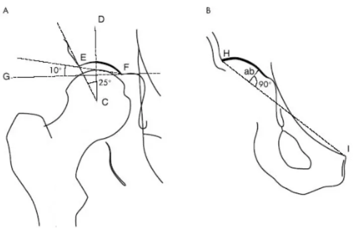
Pelvic radiographies of 320 adult patients, 170 men and 150 women, with clinically normal hip joints (640 hips) were analyzed in to determine the morphology of the acetabulum in Serbian population. The average age of patients was 47.8 years (from 21 to 65), divided by decades: the twenties 10.0% (32), the thirties 15.0% (48), the forties 19.69% (63), the fifties 28.12% (90) and over sixties 27.19% (87). The re-search was conducted as a prospective study. For each tabulum the center-edge angle of Wiberg (CEA), the tabular angle of Sharp (AA), acetabular depth (AD), ace-tabular roof obliquity (ARO) and roof angle (RA) were measured (Figure 1).
Patients with known hip disease or pain located in the hip region, including ambiguous pain probably of lumbar origin but irradiating to the region of the greater trochanter, groin, or thigh, were excluded from this study. We also ex-cluded patients with bone disorders such as Paget's disease, femoral head disease, acquired deformities, unequivocal OA
of the hip, and evident osteophytes or cysts adjacent to the hip joint cavity. Films with incorrect patient positioning (misalignment of the sacropubic symphysis vertical axis ≥ 1.5 cm) were also excluded.
Fig. 1 A – DCE (the center-edge angle of Wiberg); GFE – acetabular roof obliquity; B – acetabular depth
(segment “ab”)
Antero-posterior (AP) radiograph was used to measure the radiography of the hip joint. The patients were placed in the supine position with legs extended and internally rotated for 15°, with a distance of 100 cm between the radiographic source and the film. The central radiographic ray was aligned to be perpendicular to the cassette, entering 5 cm superior to the pubic symphysis. All measurements were made using a new Plexiglas instrument, the „arthrometer“, which com-prises a ruler and protactor appropriate for measuring hip ar-chitectural angles 3. Interpretations were performed by a sin-gle physiatrist trained in the radiographic characteristics of acetabular morphometry.
The AA was measured from the intersection of the horizontal line passing through the bottom of the “tear drop” and the line connecting the bottom of the “tear drop” to the lateral lip of the acetabulum. The greater the angle, the more dysplastic the hip (Figure 1A) 4, 5.
The CEA was defined as the angle between the line joining the center of the femoral head to the lateral margin of the acetabular roof and the line perpendicular to that joining the centers of the two femoral heads (Figure 1B) 6. The cen-ter of each femoral head was established by superimposing a circle around its margin.
The AD was defined as the greatest perpendicular dis-tance from the acetabular roof to the line joining the lateral margin of the acetabular roof and the upper corner of the symphysis pubis on the same side (Figure 1B) 6.
The ARO was defined as the angle between the line connecting the lateral edge of the acetabular roof and the lower iliacus tip of the acetabular surface and the line paral-lel to the pelvic “tear drop” (Figure 1A) 5.
Sex-related differences of anatomical parameters of ace-tabulum were assessed by paired samples t-test. A significant level of p < 0.05 was assumed. Statistical software for Win-dows version 15.0 SPSS was used for all calculations.
Results
Table 1 summarizes the means and standard deviations of the center-edge angle of Wiberg, acetabular angle of Sharp, acetabular depth, acetabular roof obliquity and roof angle of groups in Serbian population.
The following parameters for acetabular geometry were obtained (ґ ± SD): CEA – 32.5 ± 6.4° (33.6 ± 5.8° in male, 31.3 ± 6.9° in female), (ґ ± SD) AA – 38.0 ± 3.8° (37.5 ± 3.6° in male, 38.5 ± 3.9° in female) AD – 11.9 ± 2.8 mm (12.5 ± 2.7 mm in male, 11.2 ± 2.7 mm in female), ARO – 7.6 ± 5.7° (6.2 ± 4.9° in male, 9.0 ± 6.0° in female) and 18.4 ± 9.1° (19.6 ± 8.5° in male, 17.1 ± 9.5° in female).
Acetabular angle, center – edge angle of Wiberg, ace-tabular depth, aceace-tabular roof obliquity and roof angle dif-fered significantly by gender.
The average CEA was significantly greater in the men than in the women: 33.6° (SD = 5.8°) in male hips and 31.3° (SD = 6.9°) in female hips (p < 0.01, t-test).
The average AA was significantly lower in the men than in the women: 37.5° (SD = 3.6°) in male hips and 38.5° (SD = 3.9) in female hips (p < 0.01, t-test).
The average AD was significantly greater in the men than in the women: 12.5 mm (SD 2.7) in male hips and 11.2 mm (SD = 2.7) in female hips (p < 0.01, t-test).
The average ARO was significantly lower in the men than in the women: 6.2° (SD = 4.9) in male hips and 9.0° (SD = 6.0) in female hips (p < 0.01, t-test).
The Average RA was significantly greater in the men than in the women: 19.6° (SD = 8.5) in male hips and 17.1° (SD = 9.5) in female hips (p < 0.01, t-test).
Distribution of anatomical parameters of acetabulum and sex related differences within various ethnic groups are presented in Tables 1–6 7–15.
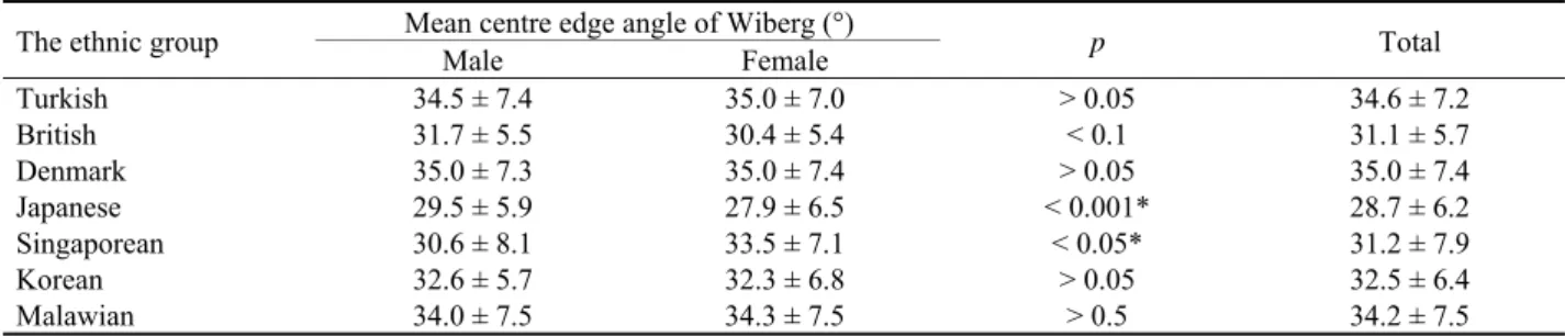
Table 2
The published values of the mean center-edge angle of Wiberg (°) by gender and ethnic group 7–15
Mean centre edge angle of Wiberg (°) The ethnic group
Male Female p Total
Turkish 34.5 ± 7.4 35.0 ± 7.0 > 0.05 34.6 ± 7.2
British 31.7 ± 5.5 30.4 ± 5.4 < 0.1 31.1 ± 5.7
Denmark 35.0 ± 7.3 35.0 ± 7.4 > 0.05 35.0 ± 7.4
Japanese 29.5 ± 5.9 27.9 ± 6.5 < 0.001* 28.7 ± 6.2
Singaporean 30.6 ± 8.1 33.5 ± 7.1 < 0.05* 31.2 ± 7.9
Korean 32.6 ± 5.7 32.3 ± 6.8 > 0.05 32.5 ± 6.4
Malawian 34.0 ± 7.5 34.3 ± 7.5 > 0.5 34.2 ± 7.5
* significant difference
Table 3
The published values of the acetabular angle of Sharp (°) by gender and ethnic group 7–15
Acetabular angle of Sharp (°) The ethnic group
Male Female p Total
Denmark 37.0 ± 3.5 39.1 ± 3.7 < 0.05* 38.0 ± 3.6
British 36.2 ± 2.8 39.0 ± 3.2 < 0.001* 37.6 ± 3.0
Japanese 39.0 ± 3.2 41.8 ± 3.4 < 0.001* 40.4 ± 3.3
Singaporean 39.9 ± 6.0 38.3 ± 5.9 > 0.05 39.1 ± 6.0
Korean 36.5 ± 3.5 37.5 ± 3.8 < 0.01* 37.0 ± 3.7
Malawian 36.9 ± 4.0 38.6 ± 4.9 < 0.05* 37.6 ± 4.5
* significant difference
Table 1 Values of each parameter of acetabulum by gender of subjects in Serbian population
Acetabular parameters Male Female t-test p Total
CEA (°) AA (°) AD (mm) ARO (°) RA (°)
33.6 ± 5.8 37.5 ± 3.6 12.5 ± 2.7 6.2 ± 4.9 19.6 ± 8.5
31.3 ± 6.9 38.5 ± 3.9 11.2 ± 2.7 9.0 ± 6.0 17.1 ± 9.5
0.51 0.28 0.35 1.57 1.02
< 0.01* < 0.01* < 0.01* < 0.01* < 0.01*
32.5 ± 6.4 38.0 ± 3.8 11.9 ± 2.8 7.6 ± 5.7 18.4 ± 10.0 *significant difference
Discussion
According to the obtained results it can be concluded that Serbian female acetabul in our study population are more dysplastic than in males using all methods of meas-urement. Regarding gender differences in the five most im-portant acetabular parameters, CEA, AA, AD, ARO, RA, we found a distinct discrepancy between our data and those re-ported in the literature 7–15.
The center-edge angle, originally described by Wiberg 16, is perhaps the most used indicator and it is in-cluded in most of the radiographic classifications. It evalu-ates the degree of lateral coverage of the femoral head in the frontal plane and a large CEA correlates with a deep ace-tabulum. Consensus seems to exist regarding the normal and pathological values of the classic CEA. A classic CEA of more than 20° between 3 and 17 years and a classic CEA of more than 25° in adults was considered “normal”, and CEA below 20° in adults and below 15° in children and adoles-cents “pathological” 16. Hips with CEA between 20° and 25° in adults and between 15° and 20° in children and adoles-cents are “intermediate” or “uncertain” hips. However, de-tailed studies are required to evaluate the normal and patho-logical values of the refined CEA. Serbian male CEA (33.6 ± 5.8°) is significantly higher (p < 0.05) than female CEA (31.3 ± 6.9°), corresponding to the findings of Lavy 13 in Japanese hips, who found a significantly higher male (29.5 ± 5.9°) than female CEA value (27.9 ± 6.5°). Umer et al. 12 reported the mean CEA in Singaporean population was 31.2 ± 7.9° (range 5–52°) and mean female CEA (33.5 ± 7.1°) that of is significantly higher (p < 0.05) than male CEA (30.6 ± 8.1°). No significant differences were
found in the CEA in Malawian, British, Korean, Turkish and Denmark population related to gender 7, 9–11, 13–15. The CEA differed significantly by gender in Japanese and Singaporean population.
The acetabular angle of Sharp is one of the most com-mon anatomical parameters of acetabulum used to assess acetabular dysplasia. Our measurements revealed the Serbian male AA value (37.5 ± 3.6°) was lower than the female one (38.5 ± 3.9°, p < 0.05) corresponding to the findings of Ja-cobsen in the Denmark population, who found a significantly higher female value (39.1 ± 3.7°) than male AA value (37.0 ± 3.5°) in the right hip. These results differ from those reported by Umer et al. 12, who observed a male AA of 39.9 ± 6.0° and a female AA of 38.3 ± 5.9° in Singaporean population. No significant differences were found in the AA in Singaporean population related to gender. The AA dif-fered significantly by gender in Malawian, British, Japanese, Korean and Denmark population 7, 8, 10, 11, 13, 14.
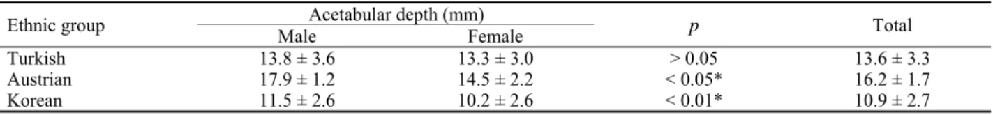
The acetabular index of depth to width evaluates the depth of the acetabulum. In comparison between normal and dysplastic hips with osteoarthritis, all normal hips were shown to have acetabular index values over 38°. Murrey 17 reported another method, which use acetabular depth to compensate for the inaccuracy of the Wiberg angle which was caused by formation of a bony spur of lateral margin of the acetabulum and displacement of the femoral head. It is considered as acetabular dysplasia if the acetabular depth is less than 9 mm. Regarding the AD we determined a signifi-cantly lower value of the Serbian female (11.2 ± 2.7°) than the male (12.5 ± 2.7°, p < 0.05). This agrees with the findings of Genser-Strobl et al. 18, who also found a significantly higher male value (17.98 ± 1.22°) than female (14.50 ± 2.21°)
Table 4
The published values of the acetabular depth (mm) by gender and ethnic group 9, 11, 18
Acetabular depth (mm) Ethnic group
Male Female p Total
Turkish 13.8 ± 3.6 13.3 ± 3.0 > 0.05 13.6 ± 3.3
Austrian 17.9 ± 1.2 14.5 ± 2.2 < 0.05* 16.2 ± 1.7
Korean 11.5 ± 2.6 10.2 ± 2.6 < 0.01* 10.9 ± 2.7
* significant difference
Table 5
The published values of the acetabular roof obliquity (°) by gender and ethnic group 11 12
Acetabular roof obliquity (°) Ethnic group
Male Female p Total
Japanese 4.6 ± 4.1 5.4 ± 4.5 < 0.05* 5.0 ± 4.3
Korean 5.2 ± 4.8 8.0 ± 5.9 < 0.01* 6.6 ± 5.6
Singaporean 7.8 ± 6.5 7.8 ± 6.8 > 0.05 7.8 ± 6.7
* significant difference
Table 6
The published values of the acetabular roof angle (°) by gender and ethnic group 11
Roof angle (°) Ethnic group
Male Female p Total
Japanese 20.9 ± 10.1 17.9 ± 10.2 < 0.01* 19.4 ± 10.2
Korean 18.6 ± 8.4 16.1 ± 9.4 < 0.01* 17.4 ± 9.0
(p < 0.05) in Austrian population. No significant differences were found in the AD in Turkish population related to gen-der 9, 19. The AD differed significantly by gender in Austrian and Korean population 11, 18.
Acetabular roof obliquity is used to evaluate the orien-tation of the acetabular roof in a coronal plane, and the supe-rior lateral coverage of the femoral head. Normal values are 10° and under, values above 10° are frequently found in acetabular dysplasia 5. Acetabular roof obliquity was normal if the angle was less than 30° under one year of age, if the angle was less than 25° between age 1 and 3, and if the angle was less than 20° from age 3 to adult 20. The mean ARO 7.6 ± 5.7° (both genders) did not differ from the value given by Han et al. 11. Furthermore, Serbian female ARO (9.0 ± 6.0°) differed significantly from the male value (6.2 ± 4.9°, p < 0.01). These values concur with the findings of Umer et al. 12 who reported a similar difference between female (7.78 ± 6.81°) and male (7.79 ± 6.46°) in Singaporean
population. The ARO differed significantly by gender in Japanese and Korean population 7, 8, 11.
Our male RA (19.6 ± 8.5°) is significantly higher (p < 0.01) than our female RA (17.1 ± 9.5°), corresponding to the findings of Nakamura et al. 21 who found a signifi-cantly higher male (20.9 ± 10.1°) than female RA value (17.9 ± 10.2°). The RA differed significantly by gender in Japanese and Korean population 7, 8, 11.
Conclusion
There are significant gender differences for all the ex-amined anatomical parameters of the acetabulum in Serbian population. There is also a clear tendency of female hips within the ethnic group to be more dysplastic than their male counterparts. We also found sex-related differences in ace-tabular morphology which were the cause for more dysplas-tic female acetabula compared with male.
R E F E R E N C E S
1. Mall G, Graw M, Gehring K, Hubig M. Determination of sex from femora. Forensic Sci Int 2000; 113(1–3): 315–21. 2. Lanyon P, Muir K, Doherty S, Doherty M. Age and sex differences
in hip joint space among asymptomatic subjects without structural change: implications for epidemiologic studies. Ar-thritis Rheum 2003; 48(4): 1041–6.
3. Lequesne M, Morvan G. Description of the potential of an ar-thrometer for standard and reduced radiographs suitable to measurement of angles and segments of hip, knee, foot and joint space widths. Joint Bone Spine 2002; 69(3): 282–92. 4. Cooperman DR, Wallensten R, Stulberg SD. Acetabular dysplasia
in the adult. Clin Orthop Relat Res 1983; 175: 79–85.
5. Delaunay S, Dussault RG, Kaplan PA, Alford BA. Radiographis measurements of dysplastic adult hips. Skeletal Radiol 1997; 26(2): 75–81.
6. Antoniades L, Spector TD, Macgregor AJ. The genetic contribution to hip joint morphometry and relationship to hip cartilage thickness. Osteoarthritis Cartilage 2001; 9(6): 593–5.
7. Fuji G, Funayama K, Benson M. Radiological measurement of the hip joint: comparison between Japanese and British. British and Japanese Orthopaedic Associations Combined Congress; 2000 Oct 3–6; London; 2000.
8. Inoue K, Wicart P, Kawasaki T, Huang J, Ushiyama T, Hukuda S, et al.. Prevalence of hip osteoarthritis and acetabular dysplasia in French and Japanese adults. Rheumatology 2000; 39(7): 745–8.
9. Goker B, Sancak A, Haznedaroglu S. Radiographic hip osteoar-thritis and acetabular dysplasia in Turkish men and women. Rheumatol Int 2005; 25(6): 419–22.
10.Jacobsen S, Sonne-Holm S, Søballe K, Gebuhr P, Lund B. Hip dys-plasia and osteoarthrosis: a survey of 4151 subjects from the Osteoarthrosis Substudy of the Copenhagen City Heart Study. Acta Orthop 2005; 76(2): 149–58.
11.Han CD, Yoo JH, Lee WS, Choe WS. Radiographic parameters of acetabular dysplasia in Korean adults. Yonsei Med J 1998; 39(5): 404–8.
12.Umer M, Thambyah A, Tan WTJ, Das De S. Acetabular mor-phometry for determining hip dysplasia in the Singaporean population. J Orthop Surg 2006; 14(1): 27–31.
13.Lavy CB, Msamati BC, Igbigbi PS. Racial and gender variations in adult hip morphology. Int Orthop 2003; 27(6): 331–3. 14.Msamati BC, Igbigbi PS, Lavy CB. Geometric measurements of
the acetabulum in adult Malawians: radiographic study. East Afr Med J 2003; 80(10): 546–9.
15.Jacobsen S, Sonne-Holm S. Hip dysplasia: a significant risk factor for the development of hip osteoarthritis. A cross-sectional survey. Rheumatology (Oxford) 2005; 44(2): 211–8.
16.Wiberg G. Studies on dysplastic acetabulae and congenital sub-luxation of the hip joint. Acta Orthop Scand (Suppl) 1939; 58: 1–132.
17.Murrey RO. The etiology of primary osteoarthritis of the hip. Br J Radiol 1965; 38(455): 810–24.
18.Genser-Strobl B, Sora MC. Potential of P40 plastination for morphometric hip measurements. Surg Radiol Anat 2005; 27(2): 147–51.
19.Aktas S, Pekindil G, Ercan S, Pekindil Y. Acetabular dysplasia in normal Turkish adults. Bull Hosp Jt Dis 2000; 59(3): 158–62. 20.Massie WK, Howorth MB. Congenital dislocation of the hip.
Part. I. Method of grading results. J Bone Joint Surg Am 1950; 32–A(3): 519–31.
21. Nakamura S, Ninomiya S, Nakamura T. Primary osteoarthritis of the hip joint in Japan. Clin Orthop Relat Res 1989; (241): 190–6.