.
Colleters in Chamaecrista (L.) Moench sect. Chamaecrista
and sect. Caliciopsis (Leguminosae-Caesalpinioideae):
anatomy and taxonomic implications
Marinalva dos Santos Silva1, Ítalo Antônio Cotta Coutinho1,2, Maicon Nascimento Araújo1
and Renata Maria Strozi Alves Meira1*
Received: September 12, 2016 Accepted: November 22, 2016
ABSTRACT
Th e genus Chamaecrista contains ca. 330 species organized into six sections, of which section Chamaecrista is the second largest (ca.75 species) distributed among six series, while the section Caliciopsis contains only two species. Colleters have been described in the genus Chamaecrista and they show potential taxonomic and phylogenetic signifi cance. Th ey are associated with lubrication, desiccation prevention and protection from microbial attacks of young developing organs. Although six types of colleters have been described for the genus Chamaecrista, there have been no studies focusing on the diversity of colleters in the sections Chamaecrista and Caliciopsis. Samples from developing leaves and fl owers of both sections were obtained from herbarium and fi eld collections and subjected to standard methodologies for both light and scanning electron microscopy. Histochemical tests were also performed to determine the nature of the exudates. Five types of non-vascularized colleters were found: short digitiform, long digitiform, club-shaped, pyriform and short bottle-shaped. Polysaccharides, pectins, lipids and proteins were detected in the exudates of all types of colleters. Among the fi ve types of colleters observed, pyriform is a novelty for Chamaecrista, reinforcing the signifi cant morphological diversity of these secretory structures in this genus.
Keywords: colleter, fl ower, histochemical analysis, leaf, polysaccharides, secretory structures, taxonomy
Introduction
Chamaecrista is one of the largest genera of the subfamily Caesalpinioideae, with ca. 330 species (Lewis 2005). According to Irwin & Barneby (1982), the genus is organized into six sections on the basis of the type of indumentum, the presence or absence of extrafl oral nectaries, the type of infl orescence and the type of venation of both sepals and leafl ets. Th e six sections (and the number of species) are: Chamaecrista sect. Absus (ca. 170 spp.), Apoucouita (ca.
20 spp.), Caliciopsis (two spp.), Chamaecrista (ca. 75 spp.), Grimaldia (one sp.)and Xerocalyx (three spp.).
Section Chamaecrista is the second most speciose section, with 2/3 of its species occurring in the America (Irwin & Barneby 1982; Rando & Pirani 2012; Rando et al. 2013). Species grouped in sect. Chamaecrista are characterized as shrubs or herbs with infl orescences with racemes reduced to either a few-fl owered fascicles or to a solitary fl ower, several pairs of pinnately-veined leafl ets, seeds obovate to rhombic or trapezoid in outline and extrafl oral nectaries commonly present (Irwin & Barneby 1982). Six series have
1Universidade Federal de Viçosa, Av. P.H. Rolfs, s/n, 36570-000, Viçosa, MG, Brazil
been described for sect. Chamaecrista: series Bauhinianae, Chamaecrista, Coriaceae, Flexuosae, Greggianae and Prostratae. Although described by Irwin & Barneby (1982) as a separate section, a phylogenetic analysis of the genus Chamaecrista, based on molecular data, has brought sect. Caliciopsis within sect. Chamaecrista (Conceição et al. 2009).
Different secretory structures with high potential for taxonomic and phylogenetic relevance have been described in Chamaecrista, including: nectaries (on leaves and/or racemes), mucilage idioblasts in the mesophyll and/or epidermis, sticky glandular hairs and colleters (Irwin & Barneby 1982; Coutinho et al. 2012; 2013; 2015; 2016; Meira et al. 2014; Francino et al. 2015). Colleters are secretory structures that are usually found on the adaxial side of vegetative and/or reproductive structures such as stipules, bracts, sepals and petals (Fahn 1979; Thomas 1991; Mayer et al. 2013; Coutinho et al. 2015). The viscous exudates secreted by colleters are said to be involved in lubrication, desiccation prevention and protection from microbial attacks of young and developing organs (Fahn 1979; Thomas 1991; Mayer et al. 2013; Coutinho et al. 2015). There are several types of colleters, which may originate from protodermal cells only or from all primary meristems (Lersten 1974; Thomas 1991; Appezzato-da-Glória & Estelita 2000; Rio et al. 2002; Silva et al. 2012; Coutinho et al. 2015). Therefore, to properly evaluate the presence of colleters in plants, their morphoanatomy, position and both time and composition of the secretion must be considered in order to avoid erroneous interpretations since such structures share morphoanatomical similarities with other secretory structures (Solereder 1908; Inamdar et al. 1986; Mohan & Inamdar 1986; Subramanian et al. 1989; Thomas 1991; Coutinho et al. 2015).
The presence and types of colleters are useful characters for taxonomic studies and have been reported for more than 60 families (Thomas 1991; Rio et al. 2005; Simões et al. 2006; Silva et al. 2012; Dalvi et al. 2013; Coutinho et al. 2015). In Chamaecrista, colleters have been described in vegetative and reproductive organs of species belonging to sections Absus, Apoucouita, Grimaldia, Chamaecrista and Xerocalyx (Coutinho et al. 2013; 2015). Six types of colleters were morphoanatomically described: short digitiform, long digitiform, short bottle-shaped, long bottle-shaped, club-shaped and racket-club-shaped (Coutinho et al. 2013; 2015). The type and distribution of colleters have provided new data that reinforce the sectional rearrangement indicated by the molecular phylogeny of Chamaecrista (Coutinho et al. 2015). The presence of club-shaped colleters on the margins of sepals was indicated as a synapomorphy for species of sect. Apoucouita. Most species of sect. Absus subsect. Absus had short bottle-shaped colleters, the same type as found in C. absus (sect. Grimaldia). Based on these data, the authors suggested that the sectional boundaries of sect. Grimaldia should be reconsidered in a future taxonomic revision of Chamaecrista to determine if this section should
be included within sect. Absus subsect. Absus. Although short bottle-shaped colleters were the most common type for sect. Chamaecrista, short digitiform and club-shaped types were also observed. However, only five species of sect. Chamaecrista were sampled by Coutinho et al. (2015), and none of the species of section Caliciopsis have been sampled in previous studies. This gap in knowledge has limited the taxonomic usefulness of colleters in sect. Chamaecrista and Caliciopsis.
In this paper, we assess the occurrence, distribution and types of colleters among species of Chamaecrista sect. Chamaecrista (including samples of 32 species not previously studied) and Caliciopsis (including samples of all species). The composition of the exudates of the colleters, as well as the taxonomic implications for the genus Chamaecrista, were also evaluated.
Materials and methods
We studied 37 species (50 taxa) of Chamaecrista (L.) Moenchsect. Chamaecrista, and two species of sect. Caliciopsis (Tab. 1). Taxonomic authorities for all taxa mentioned in this paper are given in S1 in supplementary material. Samples were obtained from field collections and herbarium material. Voucher specimens of field collections were deposited in the herbarium of the Universidade Federal de Viçosa (VIC) and Universidade do Estado da Bahia (HUNEB). Herbarium material was obtained from the collections of the following herbaria: Jardim Botânico do Rio de Janeiro (RB), Universidade Estadual de Feira de Santana (HUEFS), Universidade Federal de Viçosa (VIC), Universidade de São Paulo (SPF), and The New York Botanical Garden (NY).
Samples from herbaria were rehydrated (Smith & Smith 1942) and stored in 70 % ethanol. Samples from species that were collected in the field were fixed in FAA (formalin, acetic acid and 50 % ethanol; 1:1:18 by volume), NBF (neutral buffered formalin) (Johansen 1940) and FFS (formalin-ferrous sulphate) and stored in 70 % ethanol (Johansen 1940).
In order to assess the presence, position and micromorphology of the colleters we used a stereomicroscope (Zeiss Stemi 2000-C, Germany) and a scanning electron microscopy (SEM). For SEM, some samples stored in 70 % ethanol were subjected to critical-point drying using CO2 (CPD 030, Bal-Tec, Balzers, Liechtenstein), mounted on stubs and coated with gold (Modular Balzers Union FDU 010 with a SCA 010 sputter coating attachment, Germany) (Bozzola & Russel 1991). Examination and image capture were conducted using a LEO 1430VP SEM (Zeiss, Cambridge, United Kingdom) at the Centro de Microscopia e Microanálises at the Universidade Federal de Viçosa.
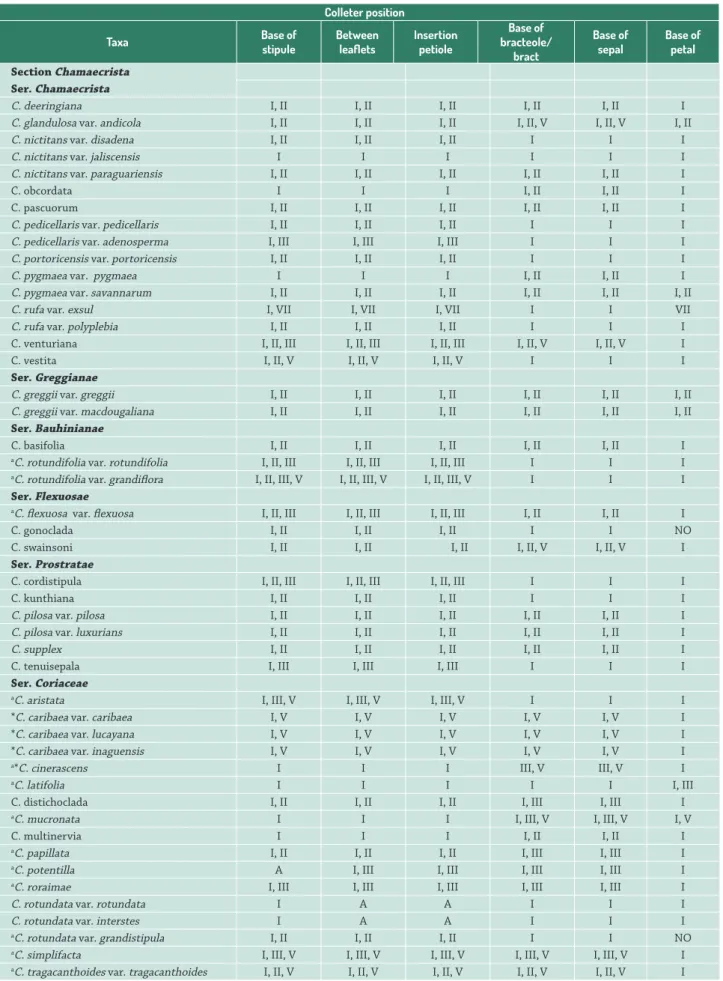
Table 1.Chamaecrista sections Chamaecrista and Caliciopsis species studied and position of the colleters.
Colleter position
Taxa Base of
stipule
Between leaflets
Insertion petiole
Base of bracteole/
bract
Base of sepal
Base of petal
Section Chamaecrista Ser.Chamaecrista
C. deeringiana I, II I, II I, II I, II I, II I
C. glandulosa var. andicola I, II I, II I, II I, II, V I, II, V I, II
C. nictitans var. disadena I, II I, II I, II I I I
C. nictitans var. jaliscensis I I I I I I
C. nictitans var. paraguariensis I, II I, II I, II I, II I, II I
C. obcordata I I I I, II I, II I
C. pascuorum I, II I, II I, II I, II I, II I
C. pedicellaris var. pedicellaris I, II I, II I, II I I I
C. pedicellaris var. adenosperma I, III I, III I, III I I I
C. portoricensis var. portoricensis I, II I, II I, II I I I
C. pygmaea var. pygmaea I I I I, II I, II I
C. pygmaea var. savannarum I, II I, II I, II I, II I, II I, II
C. rufa var. exsul I, VII I, VII I, VII I I VII
C. rufa var. polyplebia I, II I, II I, II I I I
C. venturiana I, II, III I, II, III I, II, III I, II, V I, II, V I
C. vestita I, II, V I, II, V I, II, V I I I
Ser. Greggianae
C. greggii var. greggii I, II I, II I, II I, II I, II I, II
C. greggii var. macdougaliana I, II I, II I, II I, II I, II I, II
Ser.Bauhinianae
C. basifolia I, II I, II I, II I, II I, II I
aC. rotundifolia var. rotundifolia I, II, III I, II, III I, II, III I I I
aC. rotundifolia var. grandiflora I, II, III, V I, II, III, V I, II, III, V I I I
Ser. Flexuosae
aC. flexuosa var. flexuosa I, II, III I, II, III I, II, III I, II I, II I
C. gonoclada I, II I, II I, II I I NO
C. swainsoni I, II I, II I, II I, II, V I, II, V I Ser. Prostratae
C. cordistipula I, II, III I, II, III I, II, III I I I
C. kunthiana I, II I, II I, II I I I
C. pilosa var. pilosa I, II I, II I, II I, II I, II I
C. pilosa var. luxurians I, II I, II I, II I, II I, II I
C. supplex I, II I, II I, II I, II I, II I
C. tenuisepala I, III I, III I, III I I I
Ser.Coriaceae
aC. aristata I, III, V I, III, V I, III, V I I I
*C. caribaea var. caribaea I, V I, V I, V I, V I, V I
*C. caribaea var. lucayana I, V I, V I, V I, V I, V I
*C. caribaea var. inaguensis I, V I, V I, V I, V I, V I
a*C. cinerascens I I I III, V III, V I
aC. latifolia I I I I I I, III
C. distichoclada I, II I, II I, II I, III I, III I
aC. mucronata I I I I, III, V I, III, V I, V
C. multinervia I I I I, II I, II I
aC. papillata I, II I, II I, II I, III I, III I
aC. potentilla A I, III I, III I, III I, III I
aC. roraimae I, III I, III I, III I, III I, III I
C. rotundata var. rotundata I A A I I I
C. rotundata var. interstes I A A I I I
aC. rotundata var. grandistipula I, II I, II I, II I I NO
(Historesin Leica; Leica Microsystems Nussloch, Heidelberg, Germany). Cross and longitudinal sections 5-7 μm thick were made with a rotary microtome (Spencer 820 American Optical Corporation, Buffalo, NY, USA). Sections were stained with toluidine blue at pH 4.4 (O’Brien & McCully 1981) and mounted in resin (Permount, Fisher Scientific, New Jersey, USA) for structural analysis.
Part of the fixed samples were dehydrated through tert -butanol series, embedded in histological paraffin enriched with dimethyl sulfoxide (Histosec®, Merck, Germany), cut into 7 μm thick cross and longitudinal sections (Spencer 820 American Optical Corporation, Buffalo, New York, USA), deparaffinized in xylene, rehydrated through an ethanol series (Johansen 1940) and used in histochemical tests.
The following histochemical tests were performed on 13 taxa using material previously fixed in the field with FAA or NBF (Tab. 1): for total lipids, sudan IV (Pearse 1980); for total polysaccharides, periodic acid-Schiff reagent (Maia 1979); for pectins/mucilage, ruthenium red (Johansen 1940); for acid mucopolysaccharides, alcian blue (Pearse 1980); and for total protein, xylidine Ponceau (Clark 1981). For detection of phenolic compounds, we used samples fixed in FFS (Johansen 1940). Control samples were also tested as required for each test. All observations and image captures were obtained using a light microscope (model AX70TRF; Olympus Optical, Tokyo, Japan) equipped with a U-Photo and digital camera (AxioCam HRc; Zeiss, Gottingen, Germany). The types of colleters found are described following Coutinho et al. (2015), as was the data for C. caribaea Britton and C. cinerascens (Vogel) H.S.Irwin & Barneby.
Results
Non-vascularized colleters composed of homogenous cells were found on both vegetative and reproductive organs (Tab. 1). Colleters were comprised of a stalk of varying length, and a secretory head of varying shape (Figs. 1, 2). The stalk cells were non-secretory and highly vacuolated while the cells from secretory head displayed densely stained cytoplasm (Fig. 2C-E). There was no differentiation of a
palisade epidermis since the epidermal cells are similar in appearance to the internal cells of the secretory head (Fig. 2H). The cuticle was thin and appeared distended or loose in some sections (Fig. 2B) or SEM images (Fig. 1I); no pores were observed in the cuticle. Older colleters exhibit a brownish color.
Colleters were located on the adaxial side of vegetative and reproductive structures including the base of stipules, bracts and bracteoles; the base of sepals and petals; on the rachis between pairs of leaflets; and on the stem at the insertion of the petiole (Tab. 1). Five types of colleters were observed (Tab. 1): Type I, short digitiform (Figs. 1A-B, D, G-H, 2A, F); Type II, long digitiform (Figs. 1G, 2G); Type III, club-shaped (Figs. 1F, 2E); type V, short bottle-shaped (Figs. 1C, 2D); and Type VII, pyriform (Figs. 1E, 2C). Pyriform colleters, described here for the first time, were about 230 μm long, and composed of a stalk that widened at the mid-height and had a short pointy apex (Figs. 1E, 2C).
Although colleters were found in all the species analyzed, their distribution among the series of section Chamaecrista was very distinct (Tab. 1). Type I occurred in all analyzed species and the three taxa of ser. Coriaceae (C. ulmea, C. rotundata var. rotundata, C. rotundata var. interstes); C. nictitans var. jaliscensis (ser. Chamaecrista) only possessed this type of colleter. Type VII was exclusive to C. rufa var. exsul. Types I and II were observed in 20 taxa, with 10 of them belonging to ser. Chamaecrista. Types I and III occurred in five taxa (three of ser. Coriaceae, C. tenuisepala and C. pedicelaris var. adenosperma). Five taxa from ser. Coriaceae (sect. Chamaecrista), as well as C. calycioides and C. duckeana from sect. Caliciopsis bore Types I and V. Colleters of the types I, II and III were found in five taxa of four different series (C. rotundifolia var. rotundifolia, C. flexuosa var. flexuosa, C. cordistipula, C. distichoclada and C. papillata). C. vestita, C. glandulosa var. andicola (ser. Chamaecrista), C.swainsoni (ser. Flexuosae) and C. tragacanthoides var. tragacanthoides (ser. Coriaceae) displayed colleters of types I, II and V. Types I, III and V occurred in four species of ser. Coriaceae (C. aristata, C. cinerascens, C. mucronata and C. simplifacta). C. venturiana (ser. Chamaecrista) and C. rotundifolia var. grandiflora (ser. Bauhinianae) displayed Types I, II, III and V (Tab. 1).
C. tragacanthoides var. rasa I, V I, V I, V I, V I, V I
C. ulmea I I I I I I
C. venulosa I, V I, V I, V I I V
Section Caliciopsis
C. calycioides var. calycioides I, V NO I, V I I I
C. duckeana I, V NO I, V I I I
Note: A: absent; NO: not observed. I: short digitiform; II: long digitiform; III: club-shaped; V: short bottle shaped and VII: pyriform.
aField collected material.
*Analized by Coutinho et al. 2015.
Colleter position
Taxa Base of
stipule
Between leaflets
Insertion petiole
Base of bracteole/
bract
Base of sepal
Base of petal
Figure 1.SEM images showing colleters on vegetative and reproductive organs of Chamaecrista sect. Chamaecrista. A. C. basifolia: Colletershort digitiform on stipule. B. C. gonoclada: Colleter bifurcated and short digitiform on stipule. C. C. tragacanthoides var. tragacanthoides: Colleter bottle shaped on sepal. D. C. nictitans var. paraguayensis: Colleter short digitiform on bracteole. Note the secretion (arrow). E. C. rufa var. exsul: Colleter pyriform on petal. F. C. potentilla: Colleter club shaped on sepal. G. C. rotundata var. grandistipula: Colleter short and long digitiform on stipule. H,I. C. tragacanthoides var. tragacanthoides: Colleter short digitiform on stipule and apex detail colleter, respectively. Scale bars: A, I: 100μm; B-F: 60μm; G: 200μm; H: 20μm.
Only in ser. Chamaecrista were all five types of colleters observed (Figs. 1C-I, 2A-H). The series Bauhinianae, Coriaceae and Flexuosae displayedTypes I, II, III and V (Fig. 1B), while
petiole and between leaflets than in stipules. The types of colleters varied among sepals, petals, bracteoles and bracts. Type I was present on petals of all species (Tab. 1) except in C. venulosa and C. rufa var. exsul, which only displayed
Types V and VII, respectively.
Figure 3. Histochemical tests in colleters of Chamaecrista sect. Chamaecrista. A-B. Totals polysaccharides. A. C. rotundifolia var. grandiflora: colleter short bottle shaped on stipule. B. C. rotundifolia var. rotundifolia: colleter club-shaped on stipule. C. Acid mucopolysaccharides, C. simplifacta: colleter short bottle shaped on bracteole. D.Pectins, C. potentilla: colleter club-shaped on sepal.E. Totals proteins, C. aristata: colleter bifurcated on stipule. F, G.Totals lipids, C. aristata and C. mucronata: colleter short digitiform on stipule and sepal, respectively. Scale bars: A, C, E-G: 50μm; B: 100μm; D: 40μm.
in all series studied (Tab. 1). Type VII was observed at the base of stipules and petals, between leaflets and on the stem at the insertion of the petiole of C. rufa var. exsul (ser. Chamaecrista). Bifurcated colleters were presented in seven taxa (Tab. 1) and were not considered a different type of colleter. Chamaecrista rotundata var. rotundata, C. rotundata var. interstes,C. ulmea and C. nictitans var. jaliscensis possessed only Type I colleters, while the remaining taxa
bore at least two types. Only two taxa (C. rotundata var. grandistipula and C. gonoclada)did not display colleters on petals (Tab. 1).
The presence of total polysaccharides (Fig. 3A, B), mucopolysaccharides (Fig. 3C), pectins (Fig. 3D), proteins (Fig. 3E) and total lipids (Fig. 3F, G) was confirmed by the histochemical tests for all types of colleters described. Of all the histochemical tests performed, only the secretory head of the colleters showed positive results.
Discussion
According to the position, morphology, anatomy and the composition of the exudates, the secretory structures present on the base of stipules, between leaflets, at the petiole insertion, and among sepals, petals and bracts/ bracteoles of the studied species of Chamaecrista sect. Chamaecrista and Caliciopsis are indeed colleters. Such
structures are typically related to the protection of young leaves and flowers from desiccation and attack from microorganisms since the secretion present is made of mucilage or a mixture of mucilage, resin and protein (Thomas & Dave 1989; Thomas 1991; Rocha et al. 2009).
may indicate that they play an important functional role in keeping young structures hydrated (Fahn & Cutler 1992). Lipids have been detected in the colleter secretion of species belong Apocynaceae (Appezzato-da-Glória & Estelita 2000), Leguminosae (Paiva 2009), Gentianaceae (Dalvi et al. 2013), Orchidaceae (Mayer et al. 2011) and Rubiaceae (Machado et al. 2012; Tullii et al. 2013). As claimed for polysaccharides, lipid compounds may help avoid water loss, in addition to prohibiting fungal and microorganism attack, since this hydrophobic substance lubricates the surface of young leaves and floral buds, (Fahn 1979; Thomas & Dave 1989; Thomas 1991; Evert 2006; Paiva 2009; Mayer et al. 2013). Proteins have also been reported as a component of the secretion produced by colleters from a variety of different species (Thomas & Dave 1990; Klein et al. 2004; Miguel et al. 2006; González & Tarragó 2009; Mayer et al. 2011; Dalvi et al. 2013). Some authors have suggested that such proteins may provide additional protection from fungi and parasites (Miguel et al. 2006; Vieira et al. 2006; Mayer et al. 2011). However, research aiming to unravel how exudates produced by colleters may contribute to the success of species of Chamaecrista in stressful environments is still needed.
Although cuticular pores were not observed among the colleters of the studied species, a few samples had loosely packed secretory cells at the colleter apex and a distended cuticle. This arrangement of cells and cuticle is an indication that the secretion may be released to the outside by cuticle rupture. Releasing of secretion by cuticle rupture has been suggested for colleters of Hymenaea stigonocarpa (Paiva & Machado 2006b) and demonstrated in colleters of Senna macranthera (Souza 2014), both belonging to Caesalpinioideae, as well as other families (Paiva & Machado 2006a; Tullii et al. 2013).
As observed in the species of the sect. Chamaecrista and Caliciopsis studied, non-vascularized colleters have bee previously reported for other species of Chamaecrista belonging to other sections (De-Paula & Oliveira 2007; Coutinho et al. 2013; 2015), other species of Caesalpinioideae (Paiva & Machado 2006b; Paiva 2009; Souza 2014), and even species of other families (Paiva & Machado 2006a; Paiva 2009; Vitarelli & Santos 2009; Silva et al. 2012; Dalvi et al. 2013). Anatomically, the colleters described for the species of sect. Chamaecrista and Caliciopsis studied exhibit the same homogeneous pattern of cells as reported for other Chamaecrista species (Coutinho et al. 2015).
Although Lersten (1974) pointed out that the standard type of colleter is the most widespread, variation in the morphoanatomy of these structures has since been observed such that now several different types of colleters are recognized (Mayer et al. 2011; Silva et al. 2012; Coutinho et al. 2015). Standard type colleters are comprised of a secretory palisade epidermis covering a non-secretory central axis, which may or may not be vascularized with xylem and phloem (Lersten 1974). In contrast to this
standard type of colleter, which has been described for other botanical families (Appezzato-da-Glória & Estelita 2000; Paiva & Machado 2006a; Vitarelli & Santos 2009), the most common type of colleter for the genus Chamaecrista, as shown by our data along with data provided by Coutinho et al. (2015), are comprised of homogenous cells.
As the colleters of the studied species of Chamaecrista age, they exhibit a brownish color and usually fall off when they stop secreting. Similar observations were reported for other species of Chamaecrista (Coutinho et al. 2015). This change in color may be the result of the oxidation of phenolic compounds accumulated within the cells, which is followed by the shrinking of the cytoplasm of the apical cells of colleters (Souza 2014). Similar descriptions were reported for colleters from a variety of botanical families (Thomas 1991; Paiva 2009; Souza 2014).
Colleters were observed in all of the analyzed taxa from both sect. Chamaecrista and Caliciopsis. Within subfamily Caesalpinioideae, the presence of colleters stands out when we take species from subtribe Cassinae (tribe Cassieae) into account, since five species of Senna (Souza 2014) and 55 species of Chamaecrista (De-Paula & Oliveira 2007; Coutinho et al. 2015) display these secretory structures. Bifurcated colleters, like those observed in seven of the taxa studied here, were also reported for Temnadenia violacea (Martins et al. 2010), Prestonia coalita (Rio 2001), Forsteronia (Rio 2006), Mandevilla pycnantha and M. tenuifolia (Simões et al. 2006), all species that belong to Apocynaceae, as well as in Curtia and Hockinia (Dalvi et al. 2013), species of Gentianaceae. We suspect that this bifurcation is the result of abnormalities during colleter development, and so do not correspond to a distinct type of colleter.
Although six distinct types of colleters have been described for species of Chamaecrista (Coutinho et al. 2015), only five species of ser. Coriaceae (sect. Chamaecrista) were sampled and nothing about sect. Caliciopsis was provided. The same authors reported short digitiform (I), club-shaped (III) and short bottle shaped (V) colleters for sect. Chamaecrista. Our more comprehensive sample of sect. Chamaecrista enabled us to observe two additional types of colleters for this section: long digitiform and pyriform. New information on the occurrence of colleters at the insertion of the petiole of C. aristata, C. caribaea, C. cinerascens, C. potentilla and C. simplifacta, which was overlooked by previous authors, is also provided here. The pyriform (VII) type of colleter is a novelty for Chamaecrista, and is exclusive to C. rufa var. exsul (ser. Chamaecrista), while racket-shaped (IV) and long bottle-shaped (VI) colleters were not detected in the sections studied.
are independent of the size of the bearing structure. Short digitiform (I) and short bottle shaped (V) colleters have been observed only in C. tragacanthoides var. rasa, C. venulosa, the three varieties of C. caribaea, species that belong to ser. Coriaceae, and two taxa of sect. Caliciopsis. Considering that the infrageneric relationships of Chamaecrista remain unclear, colleter type is emerging as a relevant and promising character for understanding the relationships among the species of Chamaecrista. According to both molecular and morphological data, Chamaecrista ser. Coriaceae was recognized as a monophyletic group, excluding C. caribaea, C. roraimae, and C. venulosa (Rando et al. 2016). The diversity of types and different positions reported for colleters in Chamaecrista, both by us and other authors, emphasizes how important and poorly understood this structure is. Such data promise to be useful for both taxonomic and phylogenetic studies, as has been the case for other taxa (Lersten 1975; Curtis & Lersten 1980; Thomas 1991; González 1998; Simões et al. 2006; González & Tarragó 2009; Sheue et al. 2012; Silva et al. 2012; Vitarelli et al. 2015; Fernandes et al. 2016). However, further studies are necessary to thoroughly evaluate the evolutionary history of colleters in Chamaecrista.
We showed that colleters are diverse structures in Chamaecrista sect. Chamaecrista and Caliciopsis. The short digitiform and short bottle-shaped colleters found in the two species of sect. Caliciopsis were also observed in two (C. caribaea and C. venulosa) of the three species that should be excluded from ser. Coriaceae (sect. Chamaecrista), as suggested by a recent molecular phylogenetic study. Such similar characters may suggest species of sect. Caliciopsis are closer to species of ser. Coriaceae, therefore, the position of the such species of ser. Coriaceae should be revised. However, only a comprehensive study of species of sect. Chamaecrista, including representatives of sect. Caliciopsis, will be able to confirm such a hypothesis. In addition to the types of colleters already described for sect. Chamaecrista, we added two new types of colleters (long digitiform and pyriform). Besides, the pyriform type is a novelty for the genus Chamaecrista. Our results are promising as they may be useful to future analyses combining phylogenetic and evolutionary approaches, and encourage further research on other members of Caesalpinioideae.
Acknowledgements
The authors thank CNPq, CAPES and FAPEMIG for financial support. We also thank CNPq for providing a research scholarship to R. M. S. A. Meira and CAPES for providing a PhD scholarship to M. S. Silva. We are grateful to the herbaria HUEFS, SPF, NY, RB, and VIC for kindly allowing the sampling of their voucher specimens. We thank the Plant Anatomy Laboratory and the Microscopy and Microanalysis Center of UFV and Patrícia Fonseca and Aurora Dias for technical assistance.
References
Appezzato-da-Glória B, Estelita MEM. 2000. Development, structure and distribution of colleters in Mandevilla illustris and M. velutina
(Apocynaceae). Revista Brasileira de Botânica23: 113-120. Bozzola JJ, Russel LD. 1991. Electron microscopy:principles and techniques
for biologists. New York, Jones and Bartlett Publishers.
Clark G. 1981. Staining procedures. 4th. edn. Baltimore, Willians & Wilkins. Conceição AS, Queiroz LP, Lewis GP, et al. 2009. Phylogeny of Chamaecrista
Moench (Leguminosae-Caesalpinioideae) based on nuclear and chloroplast DNA regions. Taxon 58: 1168-1180.
Coutinho IAC, Francino DMT, Azevedo AA, Meira RMSA. 2012. Anatomy of the extrafloral nectaries in species of Chamaecrista section Absus
subsection Baseophyllum (Leguminosae, Caesalpinioideae). Flora 207: 427-435.
Coutinho IAC, Francino DMT, Meira RMSA. 2013. Leaf anatomical studies of Chamaecrista subsect. Baseophyllum (Leguminosae, Caesalpinioideae): new evidence for the up-ranking of the varieties to the species level.Plant Systematics and Evolution 299: 1709-1720. Coutinho IAC, Francino DMT, Meira RMSA. 2015. New records of colleters
in Chamaecrista (Leguminosae, Caesalpinioideae S.L.): structural
diversity, secretion, functional role, and taxonomic importance. International Journal of Plant Sciences 176: 72-85.
Coutinho IAC, Rando JG, Conceição AS, Meira RMSA. 2016. A study of the morphoanatomical characters of the leaves of Chamaecrista
(L.) Moench sect. Apoucouita (Leguminosae-Caesalpinioideae). Acta Botanica Brasilica 30: 205-221.
Curtis JD, RN Lersten. 1980. Morphology and anatomy of resin glands in
Salix lucida (Salicaceae). American Journal of Botany 67: 1289-1296. Dalvi VC, Meira RMSA, Francino DMT, Silva LC, Azevedo AA. 2013.
Anatomical characteristics as taxonomic tools for the species of Curtia
and Hockinia (Saccifolieae-Gentianaceae Juss.). Plant Systematics and Evolution300: 99-112.
De-Paula OC, Oliveira DMT. 2007. Ocorrência de coléteres em embriões de três espécies de Chamaecrista Moench (Fabaceae: Caesalpinioideae). Revista Brasileira deBiociências 5: 348-350.
Evert RF. 2006. Esau’s plant anatomy: meristems, cells and tissues of the plant body: their structure function and development. 3nd. edn. New Jersey, John Wiley and Sons.
Fahn A. 1979. Secretory tissues in plants. London, Academic Press. Fahn A, Cutler DF. 1992. Xerophytes. Berlin, Gebrüder Bomtraeger. Fernandes VF, Thadeu M, Dalvi VC, Marquete R, Meira RMSA. 2016.
Colleters in Casearia (Salicaceae): a new interpretation for the theoid teeth. Botanical Journal of the Linnean Society181: 682-691. Francino DMT, Coutinho IAC, Dalvi VC, Azevedo AA, Conceição AS,
Meira RMSA. 2015. Anatomical interpretations of the taxonomy of
Chamaecrista section Absus (Leguminosae - Caesalpinioideae s.l.). Plant Systematics and Evolution 301: 2087-2103.
González AM. 1998. Colleters in Turnera and Piriqueta (Turneraceae). BotanicalJournal of the Linnean Society 128: 215-228.
González AM, Tarragó JR. 2009. Anatomical structure and secretion compounds of colleters in nine Ilex species (Aquifoliaceae) from southern South America. BotanicalJournal of the Linnean Society 160: 197-210.
Inamdar JA, Subramanian RB, Mohan JSS. 1986. Studies on resin glands of
Azadirachta indica A. Juss. (Meliaceae). Annals of Botany 58: 425-429. Irwin HS, Barneby RC. 1982. The American Cassiinae, a synoptical revision
of Leguminosae tribe Cassieae subtribe Cassiinae in the New World. Memoirs of the New York Botanical Garden35: 1-918.
Johansen DA. 1940. Plant microtechnique. New York, McGraw-Hill. Klein DS, VM Gomes, SJ Silva-Neto, M Cunha. 2004. The structure of
colleters in several species of Simira (Rubiaceae). Annals of Botany 94: 733-740.
Lersten NR. 1974. Morphology and distribution of colleters and crystals in relation to the taxonomy and bacterial leaf nodule symbiosis of
Psychotria (Rubiaceae). American Journal of Botany 61: 973-981.
Lewis GP. 2005. Tribe Cassieae. In: Lewis GP, Schrire B, Mackinder B, Lock M. (eds.) Legumes of the World.Kew, Royal Botanic Gardens. p. 111-161.
Machado SR, Barreiro DP, Rocha JF, Rodrigues TM. 2012. Dendroid colleters on vegetative and reproductive apices in Alibertia sessilis (Rubiaceae) differ in ultrastructure and secretion. Flora 207: 868-877. Maia V. 1979. Técnica histológica. São Paulo, Atheneu.
Martins FM, Kinoshita L, Castro MM. 2010. Coléteres foliares e calicinais
de Temnadenia violacea (Vell.) Miers (Apocynaceae-Apocinoideae):
estrutura e distribuição. Revista Brasileira de Botânica 33: 519-530. Mayer JLS, Cardoso-Gustavson P, Appezzato-da-Glória B. 2011. Colleters
in monocots: New record for Orchidaceae. Flora 206: 185-190. Mayer JLS, Carmello-Guerreiro SM, Mazzafera P. 2013. A functional role
for the colleters of coffee flowers. Annals of Botany 5: 1-13. Meira RMSA, Francino DMT, Ascensão L. 2014. Oleoresin trichomes of
Chamaecrista dentata (Leguminosae): structure, function, and secretory products. International Journal of Plant Sciences 175: 336-345. Miguel EC, Gomes VM, Oliveira MA, Cunha M. 2006. Colleters in Bathysa
nicholsonii K. Schum. (Rubiaceae): Ultrastructure, secretion protein composition, and antifungal activity. Plant Biology 8: 715-722. Mohan JSS, Inamdar JR. 1986. Ultrastructure and secretion of extrafloral
nectaries of Plumeria rubra L. Annals of Botany 57: 389-401. O`Brien TPE, Mccully ME. 1981. The study of plant structure principles
and selectmethods.Melbourne, Termarcarphi Pty.
Paiva EAS. 2009. Occurrence, structure and functional aspects of the colleters of Copaifera langsdorffii Desf. (Fabaceae, Caesalpinioideae). Comptes Rendus Biologies 332: 1078-1084.
Paiva EAS, Machado SR. 2006a. Colleters in Caryocar brasiliense
(Caryocaraceae): ontogenesis, ultrastructure and secretion. Brazilian Journal of Biology 66: 301-308.
Paiva EAS, Machado SR. 2006b. Ontogenesis, structure and ultrastructure
of Hymenaea stigonocarpa (Fabaceae: Caesalpinioideae) colleters.
Revista de Biologia Tropical 54: 943-950.
Pearse AGE. 1980. Histochemistry theoretical and applied.Vol 2. Edinburgh, Churchill Livingston.
Rando JG, Loeuille B, Pirani JR. 2013. Taxonomic novelties in Chamaecrista
(Leguminosae: Caesalpinioideae) from Brazil. Phytotaxa97: 17-25. Rando JG, Pirani JR. 2012. A new species of Chamaecrista sect. Chamaecrista
ser. Flexuosae (Leguminosae, Caesalpinioideae) from Serra do Cipó, Minas Gerais, Brazil.. Brittonia 64: 241-245.
Rando JG, Zuntini AR, Conceição AS, Berg C, Pirani JR, Queiroz LP. 2016. Phylogeny of Chamaecrista ser. Coriaceae (Leguminosae) unveils a lineage recently diversified in Brazilian Campo Rupestre vegetation. International Journal of Plant Sciences 177: 3-17.
Rio MCS. 2001. Estudos taxonômicos e anatômicos do gênero Prestonia
R.BR. nom. cons. (Apocynaceae). PhD Thesis, Universidade Estadual de Campinas, Brazil.
Rio MCS. 2006. Estudos anatômicos em espécies de Forsteronia G.Mey (Apocynaceae) de cerrado. PhD Thesis, Universidade Estadual de Campinas, Brazil.
Rio MCS, Castro MM, Kinoshita LS. 2002. Distribuição e caracterização anatômica dos coléteres foliares de Prestonia coalita (Vell.) Woodson (Apocynaceae). Revista Brasileira de Botânica 25: 339-349. Rio MCS, Kinoshita LS, Castro MM. 2005. Anatomia foliar como subsídio
para a taxonomia de espécies de Forstenia G. Mey (Apocynaceae) dos cerrados paulistas. Revista Brasileira de Botânica 28: 713-726. Rocha DI, Silva LC, Valente VMM, Francino DMT, Meira RMSA. 2009.
Morphoanatomy and development of leaf secretory structures in
Passiflora amethystina Mikan (Passifloraceae). Australian Journal of Botany 57: 619-626.
Sheue CR, Chen YJ, Yang YP. 2012. Stipules and colleters of the mangrove Rhizophoraceae: morphology, structure and comparative significance. Botanical Studies 53: 243-254.
Silva CJ, Barbosa LCA, Marques AE, Baracat-Pereira MC, Pinheiro AL, Meira RMSA. 2012. Anatomical characterization of the foliar colleters in Myrtoideae (Myrtaceae). Australian Journal of Botany 60: 707-717. Simões AO, Castro MM, Kinoshita LS. 2006. Calycine colleters of seven species of Apocynaceae (Apocynoideae) from Brazil. Botanical Journal of the Linnean Society 152: 387-398.
Smith FH, Smith EC. 1942. Anatomy of the inferior ovary of Darbya. American Journalof Botany 29: 464-471.
Solereder H. 1908. Systematic Anatomy of the Dicotyledons. Vol 2.Oxford, Clarendon Press.
Souza LA. 2014. Estruturas secretoras em espécies de leguminosas da subtribo Cassiinae (Fabaceae, Caesalpinioideae, Cassieae). PhD Thesis, Universidade Federal de Minas Gerais, Brazil.
Subramanian RB, Murugan V, Mohan JS, Inamdar JA. 1989. Optical microscopic studies on the structure and secretions of resin glands in some Apocynaceae. PlantSciences 99: 423-429.
Thomas V. 1991. Structural, functional and phylogenetic aspects of the colleter. Annalsof Botany 68: 287-305.
Thomas V, Dave Y. 1989. Histochemistry and senescence of colleters of
Allamanda cathartica (Apocynaceae). Annals of Botany 64: 201-203.
Thomas V, Dave Y. 1990. Structure and necrosis of stipular colleters in
Mitragyna parvifolia (Rubiaceae). Belgian Journal of Botany 123: 67-72. Tullii CF, Miguel EC, Lima NB, Fernandes KVS, Gomes VM, Cunha M.
2013. Characterization of stipular colleters of Alseis pickelii. Botany 91: 403-413.
Vieira FA, Cunha M, Klein DE, Carvalho AO, Gomes VM. 2006. Purification and characterization of beta-1, 3-glucanase from the secretion of
Simira glaziovii colleters (Rubiaceae). Brazilian Archives of Biology and Technology 49: 881-888.
Vitarelli NC, Riina R, Caruzo MBR, Cordeiro I, Fuertes-Aguilar J, Meira RMSA. 2015. Foliar secretory structures in Crotoneae (Euphorbiaceae): Diversity, anatomy, and evolutionary significance. American Journal of Botany102: 833-847.
Vitarelli NC, Santos M. 2009. Anatomia de estípulas e coléteres de