Invo lve m e nt o f the caudal raphe
nucle i in the fe e ding be havio r o f rats
Departamento de Anatomia, Instituto de Ciências Biomédicas, Universidade de São Paulo, São Paulo, SP, Brasil
L.F. Takase, J.R. Barone and M.I. Nogueira
Abstract
Involvement of the caudal raphe nuclei (raphe pallidus, RPa; raphe magnus, RMg, and raphe obscurus, ROb) in feeding behavior of adult rats was studied by measuring c-Fos protein expression, in animals submitted to the meal-feeding model of food restriction in which the rats were fed ad libitum only from 7:00 to 9:00 h, for 15 days. The experimental groups submitted to chronic fasting, named search for food (SF), ingestion of food (IF) and satiety of food (SaF) were scheduled after a previous study in which the body weight and the general and feeding behaviors were evaluated by daily monitoring. Acute, 48-h fasting (AF) was used as control. In the chronic group, the animals presented a 16% reduction in body weight in the first week, followed by a continuous, slow rise in weight over the subsequent days. Entrainment of the sleep-wake cycle to the schedule of food presentation was also observed. The RPa was the most Fos immunopositive nucleus in the chronic fasting group, followed by the RMg. The ANOVA and Tukey test (P<0.05) confirmed these results. The IF group was significantly different from the other three groups, as also was the number of labeled cells in the RPa in SF and IF groups. Nevertheless, no significant difference was observed between RMg and RPa, or RMg and ROb in the SaF and AF. However, it is interesting to observe that the groups in which the animals were more active, searching for or ingesting food, presented a larger number of labeled cells. These results suggest a different involvement of the caudal raphe nuclei in the somatic and autonomic events of feeding behavior, corroborating the functions reported for them earlier. Co rre spo nde nce
M.I. Nogueira
Laboratório de Neurociências Departamento de Anatomia, ICB, USP Av. Prof. Lineu Prestes, 2415 05508-900 São Paulo, SP Brasil
Fax: + 55-11-818-7366 E-mail: minog@ usp.br
Presented at the XIV Annual Meeting of the Federação de Sociedades de Biologia Experimental, Caxambu, MG, Brasil, August 25-28, 1999. Research supported by FAPESP (Nos. 96-0340-3, 98-2901-3 and 98-2901-0).
Received April 20, 1999 Accepted November 29, 1999
Ke y wo rds
·Feeding behavior ·Caudal raphe nuclei ·c-Fos protein expression ·Meal-feeding
Feeding behavior is a complex group of functions that comprise the search, capture and ingestion of the organic and inorganic matter necessary for the sustenance of life. This behavior consists of many different com-ponents, and different brain systems are in-volved in the regulation of its specific as-pects. As a biological rhythm, feeding be-havior is organized mainly in the diencepha-lon by the lateral and medial hypothalamus subdivisions and the paraventricular nucleus (1-3). The lateral hypothalamus seems to
walls, the vegetative components of feeding behavior (4). The exact role of these afferent signals is currently being questioned (5). The dorsomedial subdivision has been asso-ciated to gastrointestinal motility (6) and modulation of the availability of glucose and free fatty acids (3). However, rather than as specific centers, these structures must be understood as part of a neural circuit in-volved in these functions. In fact, while feed-ing may be initiated or terminated by fore-brain areas, other structures modulate this behavior at the level of the brainstem and spinal cord. Among such structures in the brainstem are the caudal raphe nuclei, namely the raphe magnus (RMg), pallidus (RPa) and obscurus (ROb) nuclei. This view is cor-roborated by the following anatomical and physiological data: the RMg has been re-lated mainly to analgesia, but together with the RPa it also seems to be involved in the regulation of respiratory frequency and body temperature (7,8). The RPa projects to the facial, hypoglossus, ambiguous and trigemi-nal motor nuclei (9,10). Both the ROb and the RPa nuclei constitute the main serotoner-gic afferent sources to the trigeminal motor nucleus (11). Some RPa neurons, probably serotonergic, tend to increase their discharge pattern during licking or grooming activities (12,13). Thyrotropin hormone releasing fac-tor administered in this nucleus promotes gastrointestinal activity in fed rats through the dorsal vagal complex (6).
The caudal raphe nuclei are located from the ventral level of the pons, anterior to the facial nerve, bregma -9.16 mm, to the pyra-midal decussation, bregma -14.30 mm, in the medulla oblongata. The RMg and the ROb nuclei lie dorsal to the RPa; thus the rostral and caudal limits of the RPa are inter-mingled, respectively, with those of the cau-dal ventral part of the RMg and with almost the whole ventral portion of the ROb. Some functional misunderstanding can be expected regarding their physical limits and the inter-pretation of results. These nuclei belong to
the brainstem reticular formation, being formed by sparse neuronal clusters crossed by fibers of passage (7). These anatomical features associated with the minute dimen-sions of the medium lateral axes of the RPa and the ROb hinder their study by means of exogenous neuronal tracers. Nevertheless, currently available immunohistochemical techniques for the detection of c-Fos protein expression have been employed to function-ally map neuroanatomical systems in re-sponse to specific and acute exogenous stimuli. The c-Fos protein, acting as an en-dogenous label, avoids the usual shortcom-ings of exogenous tracers such as possible uptake by fibers of passage and contamina-tion of adjacent areas, permitting the dis-crimination of different neuronal subsets (14). We performed experiments using c-Fos expression methods and the restriction food model, known as meal-feeding that limits feeding to a fixed period of the day to further characterize the involvement of the caudal raphe nuclei in feeding behavior (15). This model of chronic fasting uniformizes ing conditions compared to spontaneous feed-ing, and appears to constitute a suitable tool to explore the strategies employed by an organism to cope with its homeostatic re-quirements when submitted to a starvation schedule. Although these metabolic mech-anisms (16,17) and the role of the different diencephalic structures in feeding behavior have been well investigated, very little infor-mation is available concerning the role of the three raphe nuclei.
Adult male albino Wistar rats (Rattus rattus) housed under constantly controlled
environmental conditions (room tempera-ture, 23 ± 1oC, lights on from 7:00 to 19:00
h) were used. Pelleted food and water were available ad libitum. The animals were fed
To choose the experimental parameters, a preliminary study was carried out in which the alterations in body weight, the amount of food consumed, the duration of ingestion, and general behavior were monitored daily. These data formed the basis for the designa-tion of three chronic fasting groups: search for food (SF), in which food was presented to the animals, but these were prevented from reaching it; ingestion of food (IF), in which the animals were allowed to eat for 30 min, and the satiety of food (SaF) group, in which the animals ate for 2 h. These stimuli were presented on the 16th day of the meal feeding schedule. A control group of acute fasting (AF) rats, consisting of animals on a normal feeding schedule, was deprived of food for 48 h. Five animals were used in each group. A period of 90 min after the stimulus was always employed to allow peak c-Fos expression. The animals were then deeply anesthetized with chloral hydrate and trans-cardially perfused with saline and a fixative solution (4% paraformaldehyde in 4% borax and 2% acrolein, pH 9.5, at 4oC).
The central nervous system was removed, cryoprotected, frozen-sectioned as coronal sections of 40-µm thickness, and immuno-histochemically prepared using a c-Fos anti-serum (rabbit polyclonal Ab-5, 1:1,000; On-cogene Science, Inc., La Jolla, CA, USA). Incubations with the c-Fos antiserum and a biotinylated secondary antiserum (Calbio-chem-Novabiochem Co., San Diego, CA, USA) were carried out at room temperature. Sections were then incubated with the avi-din-biotin-peroxidase complex (ABC Elite Kit, Vector Labs, Burlingame, CA, USA) and reacted with diaminobenzide, a,ß d-glucose, nickel ammonium sulfate and glu-cose oxidase. The sections were then mounted on gelatin-coated slides and the intensity of immunostain was intensified with thiocar-bohydrazide and 4% osmium tetroxide. A Nissl-stained adjacent series was used as a reference, as was the atlas of Paxinos and Watson (18). The slides were analyzed
un-der bright field microscopy and c-Fos immu-nopositive neurons were detected using a VIA-170 video image maker-measurement system from Boeckeler Instruments, Inc. (Tucson, AZ, USA).
Daily observation of the animals behav-ior throughout the food-offering period showed that during the first days, when placed in the cage, the rats ate continuously after a short period of exploratory activity for about 45 min, drinking little water. They then slept or explored the environment. During this phase, they lost about 16% of body weight. After the 8th day, the rats appeared to be more adapted to the model, eating immedi-ately, more slowly and for a longer time, although most food was consumed within the first 30 min. A slow and continuous recovery of body weight was then observed. Identification of the three experimental phases of chronic feeding behavior was based on these data. The paradoxical increase in body weight despite food restriction reported earlier (16,17) illustrates the adaptation of the animals to the model during the selected period. In addition, confirming this adapta-tion, the cyclic presentation of food for a fixed period of time caused entrainment (1,19)of the circadian cycle, rendering these nocturnal animals awake and very active, expecting food, while the others in the vi-varium were sleeping.The animals in the AF group were very active and drank much more water than the other groups.
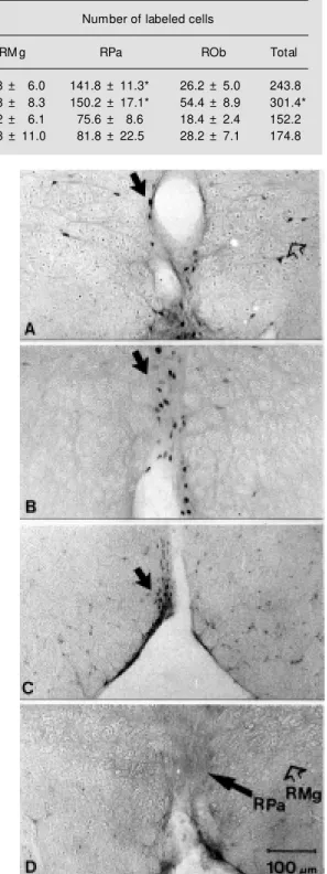
followed by AF and SaF. The RPa nucleus presented more than 50% of c-Fos-immuno-positive cells than RMg in the IF and SF groups and much more than ROb in all ex-perimental groups. ANOVA P<0.001 and the Tukey test (P<0.05) confirmed these ob-servations. The IF group was significantly different from the other three groups, as also was the RPa in the first two groups. Never-theless, there was no significant difference at this level between RMg and RPa, or RMg and ROb in the SaF and AF. However, it is interesting to observe that the groups in which the animals were more active, searching for or ingesting food, presented the highest num-ber of labeled cells. These results are illus-trated in coronal sections at bregma -10.96 in the RPa (Figure 1 A-D).
Labeling of many motor, sensory and autonomic structures related to the four phases of feeding was observed in each group, which validates the model adopted as suit-able to distinguish the differential involve-ment of the raphe nuclei in feeding behavior. The labeling observed in the following re-gions in relation to the stimuli applied sup-ports these data: in group SF, the lateral hypothalamus was densely labeled as were structures related to the olfactory system (such as the magnocellular pre-optic nuclei, the horizontal nucleus of the band of Broca, the accessory nucleus of the olfactory tract, the bed nucleus of the stria terminallis and the central anterior amygdaloid nucleus; 20); in the IF group, in addition to the prosen-cephalic regions, there was also dense label-ing in structures related to the maintenance or pattern generation like the trigeminal mo-tor and sensory complex, the dorsal vagal nucleus and nucleus of the tractus solitarius; the SaF group presented dense labeling in the dorsomedial rather than the ventrome-dial hypothalamus as reported earlier (2,10). To better understand the neuronal relations between these other labeled areas and the raphe nuclei and feeding behavior, these data are currently being analyzed in depth in Table 1 - Number of c-Fos-immunopositive cells in the raphe nuclei, magnus (RM g),
pallidus (RPa) and obscurus (ROb), in each experimental group of chronic fasting (search for food (SF), ingestion of food (IF) and satiety of food (SaF)) and of acute fasting (AF).
M ean ± SEM , N = 5. ANOVA (SF, IF, SaF and AF), F(3,16) = 8.7, P<0.001. * Tukey test (SF, IF, SaF and AF), P<0.05.
Experimental group Number of labeled cells
RM g RPa ROb Total
SF - search for food 75.8 ± 6.0 141.8 ± 11.3* 26.2 ± 5.0 243.8
IF - ingestion of food 96.8 ± 8.3 150.2 ± 17.1* 54.4 ± 8.9 301.4*
SaF - satiety of food 58.2 ± 6.1 75.6 ± 8.6 18.4 ± 2.4 152.2
AF - acute fasting 64.8 ± 11.0 81.8 ± 22.5 28.2 ± 7.1 174.8
relation to the stimulus group and will be reported soon in a full paper.
However, while some aspects require more detailed study, the present data are sufficient to conclude that the three raphe nuclei are involved differentially in the phases of feeding behavior. This conclusion derives support from electrophysiological studies in freely moving cats with implanted electrodes, which exhibit serotonergic neuron discharge related to masticatory muscle activity (13,21). Two subsets of serotonergic neurons are activated during spontaneous feeding, one by the onset of feeding that is deactivated by satiety, and another that is gradually acti-vated by onset and gradually deactiacti-vated by satiety (21). Experiments from our labora-tory on rats submitted to a normal feeding schedule and in slow-wave sleep, housed under the same conditions as those of the present experiments, revealed almost no c-Fos-immunopositive cells, thus corroborat-ing the differential involvement of the raphe nuclei in the stimuli presented to the four groups.
Comparing the present results with those reported earlier, the more intense labeling of
the RMg in the SF and IF groups appears to reflect a rise in body temperature or a change in respiratory pattern due to somatic and autonomic movements in the groups pre-senting a more intense motor activity (6,8). The more intense labeling of the RPa seems to emphasize its reported role as a pre-motor nucleus in somatic and autonomic motor events, particularly regarding mastication, swallowing and gastrointestinal contractility as well as gastric secretion through the vagal complex (6,10,13). The pattern of labeled cells in the ROb agrees with a role in gas-trointestinal activity (6) in IF and perhaps with a hemodynamic control (22) in SF or AF, due to a greater amount of water drunk by the SF or AF, probably to compensate for food deprivation.
Ackno wle dgm e nts
The authors thank Professor Jackson C. Bittencourt for laboratory facilities, Ms. Alessandra Pellegrini for collaboration with animal handling, and Mr. Wilson Roberto Campos de Azevedo for photographic assis-tance.
Re fe re nce s
1. Leal AM O & M oreira AC (1997). Food and the circadian activity of the hypothalamic-pituitary-adrenal axis. Brazilian Journal of
M edical and Biological Research, 30:
1391-1405.
2. Leibow itz SF & Stanley BG (1986). Brain peptides and the control of eating behav-ior. In: M oody TW (Editor), Neural and
Endocrine Peptides and Receptors.
Ple-num Press, New York, 333-352. 3. Zaia CTBV, Gaziri LCJ, Zaia DAM , Dellatre
E, Dolnikoff M S & Timo-Iaria C (1997). Effect of chemical stimulation of dorso-medial hypothalamic nucleus on blood plasma glucose, triglycerides and free fatty acids in rats. Brain Research Bulle-tin, 42: 195-198.
4. Timo-Iaria C (1990). Glucoreceptor sys-tems: from control of glycemia to feeding behavior. New s in Physiological Sciences, 5: 46-49.
5. Baldw in BA, Parrott RF & Ebenezer IS (1998). Food for thought: A critique on the hypothesis that endogenous cholecysto-kinin acts as a physiological satiety factor.
Progress in Neurobiology, 55: 477-507.
6. Garrick T, Prince M , Yang H, Ohning G & Taché Y (1994). Raphe pallidus stimula-tion increases gastric contractility via TRH projections to the dorsal vagal complex in rats. Brain Research, 636: 343-347. 7. Tork I & Hornung J-P (1990). Raphe nuclei
and serotoninergic system. In: Paxinos G (Editor), The Human Nervous System. Ac-ademic Press, Sydney, 1001-1102. 8. Brück K & Hinckel P (1980).
Thermoregu-latory noradrenergic and serotonergic pathw ays to hypothalamic units. Journal
of Physiology, 304: 193-202.
9. Holstege G (1997). Some anatomical ob-servations on the projections to the hypo-thalamus to brainstem and spinal cord: an
HRP and autoradiographic tracing study in the cat. Journal of Comparative Neurol-ogy, 260: 98-126.
10. Holstege G, Blok BFM & Horst GJ (1995). Brain stem systems involved in blink re-flex, feeding mechanisms, and micturi-tion. In: Paxinos G (Editor), TheRat
Ner-vous System. Academic Press, Sydney,
257-271.
11. Fort P, Luppi P-H, Sakai K, Salvert D & Jouvet M (1990). Nuclei of origin of monoaminergic, peptidergic and cholin-ergic afferents to the cat trigeminal motor nucleus. A double-labeling study w ith cholera-toxin retrograde tracer. Journal of
Comparative Neurology, 301: 262-275.
13. Ribeiro-do-Vale LE (1997). Serotonergic neurons in the caudal raphe nuclei dis-charge in association w ith activity of mas-ticatory m uscles. Brazilian Journal of
M edical and Biological Research, 30:
79-83.
14. Hoffman GE, Smith M S & Verbalis JG (1993). c-fos and related immediate early gene products as markers of activity in neuroendocrine system s. Frontiers in
Neuroendocrinology,14: 173-213.
15. Leveille GA (1966). Glycogen metabolism in meal-fed rats and chicks and the time sequence of lipogenic and enzymatic a-daptive changes. Journal of Nutrition,90: 449-460.
16. Curi R & Shinomiya H (1985). M etabolic
changes of tw enty w eeks food-restriction schedule in rats. Physiology and Behav-ior,36: 239-243.
17. Curi R, Shinomiya H, Bazotte RB & Timo-Iaria C (1984). M etabolic performance of free fed rats subjected to prolonged fast as compared to the metabolic pattern in rats under long term food restriction.
Physiology and Behavior,33: 525-531.
18. Paxinos G & Watson C (1986). The Rat
Brain in Stereotaxic Coordinates. 2nd edn.
Academic Press, San Diego.
19. Holmes M C, French KL & Seckl JR (1997). Dysregulation of diurnal rhythms of sero-tonin 5-HT2C and corticosteroid receptor gene expression in the hippocampus w ith food restriction and glucocorticoids.
Jour-nal of Neuroscience, 17: 4056-4065.
20. Shipley M T, M cLean JH & Ennis M (1995). Olfactory system. In: Paxinos G (Editor),
The Rat Nervous Syst em. Academ ic
Press, Sydney, 899-921.
21. Veasey SC, Fornal CA, M etzler CW & Jacobs BL (1995). Response of seroto-nergic caudal raphe neurons in relation to specific motor activities in freely moving cats. Journal of Neuroscience, 15: 5346-5359.