Master Thesis
The influence of menopausal status on bone turnover and
disease control in breast cancer patients with metastatic
bone disease treated with chemotherapy plus zoledronic
acid: an exploratory retrospective cohort study
Universidade Nova de Lisboa, Faculdade de Ciências Médicas
Northeastern University, College of Professional Studies
Arlindo Rebelo Ferreira
Medical Oncology ResidentMentor: Luís Costa, MD, PhD
Page 2 of 38
Abstract
Background: A recent meta-analysis showed that the adjuvant use of zoledronic acid (ZA) in postmenopausal women with early breast cancer (BC) leads to a reduction in the risk of breast cancer death by 17%. We investigated the effect of the hormonal status (pre [PrM] vs late post menopause [PoM]) on bone turnover and disease control among women with BC and bone metastases (BM) treated with ZA and chemotherapy (CT).
Methods: In this retrospective cohort study, we collected clinicopathologic variables, urinary N-terminal telopeptide (NTX) and serum tumor marker levels from women with BC and BM treated with CT and ZA. Patients were divided in PrM (<45 years) and PoM (>60 years). Study endpoints were NTX, CA15.3 and CEA variation at 3, 6 and 9 months, and time to first-line CT failure and survival. We performed multilevel mixed-effects linear regression models to assess the variation of repeated measures and cox regression models for time to event outcomes.
Results: Forty patients were eligible for analysis (8 PrM and 32 PoM).
After introduction of ZA and CT, NTX and tumor markers declined in the overall cohort. Response profile was similar between menopausal groups at month 3 and at later time points (p-value for time-hormonal status interaction at month 3=0.957). Furthermore, tumor markers response profile was also equal between groups.
Median time to first-line CT failure in PrM and PoM women was 15.2 and 17.4 months, respectively. No significant difference between groups was found, either using a univariate analysis or after controlling for visceral disease involvement (p=0.399 and 0.469, respectively). Likewise, no differences in survival were found.
Conclusions: In this cohort, no differences were found in terms of NTX or tumor markers control according to menopausal status. Similarly, no difference in time to first-line CT failure or survival was found.
Page 3 of 38
Resumo
Introdução: Uma meta-análise recente demonstrou que uso adjuvante de ácido zoledrónico (AZ) em mulheres pós-menopáusicas com cancro da mama precoce (CM) conduz a redução do risco de morte por CM em 17%. Investigámos o efeito do estado hormonal (pré [PrM] vs pós-menopausa tardia [PoM]) na remodelação óssea e controlo de doença em mulheres com CM e metástases ósseas (MO) tratadas com AZ e quimioterapia (QT).
Métodos: Neste estudo de coorte retrospetivo, colhemos variáveis clinico-patológicas e quantificámos o telopéptido N-terminal (NTX) urinário e marcadores tumorais (MT) séricos em mulheres com CM e MO tratadas com QT e AZ. As doentes foram divididas em PrM (<45 anos) e PoM (>60 anos). Endpoints do estudo: variação do NTX, CA15.3 e CEA nos meses 3, 6 e 9, tempo até falência de QT de primeira-linha e sobrevivência. Quando apropriado foram usados os testes de Wilcoxon rank-sum, modelo de efeitos lineares mistos, teste log-rank e modelo de Cox.
Resultados: Quarenta doentes foram elegíveis para análise (8 PrM e 32 PoM).
Depois da introdução de AZ e QT, os níveis de NTX e MT caíram no coorte global. O perfil de resposta não diferiu entre grupos no mês 3 ou em tempos posteriores (valor-p para interação tempo-estado hormonal no mês 3=0.957). Ademais, o perfil de resposta dos MT também não diferiu entre grupos.
O tempo mediano até falência de primeira-linha de QT em PrM e PoM foi de 15.2 e 17.4 meses, respetivamente. Não foi identificada diferença significativa entre grupos, quer em análise univariada quer após controlo para envolvimento visceral (p=0.399 e 0.469, respetivamente). Igualmente, não houve diferenças em termos de sobrevivência.
Conclusões: Neste coorte, não foram identificadas diferenças no controlo de NTX ou MT em função do estado menopausico. Igualmente, não foi identificada diferença no tempo até falência de primeira-linha de CT ou sobrevivência.
Page 4 of 38
List of figures and tables
Figures
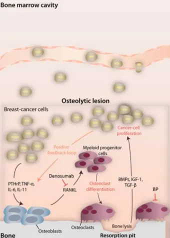
Figure 1 - Interactions between bone and cancer cells in predominantly osteolytic breast cancer lesions. Sites of action of denosumab and bisphosphonates are also noted. Bone metabolism with resorption and formation occurs. The depicted mediators emphasize the predominant pathways. Figure 2 – Study diagram.
Figure 3 – Mean (A), median (B) and mean log10 (C) variation of urine NTX at several time points
after diagnosis in the full cohort.
Figure 4 – Mean NTX at several time points after diagnosis according to menopausal status. Test of differences in mean values is shown.
Figure 5 – Mean response profile of log10(NTX) at several time points after introduction of zoledronic
acid plus chemotherapy according to menopausal status. Test of interaction time - menopausal group is shown.
Figure 6 – Mean response profile of CA15.3 and CEA at several time points after diagnosis in the full cohort (A and B, respectively) and according to menopausal status (C and D, respectively).
Figure 7 – Mean response profile of log10(CA15.3) (A) and log10(CEA) (B) at several time points after
introduction of zoledronic acid plus chemotherapy according to menopausal status. Test of interaction time - menopausal group is shown.
Figure 8 – Time to first-line chemotherapy failure according to menopausal status. Adjusted HR controlling for visceral involvement is shown.
Figure 9 – Overall survival according to menopausal status. Adjusted HR controlling for HER2 status is shown.
Tables
Table 1 – Types of bone remodeling markers. Table 2 – Bone modifying agents.
Table 3 – Cohort baseline characteristics according to menopausal status. Table 4 – Urine NTX at several time points from diagnosis.
Table 5 – Univariate association of baseline characteristics with time to first line chemotherapy failure.
Page 5 of 38
Table of contents
Introduction ……… 6
Background and significance ……….. 6
Study Objectives ………. 14
Methods ……… 15
Results ……… 19
Discussion ……… 28
Conclusion ……….. 31
Acknowledgments ………. 32
List of abbreviations ………. 33
Page 6 of 38
Introduction
This document constitutes the final thesis of the Double Master in Clinical Research from NOVA Medical School (NOVA University) and Northeastern University.
This document is an original scientific dissertation (dissertação de natureza científica original) as per
Regulamento n.º 680/2010.
This project was presented at the “Spring Meetings 2014” (Évora, Portugal) in poster format and was awarded a prize for the second best poster presented in the meeting.
Background and significance
a) Breast cancer epidemiology
Breast cancer (BC) is the most frequently diagnosed and the most common cause of women cancer related death in Europe with an estimated incidence and death rate of 108.8 and 22.4 cases per 100 000 women per year, respectively.[1] More specifically, the estimated incidence and death rate in Portugal is of 85.6 and 18.4 cases per 100 000 women per year, respectively.[1] Furthermore, breast cancer is also the most common cause of cancer related death in women both in developed and developing regions.[2]
In Europe and in the USA, the majority of patients currently diagnosed with BC present with early or locally advanced disease in terms of stages of disease progression, allowing the use of therapies with curative intent.[3] However, 5-10% of breast cancers are diagnosed with metastatic disease. Furthermore, from those treated with curative intent, around 40% will develop disease recurrence.[4] While locoregional recurrence might be eligible for curative interventions, those with distant relapse are almost universally incurable.
b) Bone metastases and skeletal related events
In patients with metastatic breast cancer bone is the most common involved site, either as de novo
disease or after disease relapse. While the 5-years incidence of bone metastases in patients treated with curative intent is around 5%, 70% of those with metastatic disease have bone involvement at some time during disease evolution.[5] Liver, lung and less frequently the brain are also typical target organs of breast cancer metastatic spread.
Page 7 of 38
cancer cell survival in the bone marrow microenvironment. This process appears to be accomplished by facilitating CXCL12-CXCR4-AKT signaling and by conferring tumor cells resistance to TNF-related apoptosis-inducing ligand (TRAIL).[6] Bone metastasis are also more common in patients whose tumors express the estrogen receptor (ER).[7] As a matter of fact, Src is active in most estrogen receptor positive and in a minority of estrogen receptor negative tumors.[6] Furthermore, Src and the ER seem to interact.[8] Despite this interaction, Src expression signature is more strongly associated with bone disease than the ER per se, an association that seems independent in nature from the ER. In line with this, even when restricting to patients with ER-negative tumors, Src expression is still highly associated with bone metastatic development, a fact that underlines the relevance of Src expression as, at least, a marker of activation of relevant pathways leading to a successful adaptation of cancer cells to bone environment.[6]
Breast cancer cells further elicit a cascade of events that lead to a profound change in bone metabolism. Bone is under continuous remodeling, a process accomplished by the coupled activity of osteoblasts (bone forming cells) and osteoclasts (bone resorbing cells). Cancer cells reshape this equilibrium to activate osteoblasts. This process is at least partially accomplished through the production of parathyroid hormone-related peptide (PTHrp), tumor necrosis factor α (TNF-α), interleukin 1 (IL-1), IL-6, IL-8 and IL-11 by cancer cells.[9] Activated osteoblasts release activator of nuclear factor κ B ligand (RANKL) that ultimately activates osteoclasts; hence, it induces bone resorption, which leads to the release of growth factors entrapped in the bone matrix (transforming growth factor-β (TGF- β), bone morphogenetic proteins (BMPs), insulin like growth factor (IGF) and fibroblast growth factor).[9, 10] This last step is the closing gap in the loop, as cancer cells will benefit from these growth stimulation factors, therefore leading to more bone turnover activation: a loop referred as the vicious cycle of bone metastases.[11]
Bone remodeling modifications ultimately conduct to increased bone fragility[12] and development of skeletal related events (SRE).
Page 8 of 38
Skeletal related events include a series of adverse outcomes derived from the presence of bone metastases, namely excessive pain requiring intervention, as radiotherapy or surgery, bone fracture, myeloid compression and hypercalcemia. The interaction between cancer and bone cells can induce the development of several types of lesions: lytic (mostly bone degrading), blastic (mostly bone forming) or mixed (lesions presenting a mixture of lytic and blastic features). Either of these lesions represent a profound change in bone architecture and therefore of bone resistance to mechanical stress. In patients without therapy aiming to control bone metabolism, a rate of approximately 4 SREs per year is expected, significantly impairing patients’ quality of life and ultimately, survival.[13, 14]
c) Markers of bone remodeling
Bone remodeling involves degradation of current bone matrix and formation of new bone matrix; in fact, both biochemical processes lead to the release of several biochemical by-products amenable of being quantified in, e.g., blood or urine.[15] NTX (amino-terminal crosslinked telopeptide of collagen type I) is a by-product of collagen degradation and one of the most commonly used markers of bone metabolism, in specific of the bone resorbing arm. On the other hand, ALP (alkaline phosphatase) is a by-product of osteoblast activity; hence, a marker of bone formation.[15] Other types of bone remodeling markers are listed in table 1.
Page 9 of 38
Table 1 – Types of bone remodeling markers.
Bone formation markers
Bone specific alkaline phosphatase (BALP) Osteocalcin (OC)
Propeptides of type I procollagen (P1NP and P1CP)
Bone resorption markers
Amino-terminal crosslinked telopeptide of collagen type I (NTX) Calcium
Carboxy-terminal crosslinked telopeptide of type I collagen (ICTP or CTX-MMP)
Carboxy-terminal crosslinked telopeptide of collagen type I (CTX)
Deoxypyridoline (DPD)
Hydroxyproline (Hyp), Hydroxylysine (Hyl)
Non collagenous bone matrix proteins: bone sialoprotein (BSP) and osteopontin (OP)
Osteoclast-derived enzymes: tartarate resistant acid phosphatase 5b (TRAP5b) and cathepsin k and L
Pyridinoline (PYD)
Parathyroid hormone related peptide (PTH-rp) Vitamin D
Regulators of bone turnover
Dickkopf-1 (DKK-1) Osteoprotegerin (OPG)
Receptor activator of NFƙB ligand (RANKL) Sclerostin
d) Bone modifying agents
Page 10 of 38
Table 2 – Bone modifying agents. NA – not applicable.
Pharmacologic Category Agents Relative potency
Bisphosphonates
Non nitrogenous-bisphosphonates
Etidronate Clodronate Tiludronate
1 10 10
Nitrogenous-bisphosphonates
Pamidronate Alendronate Ibandronate Risedronate Zoledronate
100 500 1000 2000 10000
Monoclonal antibodies Denosumab NA
BP are a group of pyrophosphate analogous with an intense tropism to the bone where, after incorporation into the matrix and subsequent absorption by osteoclasts (during the process of bone resorption), they act as potent osteoclast inhibitors (figure 1). There are two classes of BP, non-nitrogenous and non-nitrogenous. While the first act by substituting the terminal pyrophosphate moiety of adenosine triphosphate (ATP), resulting in a nonfunctional ATP molecule that competes for functional ATP in the osteoclast energy metabolism, the latter inhibit the osteoclast enzyme farnesyl diphosphate synthase (FDS). FDS is a critical isoprenyl diphosphate synthase or prenyl transferase in the mevalonate pathway that is fundamental for the synthesis of sterol isoprenoids, such as cholesterol, and non-sterol isoprenoids, such as dolichol, heme-A, isopentenyl tRNA and ubiquinone.[26] The inhibition of isoprenoid – farnesyl diphosphate and geranylgeranyl diphosphate synthesis by the inhibition of FDS prevents the post-translational prenylation of small GTPase signaling proteins such as Rho, Rac, Rab, Rap and Ras leading to disruption of osteoclast function and induction of apoptosis.[26]
On the other hand, the fully humanized monoclonal antibody denosumab acts by sequestering RANKL (figure 1). This process limits the natural action of RANKL on the osteoclast precursor surface receptor RANK, preventing osteoclast differentiation and ultimately bone resorption.
Page 11 of 38
e) Anti-tumoral action of bisphosphonates
BP are critical tools to prevent and delay SREs; however, pre-clinical and clinical studies are increasingly supporting a direct and indirect tumoral action of these agents. Some of the anti-tumor mechanisms of action of BP include a direct cytotoxic action, but also other indirect mechanisms, such as anti-angiogenesis and immune system activation.[30] While the preclinical identification of this effects seems clear, the clinical translation of these findings is less clear, and, so far, seems to be restricted to hormone-sensitive patients under an estrogen depleted environment (either due to post-menopausal status or due to ovarian suppression). Furthermore, the effect is more clear in nitrogenous-bisphosphonates, in specific for ZA.[31] Of note, more than 80% of the women with BC are post-menopausal, which opens a wide window for eventual intervention/applicability.[3] Nevertheless, at present, available clinical practice guidelines do not recommend the use of BP as a therapeutic tool in the adjuvant treatment of breast cancer.[25, 29, 32]
In the last decade, a group of trials documented relevant pieces of information regarding the anti-cancer activity of BPs in the adjuvant treatment of breast anti-cancer. The ABCSG-12 was a phase III trial that tested the adjuvant use of ZA (4 mg every 6 month for 3 years) in 1803 pre-menopausal women under ovarian suppression and tamoxifen or anastrazol.[33, 34] After a median follow-up of 8 years, those patients receiving ZA had a 33% decrease in the risk of recurrence (Hazard-ratio [HR] for disease-free survival [DFS] 0.77, 95% Confidence Interval [CI] 0.60 – 0.99; p=0.042) and a trend towards a decrease in the risk of death (HR for overall survival [OS] 0.66, 95% CI 0.43-1.02; p=0.064). Noteworthy, this effect was clearer in patients older than 40 years of age (HR for DFS 0.70, 95% CI 0.51 – 0.96 vs. 0.95, 95% CI 0.62 – 1.46 in women with less than 40 years of age; HR for OS 0.56, 95% CI 0.31 – 0.96 vs. 0.94, 95% CI 0.45 – 1.98 in women with less than 40 years of age).
The ZO-FAST and Z-FAST trials also tested the effect of BPs in terms of cancer related outcomes. Despite theirs design to test as central outcomes bone health related outcomes (as variation in bone mineral density and the occurrence of bone fracture), they also tested for cancer related outcomes as secondary outcomes (disease recurrence). In ZO-FAST (1065 women) and Z-FAST (602 women) postmenopausal women receiving adjuvant letrozole were randomized for upfront zoledronic acid (4 mg every 6 months) or zoledronic acid only after bone fracture or significant decline in bone mineral density.[35, 36] Despite de concordant results between trials in terms of improved bone mineral density for patients receiving upfront ZA, only those patients enrolled in the ZO-FAST derived a benefit in terms of disease recurrence (HR for DFS 0.66, 95% CI 0.44 – 0.97).[35, 36]
Page 12 of 38
significant benefit: 25% reduction in the risk of invasive disease or death (adjusted-HR 0.75, 95% CI 0.59-0.96) and 26% reduction in the risk of death (adjusted-HR 0.74, 95% CI 0.55-0.98).
As referred before, most of the available clinical evidence of the anti-tumor action of BPs is derived from trials that used zoledronic acid; however, other BPs were also tested. The NSABP B-34 tested the role of clodronate both as a strategy to test the action of this BP in terms of bone health related outcomes (as bone mineral density), but also in terms of disease recurrence.[38] In this study, 3323 women (pre and post-menopausal) were randomized to receive standard adjuvant therapy with or without oral clodronate (1600 mg/d for a total of 2 years). After a median follow-up of around 7 years, only those 50 years of age or older had a benefit in terms of DFS (bone and extra-bone). However, no clear improvement in terms of OS was documented for both age groups. Pamidronate was also tested as a strategy to reduce the risk of disease recurrence in breast cancer.[39, 40] In a small trial Kokufu and colleagues recruited 90 patients with node positive (≥4 positive nodes) early breast cancer treated with curative intention to receive pamidronate (n=33; 45 mg pamidronate 4 times every 2 weeks) or standard follow-up (n=57) based on personal preference.[39] After a median follow-up of five years, a reduction in the incidence of bone metastases in the group receiving pamidronate was found (12.1% vs. 40.4% in the control group; p=0.005). A non-significant numerical difference favoring pamidronate was also found in terms of distant metastases (36.4 vs. 56.1%; p=0.071) and non-osseous metastases (33.3 vs. 52.6%, p=0.077). Nevertheless, no difference regarding overall survival or disease-free survival was found. Another study testing a similar hypothesis recruited 429 perimenopausal stage I-III breast cancer patients receiving therapy with curative intent.[40] From these, 258 received pamidronate (15mg iv every 4 weeks and oral pamidronate 100mg daily during adjuvant chemotherapy) while 171 didn’t. Even though patients receiving pamidronate had a lower incidence of bone metastases (2.3% vs. 8.7% in the control group; time of endpoint evaluation not specified) no difference was found in terms of disease-free survival or overall survival.
Finally, a recent meta-analysis analyzed individual patient data from several randomized trials comparing de use of BP to no BP (either placebo-controlled or open control) in terms of the risk of recurrence and death.[31] In this study (preliminary data after analysis of 75% of 23,573 patients of interest), a statistically significant reduction of 17% in the risk of death and distant recurrence was found for post-menopausal women treated with BP only. Furthermore, a reduction of 35% in the risk of bone-specific recurrence was shown, an effect also restricted to the group of post-menopausal women. As a matter of fact, pre-post-menopausal women seem not to benefit from BPs in terms of the risk of disease recurrence and death.
Page 13 of 38
f) Framing the research question
Metastatic breast cancer survival has improved over time, mostly after the introduction of most effective therapies, as taxanes, aromatase inhibitors and anti-HER agents. Other relevant gains were achieved with more rational use of already available drugs tailored to certain risk specific groups. BPs, and most specifically ZA, seem to have antitumor properties which depend on the systemic hormonal environment. In fact, only late post-menopausal women (or premenopausal under ovarian suppression therapy) seem to benefit from zoledronic acid in terms of disease recurrence and survival. However, the available data are only derived from patients in the early breast cancer setting, and there is no consistent evidence that BPs improve survival in metastatic breast cancer patients nor was the effect of the hormonal environment tested in this circumstance.
Page 14 of 38
Study Objectives
To test whether the hormonal environment (pre vs. late post-menopausal) significantly impacts bone disease control, as measured by urinary levels of NTX, serum levels of tumor markers, time to first-line of chemotherapy failure and overall survival in patients with breast cancer affecting the bone and being treated with zoledronic acid and chemotherapy.
a) Primary objective: To determine the impact of the hormonal environment (pre vs. post-menopausal) on urinary levels of NTX at 3 months in patients receiving zoledronic acid and chemotherapy;
Hypothesis: Post-menopausal patients will present a more pronounced decline in the urinary NTX levels at 3 months when compared to pre-menopausal women.
b) Secondary objectives: To determine the impact of the hormonal environment (pre vs. post-menopausal) in patients receiving zoledronic acid and chemotherapy on the following parameters:
Urinary NTX levels at 6 and 9 months;
Hypothesis: Post-menopausal patients will present an increased decline in the urinary levels of NTX at 6 and 9 months when compared to pre-menopausal women.
Serum CA15.3 and CEA levels (tumor markers);
Hypothesis: Post-menopausal patients will present an increased decline in the serum levels of CA15.3 and CEA when compared to pre-menopausal women.
Time to first line chemotherapy failure;
Hypothesis: Post-menopausal patients will present a longer time to first line chemotherapy failure when compared to pre-menopausal women.
Overall survival;
Page 15 of 38
Methods
a) Study population
Patients with the diagnosis of metastatic breast cancer involving the bone (de novo or post-disease recurrence), receiving zoledronic acid and chemotherapy, diagnosed and treated at the Department of Medical Oncology from Hospital de Santa Maria – CHLN from 2000 and 2009 and with quantifications of urinary NTX available.
Evidence of breast cancer was obtained from the histopathology report, while evidence of bone involvement was obtained from imaging.
We will exclude patients with previous diagnosis of other oncologic disease, except contralateral breast cancer that is disease-free for more than 5 years, non-melanoma skin cancer or pre-invasive cervix cancer. We will also exclude patients with a follow-up of less than 3 months, with previous treatment for metastatic disease, previous treatment with BPs and pregnant women or women with psychiatric conditions.
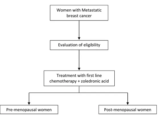
b) Study design and diagram
This study is a single center retrospective cohort study.
Figure 2 – Study diagram.
Women with Metastatic breast cancer
Evaluation of eligibility
Treatment with first line chemotherapy + zoledronic acid
Page 16 of 38
c) Data collected and study procedures
To collect the clinical variables of interest we directly extracted clinical information from medical records, both physical and electronic.
The collected variables were the following:
Demographics: age at diagnosis, race and menopausal status;
Disease and tumor characterization: date of diagnosis, WHO performance status, AJCC staging (T, N, number of involved lymph nodes and M), hormone receptor status, HER2 status, grade, histology, previous (neo)adjuvant chemotherapy regimen, (neo)adjuvant trastuzumab, (neo)adjuvant chemotherapy, date of disease relapse, metastatic disease characterization (date of metastatic diagnosis, number and localization of lesions);
Biomarkers and imaging: NTX, CEA, CA15.3, CT (thorax/abdomen/pelvis), scintigraphy,
simple radiography of the bone.
Other endpoints: date of disease progression (visceral and bone), date of introduction of
first and second line chemotherapy, date of SRE and date of death.
d) Definition of outcomes and other definitions
Biomarkers response: variation of NTX and tumor markers (CEA and CA15.3) from baseline (before introduction of BPs) and time points of interest.
Time to first line chemotherapy regimen failure: time from introduction of BPs plus first line chemotherapy to the introduction of second line chemotherapy regimen.
Menopausal status: Menopausal status will be assigned by age group. Women will be assigned as pre-menopausal if less than 45 years of age; late post-menopausal women will be those with more than 60 years of age.
e) Evaluation of urinary NTX
Page 17 of 38
f) Statistical considerations
Descriptive statistics
Tabulation of baseline demographic, clinical, pathological and treatment characteristics were performed according to menopausal status using Pearson’s χ2, Fisher’s exact test, t-test or Wilcoxon rank-sum t-test when appropriate. Graphical representations of variation of NTX and tumor markers through time were performed.
Analysis of outcomes
1) NTX and tumor markers (CEA and CA15.3) at baseline, 3, 6 and 9 months in the
full cohort and by menopausal status
a. Wilcoxon rank-sum test was performed to test differences between menopausal groups at specific time points: baseline, 3, 6 and 9 months.
b. Linear mixed effects models for longitudinal data were used to model
mean variation of NTX and tumor markers over time given the correlated nature of the outcomes (repeated measures). We used a model with random intercepts and slopes based on the hypothesis that individuals vary not only in their baseline level of response, but also in terms of their changes in the mean response over time. We further allowed the mean of intercepts and slopes to depend on the fixed effect menopausal status category (independent variable of primary interest). While the fixed effect parameter menopausal status informs us on how the sample means differ between pre and post-menopausal women, the random effect parameters represent the broad variability among subjects. We restricted analysis to 3 time intervals, from baseline to 3, 3 to 6 and 6 to 9 months.
c. NTX and tumor markers were analyzed in absolute and log transformed
values as a strategy to focus on relative changes of biomarker at different time points and obtain a normal distribution of biomarker levels. Main results will be presented using the log transformed version of the biomarkers level.
2) Time to first line chemotherapy failure and overall survival
a. The time to first line chemotherapy failure was analyzed using survival analysis techniques. The events of interest were failure of first line chemotherapy and death. We used the Kaplan-Meier estimator to estimate the survival function and Kaplan-Meier plot according to menopausal status.
Page 18 of 38
c. Cox proportional hazards model was used to test the multivariate difference between survival curves according to menopausal status. Outline of the model:
Outcome/dependent variable: failure of first line chemotherapy and death;
Independent variable: menopausal status;
Covariates: estrogen receptor status, HER2 receptor status,
histologic grade, histology and concomitant visceral involvement. Due to limitations of sample size/number of events only bivariate models were built.
Sample size calculation
This study is of exploratory in nature, so no prior knowledge is available in terms of primary outcome to perform a sample size calculation. Furthermore, we are constrained by a limited set of NTX evaluations. No further NTX evaluations were performed due to funding constrains. We will use the present results as the foundation for future assessment of feasibility of a larger study testing the same hypothesis.
Sources of confounding and strategies to avoid confounding
The following variables were selected as potential sources of confounding: estrogen receptor status, HER2 receptor status, histologic grade, histology and concomitant visceral involvement. Multivariate analysis used as a strategy to overcome confounding.
g) Ethical considerations
This study was approved by the ethics committee of the Lisbon Academic Medical Center. A waiver of the use of informed consent was granted based on the fact that urine and blood samples had already been collected for the purpose of the evaluation of NTX and tumor markers in the context of related studies and clinical practice, respectively. Moreover, the investigation
Page 19 of 38
Results
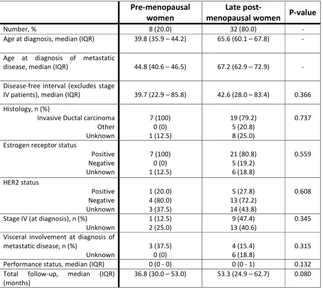
a) Cohort characteristics
A total of 40 women were included in this study, 8 (20.0%) of which pre-menopausal and 32 (80.0%) post-menopausal. Relevant patient’s clinical and pathologic characteristics are summarized in table 3. As a matter of fact, the groups were reasonably balanced regarding most of the selected variables. Noteworthy, median ages are compatible with corresponding menopausal status. Furthermore, most tumors were estrogen receptor positive, as expected from a cohort of patients with bone disease as first site of relapse, and had a fairly good performance status, which enabled the use of chemotherapy as a treatment option.
Table 3 – Cohort baseline characteristics according to menopausal status.
Pre-menopausal
women menopausal women P-value Late
post-Number, % 8 (20.0) 32 (80.0) -
Age at diagnosis, median (IQR) 39.8 (35.9 – 44.2) 65.6 (60.1 – 67.8) -
Age at diagnosis of metastatic
disease, median (IQR) 44.8 (40.6 – 46.5) 67.2 (62.9 – 72.9) -
Disease-free interval (excludes stage
IV patients), median (IQR) 39.7 (22.9 – 85.8) 42.6 (28.0 – 83.4) 0.366 Histology, n (%)
Invasive Ductal carcinoma Other Unknown 7 (100) 0 (0) 1 (12.5) 19 (79.2) 5 (20.8) 8 (25.0) 0.737
Estrogen receptor status
Positive Negative Unknown 7 (100) 0 (0) 1 (12.5) 21 (80.8) 5 (19.2) 6 (18.8) 0.559 HER2 status Positive Negative Unknown 1 (20.0) 4 (80.0) 3 (37.5) 5 (27.8) 13 (72.2) 14 (43.8) 0.608
Stage IV (at diagnosis), n (%)
Unknown 1 (12.5) 2 (25.0) 13 (40.6) 9 (47.4) 0.345 Visceral involvement at diagnosis of
metastatic disease, n (%)
Unknown 3 (37.5) 0 (0) 4 (15.4) 6 (18.8) 0.315 Performance status, median (IQR) 0 (0 - 0) 0 (0 - 1) 0.132 Total follow-up, median (IQR)
Page 20 of 38
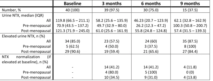
a) Urinary NTX
The majority of patients had elevated levels of urinary NTX at baseline, which is compatible with active disease in the bone. Moreover, when evaluating the full cohort, after the introduction of zoledronic acid and chemotherapy (at diagnosis - baseline) a decrease of NTX is notable (table 4 and figure 2), with 41.2% of the patients with elevated NTX at baseline reaching an NTX < 50 nmol/mmol creatinine at month 3. Furthermore, after log-transforming NTX values, a trend for an overall decline in mean log10(NTX) in the interval 0 – 3 months (p=0.125) and a statistically significant decline of
log10(NTX) in the interval 3 – 6 months (0.013) was found. Importantly, the proportion of patients
that were lost to follow-up in terms of urinary NTX measurements was high: while at month 6 75% of the patients had a valid measurement of urinary NTX, at month 9 only 37.5% had. Of note, no clear differences in terms of age, disease progression or vital status were found between lost to follow-up and retained patients; therefore, we considered this missing information as missing completely at random.
Table 4 – UrineNTX at several time points from diagnosis. NTX measured in nmol/mmol creatinine.
Baseline 3 months 6 months 9 months
Number, % 40 (100) 39 (97.5) 30 (75.0) 15 (37.5)
Urine NTX, median (IQR) All Pre-menopausal Post-menopausal
119.8 (66.5 – 211.1) 70.9 (43.5 – 137.2) 121.3 (71.9 – 245.0)
58.2 (25.6 – 135.9) 49.7 (32.9 – 80.0) 61.0 (25.6 – 161.9)
46.23 (20.7 – 123.9) 26.2 (12.3 – 47.2) 55.8 (24.8 – 124.8)
62.1 (32.8 – 162.9) 100.3 (50.8 – 200.7)
57.4 (31.5 – 139.3) Elevated urine NTX, n (%)
All Pre-menopausal Post-menopausal 34 (85.0) 5 (62.5) 29 (90.6) 23 (57.5) 4 (50.0) 19 (59.4) 24 (60) 3 (37.5) 21 (65.6) 35 (87.5) 8 (100) 27 (84.4) NTX normalization (if
Page 21 of 38
Figure 3 – Mean (A), median (B) and mean log10 (C) variation of urine NTX at several time points
after diagnosis in the full cohort.
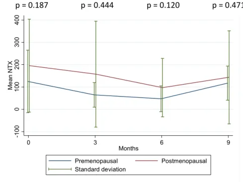
When stratified by menopausal status (figure 3), post-menopausal women present persistently absolute higher levels of mean NTX, namely at baseline and at 3 months; however, such differences do not reach statistical significance. In fact, such differences are non-significant through all the follow-up period. After log-transforming NTX values (figure 4), the early response (baseline to month 3) to zoledronic acid and chemotherapy is equal between groups (p=0.957). Both groups seem to “escape” zoledronic acid plus chemotherapy control after month 6, as demonstrated by an increase of NTX in both groups. Of note, in absolute and relative terms, urinary NTX levels seems to increase more pronouncedly in the group of premenopausal women in this interval (figure 3 and 4); however, this difference is non-significant (p=0.262). Moreover, when all the follow-up period is taken together, the response profile of urinary NTX levels to zoledronic acid and chemotherapy does not differ between pre and post-menopausal women (p=0.314).
A B
C -1 00 0 10 0 20 0 300 400 Me an NT X
0 3 6 9
Months
NTX Standard deviation
0 50 10 0 150 200 M edi an N TX
0 3 6 9
Months
NTX Interquartile range
1 1.5 2 2. 5 Mean lo g( N TX)
0 3 6 9
Months
Page 22 of 38
1
1.
5
2
2.
5
Me
an
log(
N
TX)
0 3 6 9
Months
Premenopausal Postmenopausal
Standard deviation
Figure 4 – Mean NTX at several time points after diagnosis according to menopausal status. Test of differences in mean values is shown.
Figure 5 – Mean responseprofile of log10(NTX) at several time points after introduction of zoledronic
acid plus chemotherapy according to menopausal status. Test of interaction time - menopausal group is shown.
-1
00
0
10
0
20
0
300
400
Me
an NT
X
0 3 6 9
Months
Premenopausal Postmenopausal
Standard deviation
p = 0.957 p = 0.316 p = 0.262
p = 0.314
Page 23 of 38
b) Tumor markers (CA15.3 and CEA)
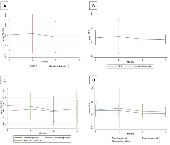
The majority of patients had elevated levels of serum CA15.3 and CEA at baseline, which is compatible with active breast cancer. After the introduction of zoledronic acid and chemotherapy (at diagnosis - baseline) a slight numerical decrease of both markers is identifiable (figure 5 and figure 6); however, only a trend for decreased values through the interval 0 – 9 months is found (p = 0.058 and 0.057, for CA15.3 and CEA respectively).
When stratified by menopausal status (figure 6), no clear differences are found between pre and post-menopausal women in the log transformed values, either at baseline or at the following time points, both for CA15.3 and CEA.
Figure 6 – Mean response profile of CA15.3 and CEA at several time points after diagnosis in the full cohort (A and B, respectively) and according to menopausal status (C and D, respectively).
-2 00 0 200 400 60 0 800 Me an CA 15.3
0 3 6 9
Months
Premenopausal Postmenopausal Standard deviation
A B
C D
-2 00 0 20 0 400 600 M ean CA 15 .3
0 3 6 9
Months
CA15.3 Standard deviation
-5 0 0 50 100 Mea n CE A
0 3 6 9
Months
CEA Standard deviation
-5 0 0 50 100 M ean C EA
0 3 6 9
Months
Page 24 of 38
Figure 7 – Mean response profile of log10(CA15.3) (A) and log10(CEA) (B) at several time points after
introduction of zoledronic acid plus chemotherapy according to menopausal status. Test of interaction time - menopausal group is shown.
0
.5
1
1.
5
Me
an log
(CEA)
0 3 6 9
Months
Premenopausal Postmenopausal
Standard deviation
1
1.
5
2
2.
5
3
Me
an l
og
(CA15.
3)
0 3 6 9
Months
Premenopausal Postmenopausal
Standard deviation
p = 0.997 p = 0.402 p = 0.531
p = 0.762
p = 0.683 p = 0.670 p = 0.315
p = 0.478
A
Page 25 of 38 0 10 20 30 40 50 60 70 80 90 100 Sw itch in g firs t l in e che m ot he ra py( % )
17 17 11 9 7 5
Post-menopause 8 6 6 5 4 1
Pre-menopause No. at risk
0 5 10 15 20 25
Follow-up time (months)
Pre-menopause Post-menopause
c) Time to first-line chemotherapy failure
When evaluating time to first-line chemotherapy failure, the full cohort presented a median follow-up time of approximately 17 months (IQR 6.3 – 23.2). A total of 30 events (90.9%) were documented, of which 8 (100%) in the premenopausal group and 22 (88.0%) in the post-menopausal group. The median time to chemotherapy failure was of 15.3 months for pre-menopausal women and 17.4 for post-menopausal women. In figure 7 a representation of the survival function according to menopausal status is depicted. When evaluating the univariate association of menopausal status and other clinical and pathological variables with change of first line chemotherapy, only the presence of visceral involvement at diagnosis of metastatic disease was significantly associated with time to chemotherapy switch (table 5). Furthermore, when testing the role of menopausal status on time to first line chemotherapy failure controlling for visceral involvement a persistent non-significant association was noted (figure 7). Despite the intention to control for other variables, due to the lack of available observations and the fact that only concomitant visceral involvement was significantly associated with first-line chemotherapy failure, only this covariate was included in the model.
Figure 8 – Time to first-line chemotherapy failure according to menopausal status. Adjusted HR controlling for visceral involvement is shown. HR – Hazard ratio; CI – Confidence interval; mo – months; n – number.
Pre-menopause Post-menop.
Median time, mo. 15.25 17.40 Events, n (%) 8 (100) 22 (88.0)
Log-rank p = 0.399
Page 26 of 38
Table 5 – Univariate association of baseline characteristics with time to first line chemotherapy failure.
P-value
Menopausal status 0.399
Age at diagnosis of metastatic disease 0.673 Time from diagnosis to metastatic disease 0.272
Histology 0.882
Estrogen receptor status 0.867
HER2 status 0.915
Stage IV at diagnosis 0.418 Visceral involvement at diagnosis of metastatic
disease 0.015
Performance status 0.240
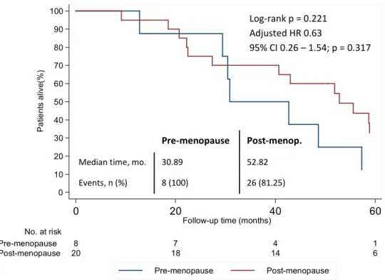
d) Overall survival
Page 27 of 38 0
10 20 30 40 50 60 70 80 90 100
Pa
tie
nt
s a
liv
e(
%
)
20 18 14 6
Post-menopause 8 7 4 1
Pre-menopause No. at risk
0 20 40 60
Follow-up time (months)
Pre-menopause Post-menopause
Figure 9 – Overall survival according to menopausal status. Adjusted HR controlling for HER2 status is shown. HR – Hazard ratio; CI – Confidence interval; mo – months; n – number.
Table 6 – Univariate association of baseline characteristics with overall survival.
P-value
Menopausal status 0.221
Age at diagnosis of metastatic disease 0.999 Time from diagnosis to metastatic disease 0.294
Histology 0.733
Estrogen receptor status 0.618
HER2 status 0.094
Stage IV at diagnosis 0.662 Visceral involvement at diagnosis of metastatic
disease 0.186
Performance status 0.377
Pre-menopause Post-menop.
Median time, mo. 30.89 52.82 Events, n (%) 8 (100) 26 (81.25)
Log-rank p = 0.221 Adjusted HR 0.63
Page 28 of 38
Discussion
Pre-clinical[30] and more recently clinical[31] evidence is accumulating in favor of an anti-cancer activity of BPs, more specifically in the adjuvant treatment of post-menopausal women with breast cancer. In the present study we intended to explore the differential anti-cancer effect of BPs according to the hormonal environment in metastatic patients receiving ZA and chemotherapy. In this subgroup of patients, no evidence of a differential anti-cancer activity of ZA between pre and post-menopausal women was found when evaluating anti-cancer activity in terms of variation of NTX (as surrogate for bone disease activity), variation of CA15.3 and CEA (as surrogate for overall disease activity), time to first line chemotherapy failure (as surrogate for overall disease progression) and overall survival.
BPs were developed and introduced in clinical practice as a tool to decrease the morbidity of bone metastases given their properties as bone remodeling modulators, in specific as osteoclast inhibitors. In this context, the primary endpoints of pivot trials were essentially centered on the rate of SREs, time to first and subsequent SRE, improvement in pain control and general quality of life. As a matter of fact, compared to placebo BPs reduced the SRE risk by 15% (RR 0.85, 95% CI 0.77-0.94), with a median 28% reduction (range, 14–48%) of SREs.[41] However, no impact on overall survival was found.[41]
Other studies tested the efficacy of BPs (zoledronic acid, pamidronate and clodronate) in the metastatic setting with a focus on their anti-cancer activity with generally inconclusive findings. However, based on the data derived from adjuvant studies, ours was the first study to test the impact of menopausal status in the anticancer activity of BPs.
Page 29 of 38
randomized to receive zoledronic acid, either as a standard fixed schedule (4mg IV over 15 minutes once every 3-4 weeks for 24 months) or under a set of rules dependent on NTX variation. Despite the accrual limitations that led to premature study closure, the NTX-directed schedule group had a higher proportion of patients presenting with an SRE, thus failing to demonstrate non-inferiority of this approach. Of note, overall survival was selected as a secondary outcome in this study. Given that patients in the standard schedule arm had more than the double of ZA administrations, this study can help us gain some insight on the anti-tumor action of zoledronic acid in terms of overall survival. No stratification based on menopausal status is planned so far.
Clodronate and pamidronate (in its oral formulation) were also tested as anti-neoplastic agents, in specific, about their ability to prevent the development of bone metastases in patients with breast cancer without clinical evidence of bone involvement and irrespective of menopausal status.[43– 45] Despite the fact that all studies were considerably underpowered to show a difference between groups, when compared to placebo, no difference was found in the incidence of new bone metastases. A recent meta-analysis summarized these findings, and found no overall effect of these agents on the incidence of bone metastases in patients with metastatic breast cancer without clinical evidence of bone metastases (HR 0.99, 95% CI 0.67 – 1.47).[41]
Page 30 of 38
The present study presents important limitations which is compatible with its exploratory nature and budget constraints. The first main limitation refers to the small sample size. Despite the preplanned design as an exploratory study, the group of premenopausal women was considerably small, which might not be representative of a conventional cohort of pre-menopausal patients. Second, a relevant lost to follow-up was found at 9 months. Regardless of the intention to perform the analysis of primary outcomes at 3 months, secondary outcomes interpretation is limited due to this fact. Indeed, the late acceleration of urinary NTX in the pre-menopausal patients cannot be properly valued in this context. Third, time to first line chemotherapy failure after the diagnosis of metastatic disease was considerably long. In fact, several of these patients stopped first line chemotherapy after a good clinical response and had a switch to hormone therapy before switching to second line chemotherapy, as common practice in patients with hormone receptor positive disease. The present analysis does not account for the type of hormone therapy received in the transition to second line chemotherapy and this fact might have been a source of heterogeneity. As a matter of fact, hormone therapy is known to interfere in bone remodeling[47, 48], and different options of hormone therapy might change bone remodeling differently. Finally, the same threshold of NTX normality was used both for pre and post-menopausal women for comparability reasons. It is long known that several factors interfere with bone remodeling markers levels, such as age, hormonal environment and gender. The persistently higher levels of NTX in post-menopausal women at baseline and after the introduction of zoledronic acid and chemotherapy (despite all of which non-significant) might be exclusively associated with this fact.
Page 31 of 38
Conclusions
In a cohort of women with breast cancer and bone metastases receiving first line chemotherapy and zoledronic acid, no difference seems to exist in terms of urinary NTX variation at 3 months according to menopausal status. Furthermore, no differences was found in terms of CA15.3 and CEA variation according to menopausal status. Finally, time to first line chemotherapy failure or overall survival seems to be similar when evaluated according to menopausal status.
Page 32 of 38
Acknowledgments
Page 33 of 38
List of abbreviations
ALP – Alkaline phosphataseBALP – Bone specific alkaline phosphatase BC/CM – Breast cancer/Cancro da mama BP – Bisphosphonates
BSP – Non collagenous bone matrix proteins: bone sialoprotein CA15.3 – Cancer antigen 15.3
CEA – Carcinoembrionic antigen CI – Confidence interval
CT/QT – Chemotherapy/Quimioterapia
CTX – Carboxy-terminal crosslinked telopeptide of collagen type I CXCL12 – chemokine (C-X-C motif) ligand 12
CXCR4 – chemokine (C-X-C motif) receptor 4 CXCR7 – chemokine (C-X-C motif) receptor 7 DKK-1 – Dickkopf-1
DTC – Dessiminated tumor cells DPD – Deoxypyridoline
ER – Estrogen receptor
HER – human epidermal growth factor receptor 2 HR – Hazard ratio
Hyl – Hydroxylysine Hyp – Hydroxyproline
ICTP – Carboxy-terminal crosslinked telopeptide of type I collagen MT – Marcador tumoral
NTX – Amino-terminal crosslinked telopeptide of collagen type I OC – Osteocalcin
OP – Osteopontin OPG – Osteoprotegerin
P1NP – Propeptides of type I procollagen PTH-rp – Parathyroid hormone related peptide PoM – Post-menopausal
PrM – Pre-menopausal PYD – Pyridinoline
RANK – Receptor activator of NFƙB RANKL – Receptor activator of NFƙB ligand SRE – Skeletal related event
TRAP5b – Tartarate resistant acid phosphatase 5b TRAIL – TNF-related apoptosis-inducing ligand USA – United States of America
Page 34 of 38
References
1. Ferlay J, Steliarova-Foucher E, Lortet-Tieulent J, et al. (2013) Cancer incidence and mortality patterns in Europe: Estimates for 40 countries in 2012. Eur J Cancer 49:1374–1403. doi: 10.1016/j.ejca.2012.12.027
2. Ferlay J, Soerjomataram I, Dikshit R, et al. (2015) Cancer incidence and mortality worldwide: Sources, methods and major patterns in GLOBOCAN 2012. Int J Cancer 136:E359–86. doi: 10.1002/ijc.29210
3. National Cancer Institute (2014) Surveillance, Epidemiology, and End Results (SEER) Program. In: SEER Stat Fact Sheets Breast Cancer. http://seer.cancer.gov/.
4. Clarke M, Collins R, Darby S, Davies C, Evans V, Godwin J, Gray R, McGale P, Peto R, Wang Y.Clarke M, Collins R, Darby S, Davies C, Evans V, Godwin J, Gray R, McGale P, Peto R WY (2005) Effects of chemotherapy and hormonal therapy for early breast cancer on recurrence and 15-year survival: an overview of the randomised trials. Lancet 365:1687–717. doi: 10.1016/S0140-6736(05)66544-0
5. Jensen A, Jacobsen J, Norgaard M, et al. (2011) Incidence of bone metastases and skeletal-related events in breast cancer patients: A population-based cohort study in Denmark. BMC Cancer 11:29.
6. Zhang XH-F, Wang Q, Gerald W, et al. (2009) Latent bone metastasis in breast cancer tied to Src-dependent survival signals. Cancer Cell 16:67–78. doi:
10.1016/j.ccr.2009.05.017
7. Rosa Mendoza ES, Moreno E, Caguioa PB (2013) Predictors of early distant metastasis in women with breast cancer. J Cancer Res Clin Oncol 139:645–652. doi:
10.1007/s00432-012-1367-z
8. Ishizawar R, Parsons SJ (2004) c-Src and cooperating partners in human cancer. Cancer Cell 6:209–14. doi: 10.1016/j.ccr.2004.09.001
9. Lee JJ, Lotze MT (2009) Molecular basis of metastasis. N Engl J Med 360:1679; author reply 1679–1680. doi: 10.1056/NEJMra0805239
10. Roato I, Ferracini R (2013) Solid Tumours Show Osteotropism: Mechanisms of Bone Metastases. Clin Rev Bone Miner Metab 11:87–93. doi: 10.1007/s12018-013-9144-3
Page 35 of 38
12. Tranquilli Leali P, Doria C, Zachos A, et al. (2009) Bone fragility: Current reviews and clinical features. Clin Cases Miner Bone Metab 6:109–113.
13. Lipton A, Theriault RL, Hortobagyi GN, et al. (2000) Pamidronate prevents skeletal complications and is effective palliative treatment in women with breast carcinoma and osteolytic bone metastases: long term follow-up of two randomized, placebo-controlled trials. Cancer 88:1082–1090.
14. Coleman RE (2006) Clinical features of metastatic bone disease and risk of skeletal morbidity. Clin Cancer Res 12:6243s–6249s. doi: 10.1158/1078-0432.CCR-06-0931
15. Lipton A, Costa L, Coleman RE (2011) Bone turnover markers: tools for prognosis and monitoring response to bisphosphonates? Breast Dis 33:59–69. doi: 10.3233/BD-2010-0327
16. Costa L, Demers LM, Gouveia-Oliveira A, et al. (2002) Prospective evaluation of the peptide-bound collagen type I cross-links N-telopeptide and C-telopeptide in predicting bone metastases status. J Clin Oncol 20:850–856. doi:
10.1200/JCO.20.3.850
17. Wada N, Fujisaki M, Ishii S, et al. (2001) Evaluation of bone metabolic markers in breast cancer with bone metastasis. Breast Cancer 8:131–137. doi:
10.1007/BF02967492
18. Lipton A, Chapman J-AW, Demers L, et al. (2011) Elevated bone turnover predicts for bone metastasis in postmenopausal breast cancer: results of NCIC CTG MA.14. J Clin Oncol 29:3605–10. doi: 10.1200/JCO.2010.31.5069
19. Body JJ, Dumon JC, Gineyts E, Delmas PD (1997) Comparative evaluation of markers of bone resorption in patients with breast cancer-induced osteolysis before and after bisphosphonate therapy. Br J Cancer 75:408–412.
20. Demers LM, Costa L, Chinchilli VM, et al. (1995) Biochemical markers of bone turnover in patients with metastatic bone disease. Clin Chem 41:1489–94.
21. Brown JE, Cook RJ, Major P, et al. (2005) Bone turnover markers as predictors of skeletal complications in prostate cancer, lung cancer, and other solid tumors. J Natl Cancer Inst 97:59–69. doi: 10.1093/jnci/dji002
22. Coleman RE, Major P, Lipton A, et al. (2005) Predictive value of bone resorption and formation markers in cancer patients with bone metastases receiving the
Page 36 of 38
23. Coleman R (2012) Randomized trial of marker-directed versus standard schedule zoledronic acid for bone metastases from breast cancer. J Clin Oncol 30:9–10.
24. Stopeck AT, Lipton A, Body J-J, et al. (2010) Denosumab compared with zoledronic acid for the treatment of bone metastases in patients with advanced breast cancer: a randomized, double-blind study. J Clin Oncol 28:5132–5139. doi:
10.1200/JCO.2010.29.7101
25. Coleman R, Body JJ, Aapro M, et al. (2014) Bone health in cancer patients: ESMO Clinical Practice Guidelines. Ann Oncol 25 Suppl 3:iii124–37. doi:
10.1093/annonc/mdu103
26. Buhaescu I, Izzedine H (2007) Mevalonate pathway: a review of clinical and therapeutical implications. Clin Biochem 40:575–84. doi:
10.1016/j.clinbiochem.2007.03.016
27. Rosen LS, Gordon D, Kaminski M, et al. (2003) Long-term efficacy and safety of zoledronic acid compared with pamidronate disodium in the treatment of skeletal complications in patients with advanced multiple myeloma or breast carcinoma: A randomized, double-blind, multicenter, comparative trial. Cancer 98:1735–1744. doi: 10.1002/cncr.11701
28. Sun L, Yu S (2013) Efficacy and safety of denosumab versus zoledronic acid in patients with bone metastases: a systematic review and meta-analysis. Am J Clin Oncol 36:399–403. doi: 10.1097/COC.0b013e31824be20e
29. Van Poznak CH, Von Roenn JH, Temin S (2011) American society of clinical oncology clinical practice guideline update: recommendations on the role of bone-modifying agents in metastatic breast cancer. J Oncol Pract 7:117–121.
30. Clézardin P (2013) Mechanisms of action of bisphosphonates in oncology: a scientific concept evolving from antiresorptive to anticancer activities. Bonekey Rep 2:267. doi: 10.1038/bonekey.2013.1
31. Coleman R, Gnant M, Paterson A, et al. (2014) Abstract S4-07: Effects of
bisphosphonate treatment on recurrence and cause-specific mortality in women with early breast cancer: A meta-analysis of individual patient data from randomised trials. Cancer Res 73:S4–07–S4–07. doi: 10.1158/0008-5472.SABCS13-S4-07
Page 37 of 38
33. Gnant M, Mlineritsch B, Schippinger W, et al. (2009) Endocrine Therapy plus Zoledronic Acid in Premenopausal Breast Cancer. N Engl J Med 360:679–691. doi: 10.1056/NEJMoa0806285
34. Gnant M, Mlineritsch B, Stoeger H, et al. (2014) Zoledronic acid combined with adjuvant endocrine therapy of tamoxifen versus anastrozol plus ovarian function suppression in premenopausal early breast cancer: final analysis of the Austrian Breast and Colorectal Cancer Study Group Trial 12. Ann Oncol 26:313–320. doi: 10.1093/annonc/mdu544
35. Brufsky AM, Harker WG, Beck JT, et al. (2012) Final 5-year results of Z-FAST trial: Adjuvant zoledronic acid maintains bone mass in postmenopausal breast cancer patients receiving letrozole. Cancer 118:1192–1201. doi: 10.1002/cncr.26313
36. Coleman R, De Boer R, Eidtmann H, et al. (2013) Zoledronic acid (zoledronate) for postmenopausal women with early breast cancer receiving adjuvant letrozole (ZO-FAST study): Final 60-month results. Ann Oncol 24:398–405. doi:
10.1093/annonc/mds277
37. Coleman RE, Marshall H, Cameron D, et al. (2011) Breast-cancer adjuvant therapy with zoledronic acid. N Engl J Med 365:1396–405. doi: 10.1056/NEJMoa1105195
38. Paterson AHG, Anderson SJ, Lembersky BC, et al. (2012) Oral clodronate for adjuvant treatment of operable breast cancer (National Surgical Adjuvant Breast and Bowel Project protocol B-34): a multicentre, placebo-controlled, randomised trial. Lancet Oncol 13:734–42. doi: 10.1016/S1470-2045(12)70226-7
39. Kokufu I, Kohno N, Yamamoto M, Takao S (2010) Adjuvant pamidronate therapy prevents the development of bone metastases in breast cancer patients with four or more positive nodes. Oncol Lett 1:247–252. doi: 10.3892/ol_00000044
40. Jung J, Hwang G, Lee Y, et al. (2005) Pamidronate as adjuvant treatment for prevention of bone metastasis in breast cancer. ASCO Meet Abstr 23:888.
41. Wong MH, Stockler MR, Pavlakis N (2012) Bisphosphonates and other bone agents for breast cancer. Cochrane Database Syst Rev 2:CD003474. doi:
10.1002/14651858.CD003474.pub3
Page 38 of 38
43. Kanis JA, Powles T, Paterson AHG, et al. (1996) Clodronate decreases the frequency of skeletal metastases in women with breast cancer. Bone 19:663–667. doi:
10.1016/S8756-3282(96)00285-2
44. Van Holten-Verzantvoort a. TM, Hermans J, Beex LV a M, et al. (1996) Does supportive pamidronate treatment prevent or delay the first manifestation of bone metastases in breast cancer patients? Eur J Cancer 32:450–454. doi:
10.1016/0959-8049(95)00564-1
45. Mardiak J, Bohunický L, Chovanec J, et al. (2000) Adjuvant clodronate therapy in patients with locally advanced breast cancer--long term results of a double blind randomized trial. Slovak Clodronate Collaborative Group. Neoplasma 47:177–80.
46. Weilbaecher KN, Guise TA, McCauley LK (2011) Cancer to bone: a fatal attraction. Nat Rev Cancer 11:411–425. doi: 10.1038/nrc3055
47. Vehmanen L, Elomaa I, Blomqvist C, Saarto T (2006) Tamoxifen treatment after adjuvant chemotherapy has opposite effects on bone mineral density in
premenopausal patients depending on menstrual status. J Clin Oncol 24:675–680. doi: 10.1200/JCO.2005.02.3515