Materials Research, Vol. 8, No. 2, 221-224, 2005 © 2005
*e-mail: [email protected]
Pechini Synthesis and Microstructure of Nickel-Doped Copper Chromites
Patrícia Mendonça Pimentel, Antonio Eduardo Martinelli*, Dulce Maria de Araújo Melo,
Anne Michelle Garrido Pedrosa, Jardel Dantas Cunha, Carlos Neco da Silva Júnior
Universidade Federal do Rio Grande do Norte, Departamento de Química, Laboratório de Análise Térmica e Materiais, C.P. 1662, 59078-970 Natal - RN, Brazil
Received: November 23, 2003; Revised: March 19, 2005
Spinel-type solid solutions were synthesized by the Pechini method and calcined between 500 and 900 °C for 4 hours and at 900 °C for 8 hours to produce ceramic pigments. The resulting powders were characterized by thermogravimetric analysis, Fourier-transform infrared spectroscopy (FTIR), X-ray diffraction (XRD), scanning electron microscopy (SEM), particle size analysis and BET surface area measurements. The formation of spinel took place upon calcination at 700 °C. IR spectroscopy revealed the presence of N1 and N2bands, typical of
spinel structures, broadened by the presence of more than one cationic species in the structure. The specific area of the resulting powder decreased from 24.7 to 1.4 m2g-1 as the calcination temperature increased from 700 to
900 °C. Microstructural analyses revealed the presence of crystalline spinel as the only phase present in powders calcined at 900 °C. Colorimetric analyses using L*a*b* coordinates and UV-visible spectroscopy revealed that the pigment was predominantly black.
Keywords:spinel, chromites, Pechini method, ceramic pigments
1. Introduction
Spinels are ternary oxides with the general formula AB2O4, where A and B are cations occupying tetrahedral and octahedral sites re-spectively. These oxides exhibit interesting electric, magnetic and catalytic properties1-4, depending on their nature, charge, and
distri-bution of ions at interstices5. CuCr
2O4 crystallizes as a tetragonally
distorted spinel structure. Its distortion is related to the cooperative Jahn-Teller effect of Cu2+ at the tetrahedral sites6. The substitution
of tetrahedral Cu2+ ions by any bivalent cation does not affect the
distribution of cations in the substituted spinel oxides7,8. Site
prefer-ence energies for oxide spinels indicate that Ni2+ and Cr3+ occupy
octahedral sites, although Cr3+ is also capable of forcing Ni2+ into
tetrahedral positions7.
The most widely used method for the preparation of spinels involves solid-state reaction of mechanically mixed metal oxides at high temperatures9,10. The Pechini method11,12, based on polymeric
precursors, can be used to prepare spinels and it does not require high temperature calcinations and permits good stoichiometric control as well as reproducibility. This method consists of the formation of a polymeric resin between a metallic acid chelate and polyhydroxide alcohol by polyesterification.
This study is focused on the preparation of solid solutions of copper chromites doped with nickel by the Pechini method, and the investigation of the effect of heat treatment on particle size and morphology. X-ray diffraction (XRD) and Fourier Transform Infra-red Spectroscopy (FTIR) patterns were used to reveal the structural properties of the materials formed.
2. Experimental
2.1. Synthesis
Nickel-doped copper chromite powders were prepared from poly-meric precursors by the Pechini method. The metal nitrate solution was mixed with a stoichiometric amount of citric acid. The resulting solution was stirred for about 1 hour on a hot plate and the temperature was stabilized at 70 °C. The mixture was heated to 90 °C, at which point ethylene glycol was added at a mass ratio of 40:60 with respect
to citric acid. The temperature was maintained constant up to resin formation, which polymerized at 300 °C. The precursor powders were then calcined for 4 hours at various temperatures, ranging from 500 to 900 °C, or at 900 °C for 8 hours.
2.2. Characterization
Chemical analyses of the metallic constituents were carried out by atomic absorption spectroscopy and a Spectra 110 Varian setup was used. Thermogravimetric analyses of the polymeric precursors were carried out in a TGA-7 Perkin Elmer balance by heating samples of 2.15 mg at 5 °C min-1 in flowing air (50 mL min-1). Particle size
distributions were obtained in a CILAS 1064 laser analyzer. Infrared spectra (FTIR) were recorded using KBr pellets in a Perkin-Elmer 16 PC instrument. X-ray diffraction patterns (XRD) were obtained from a Shimadzu XRD-6000 X-ray difractometer using CuKA radia-tion (L = 1.5418 Å). The specific surface area of the powders was measured by nitrogen adsorption on a NOVA 2000 BET system. The microstructure of the powders was observed by examining Au-coated samples in a Philips ESEM-XL30 scanning electron microscope that was set in the high-vacuum mode. The diffuse reflectance of the pow-ders was measured in the 300-800 nm range using a Gretac Macbeth 2180-2180 UV spectrophotometer (D-65 light). The color of the pig-ment was evaluated from L*a*b* colorimetric coordinates according to CIE (Commission Internationale I´Eclairage) standards using
$E2 = L2 + a2 + b2 (1)
where L* varies from black (0) to white (100), a* from green (-) to red (+), and b* from blue (-) to yellow (+).
3. Results and Discussion
222 Pimentel et al. Materials Research
the material lost 2% of its mass upon heating to 500 °C, due to de-composition of organic compounds. Samples calcined from 500 to 900 °C revealed no additional mass loss.
X-ray diffraction patterns of the calcined powders are shown in Figure 2. The presence of a distorted tetragonal spinel phase along with other crystalline phases, such as CuO and Cr2O4, could be observed. Although spinel was formed even at relatively low calcin-ing temperatures, it is possible to observe the steady increase in the
Figure 1. Thermogravimetric plot of the spinel polymeric precursor.
Q
s
Figure 2. X-ray diffraction patterns of the polymeric precursor and powders
calcined at 500 °C, 700 °C, and 900 °C for 4 hours.
relative peak intensity of spinel with respect to CuO and Cr2O4, with increasing synthesis temperature. This indicated ongoing crystalliza-tion of spinel from its precursors. The synthesis of spinel also affected particle size distribution in the resulting powder (Figure 3). Although slight, it is possible to notice that the average particle size (D50) of the powder, representative of powder agglomerates, steadily decreased from ~10 Mm to ~ 6 Mm as the calcining temperature increased from 500 to 900 oC and time from 4 to 8 hours at 900 oC. In addition, the
Figure 3. Particle size distribution of powders calcined at: a) 500 oC; b) 700 oC; c) 900 °C for 4 hours; d) 900 oC for 8 hours.
(a) (b)
Vol. 8, No 2, 2005 Pechini Synthesis and Microstructure of Nickel-Doped Copper Chromites 223
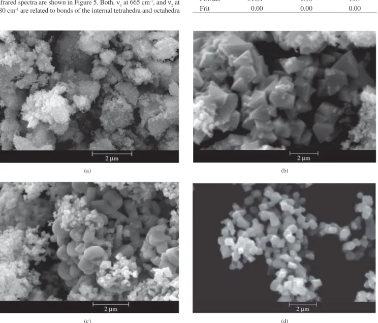
fraction of fine loose particles (equivalent diameter < 1 Mm) also increased with crystallization of spinel. This can be confirmed from the SEM images shown in Figure 4. Along with changes in particle morphology as a function of calcining temperature, heat treatment at 500 °C (Figure 4a) resulted in substantial agglomeration of the powder, which is typical of the presence of unreacted CuO and Cr2O4. Calcining at higher temperatures (700 or 900 °C) or longer times (8 hours) resulted in loose and fine octahedron-shaped crystals characteristic of spinel and agglomerates. The values summarized in Table 1 reveal that the formation of spinel was accompanied by a steady decrease in specific surface area, a consequence of consump-tion of CuO - Cr2O4 irregular agglomerates to form regular-shaped spinel nanocrystals. The relatively low values encountered are typical of Pechini synthesis, and can be attributed to the M:AC (M = Metal; AC = citric acid) molar ratio used in the synthesis, and consequently, to the chain polymer size used in pigment preparation.
According to group theory, spinel type oxides should exhibit four IR bandsN1-N413-14. In this investigation measurements were carried out
up to 500 cm-1, thus limiting the study to the high frequency bands (N 1
and N2) of the IR spectrum. Since these bands are nearly insensitive to
changes in the bivalent cation9, they should not be significantly affected
when Cu2+ from CuCr
2O4 is substituted by another bivalent cation. The
infrared spectra are shown in Figure 5. Both,N1 at 665 cm-1, and N2 at
580 cm-1 are related to bonds of the internal tetrahedra and octahedra
of the structure of Cu0.8Ni0.2Cr2O4. The broadening of these bands is probably due to the presence of more than one type of cation14.
Results from UV- visible spectroscopy (Figure 6) revealed an absorption band in the 400-700 nm range as a result of complete light absorption, typical of highly reflective systems. This was further confirmed by the colorimetric coordinates (Table 2). The relatively high values of L* (31.81) along with low a* and b*, 0.10 and 1.07 respectively, suggested full light absorption, and consequently a black pigment. When applied to ceramic frits the pigment remained
Table 1. Specific surface area of calcined powders. Calcination profile
500 °C/4 h 700 °C/4 h 900 °C/4 h 900 °C/8 h Specific
surface area (m2 g-1)
24.7 7.2 2.0 1.4
Table 2. Colorimetric coordinates of powder calcined at 900 °C for 8 hours.
L* a* b*
Powder 31.81 0.10 1.07
Frit 0.00 0.00 0.00
Figure 4. SEM images of powders calcined at: a) 500 °C; b) 700 °C; c) 900 °C for 4 hours; d) 900 °C for 8 hours.
(a) (b)
224 Pimentel et al. Materials Research
black although it changed its hue slightly. The values L*, a* and b* changed to zero as a consequence of the interaction of the pigment with the glassy material.
4. Conclusions
Cu0.8Ni0.2Cr2O4 spinel was synthesized from polymeric precursors using the Pechini method. The crystallization of the spinel structure upon calcining at 700 °C. Cu0.8Ni0.2Cr2O4was the only phase present upon calcination at 900 °C. Both, loose particles and agglomerates were formed. The average size of the powder was 8 Mm. Black pig-ments were obtained from the synthesis. Despite slight variations in the colorimetric coordinates, no significant change in color was observed when the pigments were applied to ceramic frits.
Acknowledgments
The authors would like to thank CNPq as well as the National Petroleum Agency (ANP-Brazil) for providing the financial support necessary to carry on the present research work through its Human Resource Program PRH-30-UFRN.
References
1. Nathawan P, Darshane VS. Structural. Transport, magnetic and infrared studies of the oxidic spinels Co2-XTi1-XFe2XO4.Journal of Physics. 1988; 21(6):3191-3203.
2. Vlasenko VM, Chernobrivets VL. Methane chlorination on spinel copper-chromium catalyst in the presence of oxygen. Russian Journal of Applies Chemistry. 1998; 71(8):1393-1396.
3. Erran E, Trifino F, Vaccari A, Richter M. Structure and reactivity of Zn-Cr mixed oxides Role of non-stoichiometry in the catalytic synthesis of methanol.Catalysis Letter. 1989; 3(1):65-72.
4. Câmara MSC, Lisboa-Filho PN, Cabrelon MD, Gama L, Ortiz WA,
Paiva-