http://dx.doi.org/10.1590/jvb.2013.052
Emergency embolization after resection
of a laryngeal Schwannoma
Embolização de urgência após ressecção de schwanoma de laringe
Fábio Augusto Cypreste Oliveira1,2,3, Carlos Eduardo de Sousa Amorelli1,2,3, Fábio Lemos Campedelli1,2,3,
Werther Sales4, Maria Cunha Ribeiro Amorelli4, Juliana Caetano Barreto5,
Mariana Caetano Barreto5, Philippe Moreira da Silva4
Abstract
Schwannoma is a rare cause of benign tumors of the larynx. he irst-choice treatment is surgical resection. he objective of this paper is to report on a rare case of a young female patient who sufered severe intraoperative hemorrhaging during surgical resection of a laryngeal Schwannoma and needed emergency embolization.
Keywords: therapeutic embolization; schwannoma; larynx.
Resumo
O schwanoma representa etiologia rara de tumor benigno de laringe, tendo como principal tratamento a ressecção cirúrgica. O objetivo deste trabalho é relatar um caso raro de paciente jovem submetido à ressecção cirúrgica de schwanoma laríngeo, evoluindo, no transperatório, para complicação hemorrágica grave e necessitando de embolização de urgência.
Palavras-chave: embolização terapêutica; schwanoma; laringe.
1Sociedade Brasileira de Angiologia e Cirurgia Vascular – SBACV, São Paulo, SP, Brazil. 2Colégio Brasileiro de Radiologia e Diagnóstico por Imagem – CBR, São Paulo, SP, Brazil. 3Associação Médica Brasileira – AMB, São Paulo, SP, Brazil.
4Angiogyn, Goiânia, GO, Brazil.
5Hospital das Clínicas – HC, Universidade Federal de Goiás – UFG, Goiânia, GO, Brazil.
Financial support: None.
Conlicts of interest: No conlicts of interest declared concerning the publication of this article. Submitted on: 05.11.13. Accepted on: 05.19.13.
he study was carried out at Angiogyn – Service of Vascular and Endovascular Surgery, Hospital São Francisco de Assis, Goiânia-GO, Brazil.
312 J Vasc Bras. 2013 Out.-Dez.; 12(4):312-314
Fábio Augusto Cypreste Oliveira, Carlos Eduardo de Sousa Amorelli et al.
bleeding via the oral cavity, presenting hemodynamic instability, even after careful review of hemostasis and tests for intraoperative coagulation disorder. The vascular surgery team were called and requested to make an emergency assessment.
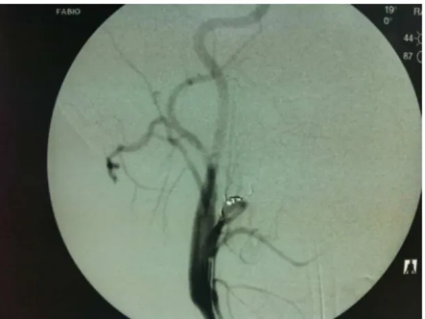
After hemodynamic stabilization, selective arteriography of the right carotid arteries was conducted, showing injury to branches of the right external carotid artery, with considerable contrast leakage (Figure 2).
Superselective catheterization was performed using an Echelon 14
microcatheter (eV3 Neurovascular, Inc.) in a coaxial system, and embolization was achieved using controlled release 2 mm × 2 mm and 2.5 mm × 2 mm Micrus
microcoils (Micrus Endovascular Corporation. CA/ USA), with immediate control of bleeding (Figure 3). The patient recovered in the intensive care unit for 24 hours without further intercurrent conditions and without requiring blood transfusion, with hemodynamic stability maintained and no further bleeding. She was discharged from hospital 48 hours after embolization. The material removed was sent for pathoanatomical assessment and histological
analysis conirmed a laryngeal Schwannoma. The
patient attended outpatients follow-up for 30 days with the vascular surgery team and showed no sign of relapse of bleeding, laryngeal symptoms exhibited improvement and there was no evidence of neurological damage.
DISCUSSION
Schwannomas of the larynx present as tumors with extensive vascularization and slow growth and symptomology is caused by their growth and mass
INTRODUCTION
Schwannomas are benign tumors that typically grow slowly, are encapsulated and have extensive vascularization. They get their name from the fact that they originate in the Schwann cells of motor, sensory or cranial nerves, but do not contain nervous elements. The most common site is the side of the neck, originating in the cervical nerve roots and the cervical sympathetic trunk1. They are rarely found
in the larynx and there are only sporadic reports of this presentation in the literature. Laryngeal Schwannomas account for less than 0.5% benign
tumors of the larynx and the irst choice treatment
is surgery, despite the fact that degeneration to malignancy is extremely rare2-7.
CASE DESCRIPTION
The patient was a 29-year-old female (S.R.X.) with a history of progressive solids dysphagia and a sensation of pharyngeal bolus. After initial evaluation by the otorhinolaryngology team, videolaryngoscopy
and computed tomography of the neck identiied
an encapsulated solid tumor of the larynx with an intimate relationship with the right external and internal carotid arteries (Figure 1).
T h e p a t i e n t w a s o p e r a t e d o n b y t h e otorhinolaryngology team under general anesthetic. A transoral approach was used to achieve total resection of the tumor. However, after removal of the tumor, the patient exhibited massive and persistent
Figure 1. Computed tomography with venous contrast and axial cross-section, showing laryngeal tumor with an intimate relationship with the right-side carotid arteries.
Figure 2. Selective arteriography with digital subtraction, showing contrast leaking via branches of the right external carotid artery.
Emergency after resection of a laryngeal Schwannoma
effects. The treatment of choice is surgical resection and access can be intraoral, by direct laryngoscopy, or via an external approach (lateral pharyngotomy or
laryngoissure)8,9, depending on the tumor’s anatomic
characteristics. The transoral approach is associated with a lower incidence of recurrent paralysis of the laryngeal nerve during the postoperative period, but
complete resection of the tumor can prove dificult
using this approach4.
Arteriographic assessment and preoperative embolization are described for management of head and neck Schwannomas10, but no cases of
embolization of a laryngeal Schwannoma, whether preoperative or postoperative, were found in the literature, probably because of the rarity of the presentation.
This paper described a rare case of a patient with a Schwannoma of the larynx who required immediate emergency embolization to control severe hemorrhage.
ACKNOWLEDGEMENTS
We are grateful to Dr. Gustavo Jorge of the otorhinolaryngology team at the Hospital São Francisco de Assis for the referral and for following the case in conjunction.
REFERENCES
1. Fini-Storchi I, Frosini P. Laryngeal neurinomas (a case report and review). ORL. 1997;59:182-5. PMid:9186976. http://dx.doi. org/10.1159/000276935
2. Palva T, Jokinem K, Karja J. Neurilemmoma (schwannoma) of the larynx. J Laryngol Otol. 1975;89:203-7. PMid:1123570. http:// dx.doi.org/10.1017/S0022215100080270
3. Phang WK, Raman R, Jayalaksmi E. Neurogenous tumour of the larynx (a case report). J Laryngol Otol. 1987;101:1209-10. PMid:3694036. http://dx.doi.org/10.1017/S0022215100103512
4. Takumida M, Taira T, Suzuki M, Yajin K, Harada Y. Neurilemmoma of the larynx (a case report). J Laryngol Otol. 1986;100:847-50. PMid:3734607. http://dx.doi.org/10.1017/S0022215100100180
5. Barnes L, Ferlito A. Soft tissue neoplasms. In: Ferlito A. Neoplasms of the larynx. Edinburgh: Churchill Livingstone; 1993. p. 277-9.
6. Supance JS, Quenelle DJ, Crissman J. Endolaryngeal neuroibromas. Otolaryngol Head Neck Surg. 1980;88:74-8. PMid:7393604.
7. Delozier HL. Intrinsic malignant schwannoma of the larynx: a case report. Ann Otol Rhinol Laryngol. 1982;91:336-8. PMid:7092059.
8. Schaefer BT, Som PM, Biller HF, Som ML, Arnold LM. Schwannoma of the larynx: review and computed tomographic scan analysis. Head Neck Surg. 1986;8:469-72. PMid:3721890. http://dx.doi. org/10.1002/hed.2890080613
9. Whittam DE, Morris TMO. Neurilemmoma of the larynx. J Laryngol Otol. 1970;84:747-50. http://dx.doi.org/10.1017/ S0022215100072480
10. Abramowitz J, Dion JE, Jensen ME, et al. Angiographic diagnosis and management of head and neck schwannomas. AJNR. 1991;12:977-984. PMid:1950934.
Correspondence
Fabio Augusto Cypreste Oliveira Av. Alphaville Flamboyant, 3900, casa 283 CEP 74884527 - Goiânia (GO), Brasil Fone: +55 (62) 81475111 E-mail: [email protected]
Author’s information
FACO, CESA and FLC are physicians with a Lato Sensu degree in Vascular Surgery – Angioradiology and Endovascular Surgery from Sociedade Brasileira de Angiologia e Cirurgia Vascular (SBACV), Colégio Brasileiro de Radiologia (CBR), and Associação Médica Brasileira (AMB). WS is vascular surgeon at Angiogyn. MCRA is hematologist at Angiogyn. JCB is resident physician of infectology at Hospital das Clínicas da Universidade Federal de Goiás. MCB is resident physician of intensive care at Hospital das Clínicas da Universidade Federal de Goiás. PMS is nurse technician and surgical technician at Angiogyn.
Author’s contributions
Conception and design: FACO Analysis and interpretation: FACO, CESA, FLC, WS Data collection: FACO, MCRA, JCB, MCB, PMS Writing the article: FACO, MCRA, JCB, MCB,WS Critical revision of the article: FACO, CESA, FLC Final approval of the article*: FACO, CESA, FLC Statistical analysis: FACO, CESA, FLC Overall responsibility: FACO, CESA, FLC Obtained funding: None.
*All authors should have read and approved of the inal version of the article submitted to J Vasc Bras.
Figure 3. Control arteriography after selective embolization. No sign of contrast leakage.