Measurement of the optical density of packable composites –
comparison between direct and indirect digital systems
Mensuração da densidade óptica de compósitos compactáveis –
comparação entre sistemas digitais direto e indireto
Luiz Felipe Rodrigues Graziottin* Nilza Pereira da Costa**
Ivori Dutra da Silveira* Elaine Bauer Veeck**
ABSTRACT: The aim of this study was to me a su re the op ti cal den sity of four pac ka ble com po si te re sins with widths of 1, 2, 3 and 4 mm, by me ans of Di go ra® (di rect) and Dent Scan Dent Vi ew® (in di rect) di gi tal ima ging systems, in or der to com pa re both methods. Twenty acr ylic pla tes, with the pro po sed thick nes ses, were used, each one con ta i ning a sam -ple of each re sin. Each acr ylic pla te was ra di o grap hed three ti mes, un der a stan dar di zed tech ni que. For the Di go ra® system, an op ti cal pla te was used un der each re sin sam ple, and, for the Dent Scan Dent Vi ew® system, oc clu sal films were em plo yed, to ta li zing 60 ex po su res for each system. Opti cal pla tes and films were scan ned and three con se cu ti ve op ti cal re a douts were car ri ed out, to ta li zing 1,440 re a douts. The re sults were sub mit ted to sta tis ti cal analy sis and re ve a led that the ave ra ge op ti cal den sity of the four re sins al ways in cre a sed as thick ness in cre a sed. Re gar ding the com -pa ri sons bet we en the com po si te re sins, in both analy sis the re sin with the gre a ter op ti cal den sity was Su re fi l fol -lowed by Pro dig y Con den sa ble, Aler t and So li ta i re®. The cor re la ti ons bet we en the re sults of Di go ra® and Dent Scan Dent Vi ew® were sig ni fi cant for the dif fe rent thick nes ses and ma te ri als. The ob ser ved ten dency is that as the va lu es ob -ta i ned with the Di go ra® system in cre a se, so do the va lu es ob ta i ned with Dent Scan Dent Vi ew®. Whi le thick ness in cre a -sed, the va lu es of op ti cal den sity in both Di go ra® and Dent Scan Dent Vi ew® ten ded to ap pro ach each ot her. The Di go -ra® system pre sen ted smal ler am pli tu de bet we en the re sults ob ta i ned in ad ja cent thick nes ses.
DESCRIPTORS: Com po si te re sins; Ima ge pro ces sing, com pu ter-as sis ted; Ra di o grap hic image enhan ce ment.
RESUMO: O objetivo deste estudo foi mensurar a densidade óptica de quatro resinas compostas compactáveis nas espessuras de 1, 2, 3 e 4mm utilizando os sistemas de digitalização de imagens Digora® (direto) e DentScan DentView® (indireto) e compará-los en tre si. Foram utilizadas 20 placas de acrílico nas espessuras propostas, cada uma contendo uma amostra de cada resina. Cada placa acrílica foi radiografada três vezes, sob técnica padronizada, usando, para o sistema Digora® uma placa óptica sob cada amostra de resina, e para o sistema DentScan DentView®, filmes oclusais, totalizando 60 exposições para cada sistema. Após, as placas ópticas e os filmes foram escaneados, e fez-se três leituras ópticas consecutivas de cada imagem, totalizando 1.440 leituras. Os resultados submetidos à análise estatística, permitiram concluir que, foi sempre crescente a média de densidade óptica das quatro resinas com o aumento da espessura. Quanto à comparação en tre as resinas compostas, nas duas análises a de maior densidade óptica foi a Surefil, seguida em ordem decrescente, pelas resinas Prodigy Condensable, Alert e Sol i taire®. As correlações en tre os resultados do Digora® e DentScan DentView® foram significativas para as diferentes espessuras e materiais. A tendência observada é que à medida que os valores do Digora® aumentam, os valores do DentScan DentView® também aumentam. Com o aumento das espessuras, os valores das médias de densidade óptica das análises efetuadas com o Digora® e com o DentScan DentView® tenderam a se aproximar. O sistema Digora® apresentou, comparado com o DentScan DentView®, uma menor am pli tude en tre os valores das espessuras próximas.
DESCRITORES: Resinas compostas; Processamento de imagens assistido por computador; Intensificação de imagem radiográfica.
INTRODUCTION
Re cently, the in dus try has in tro duced packable com pos ite res ins of high vis cos ity to the mar ket. It aimed at join ing the char ac ter is tics of amal gam with the aes thetic and ad he sive fea tures of com
-pos ite res ins. Packable res ins are ma nip u lated in the same way as the well-known sil ver amal gam. Christensen3
(1998) states that the dif fer ences for merly faced by pro fes sion als in confecting res to ra tions with stan dard com pos ite res ins – such as de -fec tive prox i mal con tour, blank spaces or voids in
the ma te rial and lack of con tact point – can be by -passed with the uti li za tion of packable com pos ite res ins and im proved ma tri ces. Nash et al.10
(1998) sus tain that den tists have now avail able an aes -thetic re stor ative ma te rial whose tech nique they are fa mil iar with. Ac cord ing to Iório8
(1999), ma te -ri als of this na ture will re place hy b-rid and microhybrid com pos ite res ins in pos te rior teeth res to ra tions, and the uti li za tion of cur rent multi-pur pose res ins will be re stricted to an te rior teeth.
Muñoz Chávez et al.9
(1999) and Porto Neto, Machado11
(1999) con sider that, in or der to ob tain a com plete suc cess in the re stor ative treat ment with com pos ite res ins, a pe ri odic fol lowup is re -quired for clin i cal and, if nec es sary, ra dio graphic eval u a tions of pa tients.
How ever, to as sess the sit u a tion of res to ra tions in pe ri odic ra dio graphic con trols, be sides good-qual ity ra dio graphs, it is nec es sary that the re stor ative ma -te rial pres ents good radiopacity. Radiopacity must be ad e quate so that one can: dis tin guish the re stor -ative ma te rial from ad ja cent den tal struc tures, as well as from sec ond ary car ies or decalcified dentin; eval u ate mar ginal ad ap ta tion in the cer vi cal mar gin; eval u ate the prox i mal con tour of the res to ra tion and iden tify in ter nal voids within the ma te rial, as stated by Figueredo et al.5
(1999) and Iório8
(1999).
Farman et al.4 (1996) men tion that the search for aes thet i cally sat is fac tory res to ra tions with out mer cury jus ti fies the im por tance of the im age qual ity of new re stor ative ma te ri als.
In 1987, Watts14
rec om mended that, when data on the radiopacity of dif fer ent ma te ri als are com -pared, val ues should be ex pressed “in equiv a lent alu mi num thick ness” (pu rity of 99.5%). How ever, Figueiredo et al.5
(1999) con sider that the dig i tized im age ap pears as a new re source for the eval u a tion of op ti cal den sity – its unit, as sessed by means of the com puter, is called pixel.
The struc tural com po si tion of packable com pos ite res ins arises sci en tific cu ri os ity as to dis -tinc tions be tween the var i ous com mer cial brands, es pe cially re gard ing ra dio graphic den sity and its ex pres sion. The re sult has great clin i cal im por -tance, since it is a mat ter of con cern not only for ra di ol o gists but also for cli ni cians, who must dis -tin guish com pos ite res ins from other ma te ri als, den tal struc tures, car ies le sions and other al ter -ations.
Com paring di rect and in di rect dig i tal sys tems, we aimed at un der stand ing a bit more about this
new tech nol ogy, and also to know more about the pos si bil ity of in te grat ing both sys tems in the anal -y sis of op ti cal den sit-y.
Con sidering the ex posed rea sons, the sur vey is jus ti fied. It aims to eval u ate and com pare the op ti cal den si ties of dif fer ent packable com pos ite res -ins, in spec i mens of 1, 2, 3 and 4 mm, by measn of dig i tal ra dio graphic im ages, also com par ing the re -sults ob tained by means of two dif fer ent sys tems of dig i tal ra di ol ogy.
MATERIALS AND METHOD
Dur ing this sur vey, four brands of light-cur ing packable com pos ite res ins were used: Alert (Je -neric/Pentron) shade clear, Prodigy Condensable (Sybron/Kerr) shade A1, Sol i taire®
(Heraeus Kulzer) shade I and Surefil (Dentsply/Caulk) shade A, all within their valid terms.
Preparation of samples
Twenty trans lu cent acrylic plates of 1, 2, 3 and 4 mm were confected with 5.7 cm x 7.1 cm. They were cal i brated us ing a 1-mm-ac cu racy thick ness
gauge (Mitutoyo), and there were five plates of each thick ness.
Each plate was di vided in four quad rants. A hole with the di am e ter of 4 mm was confected at the cen tre of each quad rant. The depth of each hole cor re sponded to the to tal thick ness of each plate. The hole served as a site for the in ser tion and com pac tion of the ex am ined res ins.
At the bor der of each hole of the same plate, a round clock wise niche was confected, each one cor re spond ing to the po si tion of clock fig ures 3, 6, 9 and 12. These niches served as ref er ences for the in ser tion of the re spec tive resin and were used to drain the ex cess of ma te rial.
To iden tify the thick ness of plates, each one re ceived on its left up per edge the cor re spond ing num -ber of per fo ra tions with a num -ber 2 spher i cal drill. The per fo ra tions were then filled with con ven tional com pos ite resin, to im prove their vi su al iza tion. Like -wise, with the in ten tion of in di vid u al iz ing each plate, 1 to 5 ver ti cal in ci sions were confected in the up per edge of each plate of equal thick ness, which were also filled with con ven tional com pos ite resin.
The re stor ative ma te ri als were in serted and com pacted in their re spec tive or i fices, with out hand con tact, since the op er a tor’s hands were cov -ered with rub ber gloves (Polymed®
) for clin i cal pro ce dures. Resins were in serted and com pacted fol -low ing the al pha betic or der of the ma te ri als’ com mer cial names, in a clock wise se quence, start -ing at the up per left quad rant. Thus, the Alert resin was al ways in serted in the or i fice lo cated in the up per left quad rant, Prodigy Condensable in the or i fice of the up per right quad rant, Sol i taire®
in the or i fice of the lower right quad rant and Surefil in the or i fice of the lower left quad rant.
In ser tion was car ried out with a plas tic amal -gam con tainer, in in cre ments of 2mm. Com pac -tion was car ried out with a spat ula for com pos ite resins (num ber 6 – Thomp son). Each in cre ment was light-cured for 40 sec onds (Gnatus Opt light 600), with the in ten sity of light checked through a radio meter (Cure Rite Efos), which re vealed the value of 420 mW/cm².
Af ter the com pac tion of the last or sin gle in cre -ment, the ma te rial was pressed by means of a trans lu cent smooth glass plate cov ered with a plas tic film for trans lu cency (Maxprint®
) cut in the same size, so as to limit the in serted resin thick -ness and to level its sur face. Spe cial care was taken to avoid to tal seal ing of the niche area, so that the ex cess of resin could flow with out re sis -tance. Light-cur ing was than car ried out for 10 sec onds over the kit glass plate/plas tic film. Once ceased this first light-cur ing step, the glass plate was re moved and light-cur ing was com pleted for an other 30 sec onds.
Af ter light-cur ing, plates were pro tected with foodpack ing PVC film, in or der to pre vent con tam -i na t-ion.
Ob tain ment of ra dio graphs by means of the
Digora
®dig i tal sys tem
Four smallsized op ti cal plates, with the di men -sions of 26 x 35 x 1.6 mm, from the di rect dig i tiz ing sys tem Digora® were used (Soredex, Orion Cor po -ra tion, Hel sinki, Fin land).
A pre lim i nary test was car ried out. It var ied the ex po sure time in or der to de ter mine which dig i tal im age showed the best den sity and con trast, ac cord ing to the agree ment of two spe cial ists in den tal ra di ol ogy. This eval u at ing mo dal ity is a rule of the Dis ci -pline of Ra di ol ogy, School of Den tistry, PUC-RS.
The stan dard iza tion of the dig i tal im ages was ob tained ac cord ing to the fol low ing de scrip tion:
• Uti li za tion of an x-ray de vice (Dabi-Atlante® ) with elec tri cal re gime of 70 kVp, 50/60 Hz and 8 mA;
• Ex po sure time of 0.05 s;
• main te nance of 40 cm of fo cal dis tance;
• x-ray cen tral beam per pen dic u lar to the cen ter of the acrylic plate.
Each set of plates (five plates) was radiographed in the same ses sion. The op ti cal plates, be fore each ex po sure, were prop erly packed in their in di vid ual plas tic en ve lopes that ac com pany the sys tem.
Each acrylic plate was radiographed as fol lows: on a Formica sur face ref er ences were drew so as to al low stan dard ized po si tion ing of the acrylic plates/op ti cal plates set. Each op ti cal plate was placed un der each acrylic plate quad rant so that each resin spec i men was as cen tral ized as pos si -ble. Af ter a 0.05-sec ond ex po sure, the op ti cal plates were sub mit ted to scan ning and dig i tiz ing.
Three ra dio graphs of each plate were ob tained, totalizing 15 ra dio graphs of each thick ness in or der to min i mize the prob a bil ity of er ror due to pos si ble vari a tion of the elec tric cur rent dur ing the ob -tain ment of ra dio graphs or dur ing the scan ning pro cess.
Ob tain ment of ra dio graphs with the Dentscan
Dentview
®dig i tal sys tem
Occlusal Ko dak®
films (num ber 4, Ektaspeed Plus) were uti lized. They had the same emul sion num ber (2100790 2000-10) and were within their va lid ity term.
A pre lim i nary test was car ried out by vary ing the ex po sure time, in or der to ver ify which ra dio -graph would pres ent the best vi sual con trast and den sity, ac cord ing to the agree ment of two spe cial -ists in den tal ra di ol ogy.
The stan dard iza tion of ra dio graphs was ob -tained by means of the fol low ing pa ram e ters:
• uti li za tion of an x-ray de vice (Dabi-Atlante®) with an elec tri cal re gime of 70 kVp, 50/60 Hz and 8 mA;
• ex po sure time of 0.4 s;
• main te nance of fo cal dis tance in 40 cm; • au to matic pro cess ing in an AT 2000 Air Tech
-niques Inc. ma chine, stan dard ized at 29ºC, with a pro cess ing time, of 4 min utes and 30 sec -onds (dry to dry) and uti li za tion of a Ko dak® X-OMAT fixer and de vel oper;
• x-ray cen tral beam per pen dic u lar to the cen ter of the film.
-tioned codes. Three ra dio graphs of each plate were ob tained, totalizing 15 ra dio graphs of each thick -ness, in or der to min i mize the prob a bil ity of er ror due to pos si ble vari a tion of the elec tric cur rent dur ing the ob tain ment of ra dio graphs, film de fects or fail ure dur ing film pro cess ing.
Image digitizing with the Digora
®digital system
Im age dig i tiz ing was car ried out in a room with poor light ing, by means of the Digora®
di rect im age dig i tiz ing sys tem (Soredex Orion Cor po ra tion, Hel -sinki, Fin land).
Each op ti cal plate was un packed and in tro -duced in the la ser read ing. The op ti cal plate that held the Alert resin im age was al ways the first to be in tro duced in the scan ner, al ways fol low ing the al pha bet i cal or der.
Each ra dio graph was iden ti fied, within the sys tem, by means of three dig its: the first cor re -sponded to the acrylic plate thick ness, the sec ond stood for the plate num ber and the third in di cated the num ber of the ra dio graphic shot. For ex am ple, num ber 432 meant: 4 mm of thick ness, plate num ber 3, 2nd
ra dio graphic shot.
For op ti cal read ing, an area of 20 x 20 pix els was se lected, with x and y co or di nates, lo cated near the cen ter of each resin spec i men. For each resin, three read ings were car ried out on each ra dio graph. Since there were three ra dio graphs from each plate, a to tal of 36 read ings by plate were ob tained. Once we worked with 20 plates, (5 for each thick ness), a to tal of 720 op ti cal read ings were ob tained.
Both im age dig i ta li za tion and op ti cal read ings were al ways car ried out by the same pro fes sional. The mean value of op ti cal den sity for each com -pos ite resin was writ ten down, and the data were an a lyzed by means of sta tis ti cal tests – anal y sis of vari ance (ANOVA) and Tukey mul ti ple com par i -sons tests at a sig nif i cance level of 1% – gen er at ing re sults which are pre sented in graphs.
Image digitizing with the DentScan DentView
®digital system
The APICA Eng. Ltda. DentScan DentView® Sys -tem - Den tal Tech nol ogies (1994) was uti lized.
The dig i tized ra dio graphic im age was ob tained by scan ning each ra dio graph, one at a time, with the aid of a proper sheet sup plied by the man u fac turer. The hands of the op er a tor were al ways pro -tected by gloves. The dig i tized ra dio graphic im age was en larged three times for better im age res o lu -tion. A dig i tal mil li me ter grid was ap plied over this
im age on the mon i tor, which al lowed to es tab lish den sity. The “avanzados” func tion and the den -sity-measuring tools were ap plied, which al lowed se lect ing the im age ar eas where op ti cal read out was car ried out through the dis play of num bers, af ter a click with the mouse.
For each of the 60 ra dio graphic im ages cor re spond ing to a packable com pos ite resin, three op -ti cal read outs were car ried out. Thus, for each resin there were 45 read outs of each thick ness, totalizing 180 op ti cal read outs from the same resin for the four stud ied thick nesses. Con sidering that four packable com pos ite res ins were stud ied, 720 op ti cal read outs were ob tained.
Both im age dig i ta li za tion and op ti cal read ings were al ways car ried out by the same pro fes sional. The out comes were ex pressed in graphs and an a lyzed by means of the sta tis ti cal test ANOVA.
RESULTS
Based on the ob jec tives and meth od ol ogy em -ployed in the pres ent re search, whose aim was to eval u ate and com pare the op ti cal den sity of packable com pos ite res ins in dif fer ent thick nesses by means of the Digora®
and DentScan DentView® dig -i t-iz -ing sys tems, -it was pos s-i ble to gather data, which were an a lyzed by means of the fol low ing sta tis ti cal tests: anal y sis of vari ance (ANOVA), mul ti -ple com par i sons Tukey test with the sig nif i cance level of 1%, as well as cor re la tion anal y sis (Pearson cor re la tion co ef fi cient).
Comparison between thicknesses by means of
the DentScan DentView
®digital system
Through the anal y sis of vari ance the re sults re -vealed that for the four packable res ins – Alert, Prodigy Condensable, Sol i taire®
and Surefil –, the thick ness of 1mm showed the small est val ues of op ti cal den sity, fol lowed by 2 , 3 and 4 mm. The two lat ter thick nesses did not dif fer from each other (p = 0.01) (Graph 1).
Comparison between thicknesses by means of
the Digora
®digital system
Through the re sults of the anal y sis of vari ance, we could ob serve that, for the four packable res ins – Alert, Prodigy Condensable, Sol i taire®
all the oth ers, pre sent ing the high est value of op ti -cal den sity (p = 0.01) (Graph 2).
Comparison between resins by means of the
DentScan DentView
®digital system
By means of the anal y sis of vari ance, it was ob -served that, with the thick ness of 1mm, the Sol i -taire® resin shows the small est val ues of op ti cal den sity, fol lowed, in in creas ing or der, by Alert and Prodigy Condensable, and, fi nally, by Surefil, which pres ents a higher value, al though it was not sta tis ti cally dif fer ent from Prodigy Condensable (p = 0.01) (Graph 3).
It was ob served that, for the thick nesses of 2, 3 and 4 mm, Sol i taire®
pre sented the low est val ues,
fol lowed, in in creas ing or der, by Alert and Prodigy Condensable, and, fi nally, by Surefil. There were sta tis ti cal dif fer ences be tween all of them (p = 0.01) (Graph 3).
Comparison between resins by means of the
Digora
®digital system
By means of the anal y sis of vari ance, it was ob -served that, for the thick nesses of 1 and 3 mm, Sol i taire®
pre sented the low est value of op ti cal density, fol lowed by Alert and Prodigy Con -densable, which did not dif fer from each other, and fi nally, by Surefil, which pre sented the high est value, al though it was not sta tis ti cally dif fer ent from Prodigy Condensable (p = 0.01) (Graph 4).
GRAPH 1- Mean va lu es of op ti cal den sity of each of the four pac ka ble com po si te re sins, using the Dent Scan Dent Vi ew® di gi tal system. Por to Ale gre, 2002.
GRAPH 2 - Mean va lu es of op ti cal den sity of each of the four pac ka ble com po si te re sins, using the Di go ra® di gi tal
system. Por to Ale gre, 2002.
GRAPH 3 - Mean va lu es of op ti cal den sity of each of the four pac ka ble com po si te re sins, in the dif fe rent analy zed thick nes ses, using the Dent Scan Dent Vi ew® di gi tal
system. Por to Ale gre, 2002.
GRAPH 4 - Mean va lu es of op ti cal den sity of each of the four pac ka ble com po si te re sins, in the dif fe rent analy zed thick nes ses, using the Di go ra® di gi tal system. Por to Ale
It was ob served that there are sig nif i cant dif fer -ences be tween the av er ages of op ti cal den sity of the four stud ied res ins for the thick ness of 2 mm (p = 0.01). Sol i taire® pre sented the low est value, fol lowed by Alert, Prodigy Condensable, and Surefil (p = 0.01) (Graph 4).
It was also ob served that, for the thick ness of 4mm, Sol i taire® pre sented the low est value of op ti -cal den sity, fol lowed by Alert, and Prodigy Condensable, which dif fered from each other, and fi nally, by Surefil resin, which pre sented the high est value, al though with no sta tis ti cal dif fer
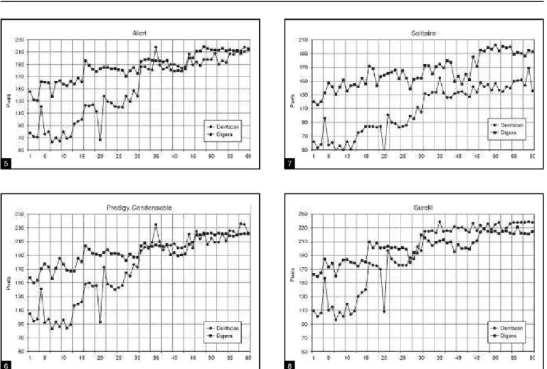
GRAPHS 5, 6, 7 and 8 Com pa ri son bet we en the va lu es of op ti cal den sity ob ta i ned by me ans of the Dent Scan Dent -View® and Di go ra® di gi tal systems, for each of the four pac ka ble com po si te re sins. Por to Alegre, 2002.
T ABLE 1 Re sults of the analy sis of cor re la ti on, con si -de ring thick nes ses. Por to Ale gre, 2002.
Thickness Pearson correlation coefficient between Digora®
e DentScan DentView® p
1 mm 0.748 0.001
2 mm 0.765 0.001
3 mm 0.873 0.001
4 mm 0.927 0.001
TABLE 2 Re sults of the analy sis of cor re la ti on, con si -de ring ma te ri als. Por to Ale gre, 2002.
Material Pearson correlation coefficient between Digora®
e DentScan DentView® p
Alert 0.868 0.001
Pro digy
Con den sa ble 0.861 0.001
So li ta i re®
0.792 0.001
ence in re la tion to Prodigy Condensable (p = 0.01) (Graph 4).
Comparison between the DentScan
DentView
®and the Digora
®digital systems
In or der to as sess the oc cur rence of sig nif i cant cor re la tion be tween the mea sure ments ob tained by means of DentScan DentView®
and Digora® , anal y sis of cor re la tion was ap plied to the vari ables thick ness and ma te rial (Ta bles 1 and 2).
The cor re la tions be tween the re sults ob tained with Digora®
and DentScan DentView®
were sig nif i -cant, for dif fer ent thick nesses and ma te ri als. The ob served ten dency was that as the Digora®
val ues in creased, so did the DentScan DentView®
val ues (Graphs 5, 6, 7 and 8).
DISCUSSION
The uti li za tion of dig i tal im age in this work was due to the fact that this tech nol ogy di min ishes the vari ables that cor re spond to hu man lim i ta tions, and also to the great re per cus sion of this tech nol -ogy in dif fer ent den tal spe cial ties. It of fers a great num ber of tools and ben e fits, such as the dra matic de crease in the ra di a tion dose, the de creased work ing time, the pos si bil ity of work ing on-line (im age trans mis sion), among oth ers.
Bustamante et al.2
(1998) stated that, in a near fu ture, dig i tal sys tems will be part of the den tist’s in stru ments, for pa tients’ safety and pro tec tion. Thus, it is said that within a short pe riod of time, dig i tal im ag ing will be the cho sen meth od ol ogy for the eval u a tion of the op ti cal den sity of ma te ri als.
The method for the val i da tion of read ings em -ployed in this study was also uti lized by Farman et al.4 (1996); Gürdal, Akdeniz6 (1998); Figueiredo et
al.5 (1999) and Ramalho et al.12 (1999), who also used the dig i tal im age re source for the eval u a tion of the op ti cal den sity of other den tal ma te ri als.
In the com par i son be tween the dif fer ent an a lyzed thick nesses, the re sults ob tained by means of Digora® were sim i lar to the those ob tained with DentScan DentView®, for, in both, there was in creas -ing op ti cal den sity as thick ness in creased. There was no sta tis ti cally sig nif i cant dif fer ence be tween the thick nesses of 2 and 3 mm with Digora®, while the same was ob served be tween the thick nesses of 3 and 4 mm, when DentScan DentView® was em -ployed. As to the com par i son be tween the eval u ated com pos ite res ins, in both anal y ses, the great est op ti -cal den sity was ob served for Surefil, fol lowed by
Prodigy Condensable, Alert and Sol i taire® . The lat ter, in both anal y sis, al ways pre sented the smal lest op ti cal den sity. How ever, with DentScan Dent -View® there was no sta tis ti cally sig nif i cant dif fer ence be tween Surefil®
and Prodigy Condensable, for the thick ness of 1 mm. With Digora®
, the mean val ues of op ti cal den sity were sim i lar for Alert and Prodigy Condensable, and also for Prodigy Condensable and Surefil, for thick nesses of 1 and 3 mm. No statistically sig nif i cant dif fer ence was ob served be -tween the mean val ues of op ti cal den sity of Prodi gy Condensable and Surefil, for the thick ness of 4 mm, at the level of sig nif i cance of 1%. The de vi a tion stan dards of the val ues of op ti cal den sity an a -lyzed by Digora® were a lot in fe rior to the de vi a tion stan dards of the val ues of op ti cal den sity ob tained by means of DentScan DentView®
. The high op ti cal den sity of Prodigy Condensable and Surefil sug -gests that their in or ganic fill ing com po nents pres ent che mical el e ments with high atomic num ber, or that such com po nents are pres ent in greater con -centration, which pro vides them with higher radiopacity. Alert and Sol i taire®
pres ent lower val -ues of op ti cal den sity since they do not con tain chem i cal el e ments with high atomic num bers and pres ent lower con cen tra tions of in or ganic fil ling com po nents. These re sults are in ac cor dance with the in for ma tion pro vided by Leinfelder et al. (1999)
as to packable com pos ites – the au thors pres ent Surefil as a high-radiopacity resin, and Sol i taire®, as a low-radiopacity resin. How ever, they do not classificate the Prodigy Condensable resin.
Within the same thick ness, for each one of the res ins, Digora®
pre sented higher and more ho mog -e nous val u-es, which m-eans that th-er-e was low-er am pli tude. The same was re ported by Bustamante
et al.2
(1998), when they tested the va lid ity of both sys tems in a study on osseointegrated im plants, com par ing the ob tained re sults as to their dis tri bu tion stan dard and eval u at ing the in flu ence of ti -ta nium im plants on the op ti cal den sity of the perimplant area. Barros1
(2000) eval u ated the op ti -cal den sity of bone by means of dig i tized im ages, in the retromolar re gion of ten corpses, us ing the dig -i tal sys tems DentScan DentV-iew®
and Digora® (both in an in di rect way), and also re ported the lower am pli tude of the re sults ob tained by means of the lat ter sys tem.
The cor re la tions be tween the re sults ob tained with Digora®
sig nif i cant for dif fer ent thick nesses and ma te ri als. The ob served ten dency was that, as Digora val ues in creased, so did the val ues ob tained with DentScan DentView. This ten dency had al ready been re ported by Henkin7
(1999), in the in vivo
eval u a tion of the dif fer ences be tween the op ti cal den sity of the dentin ad ja cent to decalcified and sound im ages, in up per and lower pre mol ars, by means of DentScan DentView and Digora (both in an in di rect way). The au thor at trib utes the nu mer i cal dif fer ences be tween the sys tems to the dif -fer ent types of equip ment for im age cap tion.
Ramalho et al.12
(1999) also com pared the same gray level ob tained through dif fer ent dig i tal sys tems. In that work, in di rect dig i tized ra dio graphs were obatined by means of the DentScan DentView®
sys -tem and cap tured im ages were ob tained by means of the op ti cal plates of the Digora®
sys tem op ti cal plates. They were an a lyzed as to the mean gray level ob tained for root ca nals, be fore and af ter fill ing. The re sults re vealed sta tis ti cally sig nif i cant dif fer ences be tween the val ues, prov ing that each sys tem has its own scale of bright ness and con trast, and that the data ob tained in a spe cific sys tem should not be ex -tended to oth ers. The au thors be lieve that the main re spon si ble for the dif fer ences be tween the gray level val ues was the method of dig i tiz ing uti lized in each sys tem. In di rectly dig i tized ra dio graphs tend to pres ent higher vari a tion of the gray level, since read out de pends on the spe cific char ac ter is tics of the scan ner. In di rect dig i tiz ing sys tems of the CR type, such as Digora®
, the gray lev els of each pixel cor re spond to the op ti cal plate par ti cles of phos pho rous due to xra di a tion, and per haps, the re -sult of this con ver sion is more ex act. Any way, it is un der stood that di rect and in di rect dig i tiz ing sys -tems must have their own gray scale ref er ences for pos si ble dif fer ent anal y ses.
One can no tice that, with the in crease in thick nesses, the mean val ues of op ti cal den sity ob -tained by means of Digora®
and DentScan DentView®
tended to ap prox i mate. Versteeg et al.13
(1997) con sider that direct dig i -tal im ag ing is more ef fi cient than in di rect dig i -tal
im ag ing. Gürdal, Akdeniz6
(1998) stated that di rect dig i tal im ag ing de creases the loss of in for ma tion that can oc cur in in di rect dig i tiz ing. More works must be de vel oped in or der to con firm this state -ment.
CONCLUSIONS
Once the re sults on the op ti cal den sity of packable com pos ite res ins were an a lyzed by means of the Digora®
and DentScan DentView®
dig i tal im -age sys tems, it was pos si ble, with the meth od ol ogy pro posed in this study, to con clude that:
• the mean op ti cal den sity of the four packable com pos ite res ins al ways in creased as thick ness in creased, in both sistems;
• as to the com par i son be tween the com pos ite res ins, in both anal y ses, the resin with the great est op ti cal den sity was Surefil, fol lowed by Prodigy Condensable, Alert and Sol i taire® – the lat ter was al ways the one with the low est op ti cal den sity, in both anal y ses;
• in spite of the re li abil ity of the dig i tal meth ods uti lized here to mea sure the op ti cal den sity of packable com pos ite res ins, they should not be used in a same case, in dif fer ent stages. On the con trary, com par i sons should al ways be car -ried with the same method, since the nu mer i cal val ues that re fer to op ti cal den sity are dif fer ent in these sys tems. Even so, it was ob served that the cor re la tions be tween the re sults of Digora® and DentScan DentView®
were sig nif i cant for dif fer ent thick nesses and ma te ri als. The ob -served ten dency is that, as the val ues ob tained with Digora®
in crease, so do the val ues ob tained with DentScan DentView®
;
• with an in crease in thick ness, the val ues of op -ti cal den sity ob tained by means of Digora®
and DentScan DentView®
tended to ap prox i mate; • when com pared to DentScan DentView®
, the Digora®
sys tem pre sented a lower am pli tude be tween the val ues ob tained for ad ja cent thick -nesses.
REFERENCES
1. B ar ros FJB C. Ava li a ção óp ti ca da den si da de ós sea na re gião re tro mo lar em man dí bu las de ca dá ve res com te ci dos mo les in tac tos, atra vés do u so de ima gens di gi ta -li za das [Tese] Por to Ale gre: Fa cu l da de de Odon to lo gia, Pon ti fí cia U ni ver si da de Ca tó li ca do Rio G ran de do Su l; 2000.
2. Bus ta man te NP, Ve eck EB, Cos ta NP. Aná li se óp ti ca da den si da de de im plan tes de ti tâ nio e sua in fluên cia na re -gião pe ri im plan tar em di fe ren tes sis te mas de ra di o gra fi as di gi ta is. Rev Bras Cir Implant 1998;5(4):67-78.
4. Far man TT, Far man AG, Scar fe WC, Gold smith LJ. Opti cal den si ti es of den tal re sin com po si tes: a com pa ri son of CCD, sto ra ge Phosp hor, and Ektas pe ed Plus ra di o grap hic film. Gen Dent 1996;44:532-7.
5. Figueiredo JAP, Vidor M, Bento LW, Boucinha ACD, Leipelt K, Silveira BT, Barbisan AO, Caminha JAN. Avaliação da radiopacidade de quatro marcas de resinas compostas fotopolimerizáveis através de imagem digitalizada. Stomatos 1999;8:15-22.
6. Gür dal P, Akde niz BG. Com pa ri son of two met hods for ra -di o me tric eva lu a ti on of re sin-ba sed res to ra ti ve ma te ri als. Den to-Ma xil lo-Fa ci al Ra di o logy 1998;27(4):236-9. 7. Hen kin IMT. Ava li a ção da den si da de óp ti ca da den ti na, em
fa ces pro xi ma is de prémo la res hí gi dos e com cá rie de es mal te, atra vés da uti li za ção de sis te mas in di re tos de aná li se di gi tal. [Dis ser ta ção de Mes tra do] Por to Ale gre: Fa cul da -de -de Odon to lo gia, Pon ti fí cia Uni ver si da -de Ca tó li ca do Rio Gran de do Sul; 1999.
8. Iório PAC. Resinas Compostas. In: Iório PAC. Dentística Clínica: Adesiva e Estética. São Paulo: Santos; 1999. p. 88-124.
9. Muñoz Chávez OF, Reis JIL, Santos LM, Andrade MF. Resinas compostas compactáveis – relato de caso clínico. J Bras Clín Estét Odontol 1999;3(16):11-7.
10. Nash RW, Radz GM, Le in fel der KF. A re port on a new con den sa ble com po si te re sin. Com pen di um of con ti nu ing edu -ca ti on in den tistry 1998;16(3):230-7.
11. Porto Neto ST, Machado CT. Resinas condensáveis. J Bras Odontol Clín 1999;3(13):35-9.
12. Ra ma lho LMP, Sar men to VA, Spohr AM, Löf AS, Cos ta NP. Men su ra ção da den si da de óp ti ca de áre as de ima gens ra -diográficas – com pa ra ção en tre um sis te ma di gi tal di re to e um in di re to. Re vis ta Odon to ló gi ca da Uni ver si da de de San -to Ama ro 1999;4(2):48-50.
13. Versteeg CH, Sanderink GCH, Van Der Stelt PF. Ef fi cacy of dig i tal intra-oral ra di og ra phy in clin i cal den tistry. J Dent 1997;25(3-4):215-24.
14. Watts DC. Cha rac te ri za ti on of alu mi num ra di o pa city stan -dards for res to ra ti ve ma te ri als. J Dent 1987;15(4):175-7.