Rehabilitation after partial brachial plexus palsy
Reabilitação na paralisia parcial do plexo braquial
This study was performed at the Rede Sarah de Hospitais de Reabilitação, Brasília, DF, Brazil.
Submitted to SGP (Sistema de Gestão de Publicações/Manager Publications System) of RBCP (Revista Brasileira de Cirurgia Plástica/Brazilian Journal of Plastic Surgery).
Article received: August 3, 2010 Article accepted: October 18, 2010
Kátia torres Batista1
Hugo Joséde araúJo2
1. Plastic Surgeon at the Hospital Sarah Brasília, Full Member of the Sociedade Brasileira de Cirurgia Plástica (Brazilian Society of Plastic Surgery – SBCP), President of the Regional do Distrito Federal da SBCP, Brasília, DF, Brazil
2. Plastic Surgeon at the Hospital Sarah Brasília, Full Member of the SBCP, Brasília, DF, Brazil. ABSTRACT
A variety of muscle transfer techniques have been proposed to restore motion of the upper extremities following severe brachial plexus palsy. Paralysis of the deltoid and supraspi natus muscles can be treated with transfer of the trapezius muscle. Paralysis of the wrist,
hand, and digital extensor muscles can be corrected using the pronator teres, lexor carpi
ulnaris, and palmaris longus muscles if the median nerve is preserved. Here we describe the rehabilitation of a patient with an old partial injury to the right brachial plexus that primarily involved the upper trunk from the C6 root to the posterior cord. Weakness of the deltoid muscle, wrist, and digital extensor muscles was observed. Microsurgical repair of the brachial plexus had not been performed. Tendon transfer surgery was performed to improve wrist, hand, and digital extension. One year later, transfer of the trapezius muscle was performed to stabilize the shoulder. The success of muscle transfer in the treatment of the brachial plexus palsy required the surgeon’s specialization, the patient’s motivation, and a rehabilitation program.
Keywords: Brachial plexus/injuries. Muscle, skeletal/physiopathology. Muscle, skeletal/ transplantation.
RESUMO
Muitas transferências musculares têm sido defendidas para restaurar os movimentos do membro superior após paralisia grave do plexo braquial. A paralisia dos músculos deltoide e supraespinal pode ser tratada por meio de transferência do músculo trapézio. A paralisia dos músculos extensores de punho, mão e dedos, quando o nervo mediano está preservado,
pode ser corrigida com emprego dos músculos pronador redondo, lexor ulnar do carpo e
palmar longo. Os autores descrevem um caso de reabilitação de paciente portador de lesão parcial antiga do plexo braquial à direita, de predomínio em tronco superior, principalmente da raiz de C6 e de fascículo posterior. Foi evidenciada fraqueza dos músculos deltoide e extensores do punho e dos dedos, sem antecedentes de reparo microcirúrgico do plexo bra quial. Foi realizada, inicialmente, cirurgia de transferência tendínea para ganho de extensão de punho, mão e dedos e, após um ano, transferência do músculo trapézio, para estabiliza ção do ombro. O sucesso na transferência para tratamento de paralisia do plexo braquial requereu especialização do cirurgião, motivação do paciente e programa de reabilitação.
INTRODUCTION
Most traumatic lesions of the brachial plexus result from motorcycle accidents. The lesions can be located at the su praclavicular level (affecting roots and trunks) or the in fraclavicular level (affecting cords and terminal branches) and may be total or partial.
Surgical exploration may be acute, i.e., performed at the moment of trauma in the case of an open injury, an injury
associated with a vascular lesion, or total avulsion (C5‒
T1). Cases in which recovery of the complete lesion is not observed on electroneuromyography examination between
the irst 2 to 3 months and 12 months after the injury or
in which there is persistent shoulder and elbow paralysis are candidates for investigation. Treatment options include microsurgical exploration with neurolysis, repair, grafting, or nerve transfer, depending on root avulsion. Transfer options include moving the accessory nerve to the suprascapular nerve or the musculocutaneous nerve to the ulnar and inter costal nerves. Good functional results have been reported in the literature1,2.
The primary goals of brachial plexus reconstructive surgery include stabilization of the shoulder, restoration of the ability to hold objects between the arm and the thorax,
restoration of active lexion of the elbow against gravity,
and recovery of the protective sensitivity of the palm. From a functional point of view, reconstructive surgery of the brachial plexus restores elbow and shoulder function, wrist
and digital lexion, and digital and intrinsic muscle extension.
The occurrence of gradual changes in the shoulder joint,
which are classiied according to Birch3, Pearl4 and Waters et
al.5, and, should be noted. These 3 authors classify the injury
according to increasing severity of subluxation of the humeral head of the joint. Waters’ et al.5 classiication describes the
following 5 types of injury: normal (type I); deformity of the posterior glenoid cavity (type II); subluxation of the humeral head and glenoid plus dysplasia (type III); development of a
false glenoid cavity (type IV); and lattening of both the glenoid
cavity and the humeral head (type V). The degree of joint deformity can help the surgeon select the surgical procedure5.
In the case of avulsion, unsuccessful neurorrhaphy, or untreated and lateadmitted patients with injuries of the brachial plexus, tendon transfers are indicated for the reco very of upper extremity function1,2,68. Although various pro
cedures have been described, rehabilitation continues to be a challenge for surgeons, and the reporting of results remains scarce. However, it is worth noting that the goal of tendon transfer is to improve tendon balance and upper extremity function. Transfer of the trapezius muscle has been described for the treatment of paralysis of the deltoid and supraspinatus muscles13,6,7. In the case of paralysis of the forearm extensor
muscles in which elbow extension is preserved, transfer of
the pronator teres, lexor carpi ulnaris, and palmaris longus
muscles yields satisfactory results for shoulder stabilization as well as wrist, hand, and digital function815.
In the present report, we describe the surgical techniques and the rehabilitation program that were used in the treatment of a patient with an old brachial plexus injury that involved mainly the upper trunk, in particular from the C6 root to the posterior cord. Weakness of the deltoid muscle, wrist, and digital extensor muscles was observed. No evidence of previous microsurgical repair of the brachial plexus was observed.
CASE REPORT
A 34yearold man was admitted in 2003 with a lesion of the right brachial plexus caused by a fall from a tree that had occurred 18 months previously. No microsurgical repair had been performed.
On physical examination, the patient presented with hy potrophy and hypoesthesia to deep pressure in the entire right upper extremity from C4 to T1. His inability to abduct the
shoulder or actively extend the wrist, hand, and ingers as
well as evidence of subluxation revealed the involvement of the scapular region; the deltoid and supraspinatus muscles; and the wrist, hand, and digital extensor muscles (Figures 1
and 2). Elbow lexion-extension was preserved.
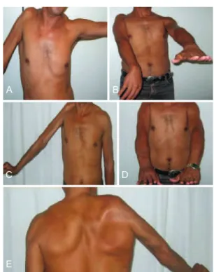
Figure 1 – In A and B, preoperative appearance showing shoulder
limitation and the inability to extend the wrist, hand, and ingers.
In C, D, and E, appearance in the 5th postoperative year with
abduction to 70° and wrist, hand, and digital extension.
A B
C D
Figure 2 – In A, preoperative appearance.
In B, appearance in the 7th postoperative year.
A B
Table 1 – Physiotherapy program performed for the radial paralysis transfer.
Time Program
Preoperative
EMG; sensorimotor map; maintenance of AOMs and strengthening of the muscles to be transferred; transfer training
Postoperative – 4th week
Immobilization in a plaster cast: elbow lexion of 90°, pronated forearm, wrist in semi-extension, MFJ lexion of 15°, extended IFJ; use of a sling; shoulder mobilization
Postoperative – 5th week
Transform the plaster cast into a splint with a free elbow; scar massage; desensitization with textures; use of turbulence, if necessary, to reduce joint rigidity (avoid proximity to the water jet); passive exercises and AOMs up to maximum muscle tension
Postoperative – 6th week
Maintain the splint, remove for training of the transferred muscles
Postoperative – 7th week
Remove the splint for exercises, bathing, and dressing; perform light manual activities; do not lex the wrist and
ingers simultaneously
Postoperative – 8th week
Stop the use of the splint; start light counter resistance exercises; allow effortless everyday activities
Postoperative –
6 months Control sensorimotor map
AOM = amplitude of movements; IFJ = interphalangeal joint; MFJ = metacarpo phalangeal joint; EMG = electroneuromyographic study.
An electroneuromyographic (EMG) study revealed an incomplete axonal injury of the right brachial plexus with severe involvement of the shoulder, wrist, and digital extensor muscles. There were severe signs of recent and chronic dener vation, an axonal injury of the lateral cutaneous nerve of the forearm, and a preganglionic injury (radiculopathy) at C6 with signs of moderate to severe denervation intensity.
In 2004, to restore wrist, hand, and digital extension, transfers were performed of the pronator teres muscle to the
extensor carpi radialis brevis muscle, the lexor carpi ulnaris
muscle to the extensor digitorum communis muscle, and the palmaris longus muscle to the extensor pollicis longus muscle
according to the modiied Burkhalter13 technique (Figure 3).
A physiotherapy program was started on the 30th postopera
tive day (Table 1).
In 2005, a transfer of the trapezius muscle with a segment of the acromion to the lateral surface of the humerus using the modi
ied Saha technique was performed to stabilize the shoulder
and permit arm abduction6,11 (Figure 4). The quality of the
glenohumeral joint was assessed via a radiological exam. Pre and postoperative physiotherapy programs were conducted (Table 2 and Figure 5). In the evaluation performed 7 years postoperatively, functional improvement and a muscle map
Figure 3 – Intraoperative view of transfer for wrist extension. In A, identiication of the pronator teres and extensor carpi radialis
brevis muscles. In B, release of the lexor carpi ulnaris muscle. In C,
transfer of the lexor carpi ulnaris muscle to the common extensor muscle of ingers. In D, palmaris longus muscle transferred to the
long extensor pollicis longus muscle.
A
C
B
D
Figure 4 – In A, patient in lateral decubitus position with a free
hand and arm. In B, curvilinear incision over the spine reaching the lateral margin of the scapula with exposed trapezius and deltoid
muscles, and lateral proximal third of the humerus. In C, release of the trapezius muscle insertion with a segment of the acromion and
ixation of the humerus with abduction to 90° using 2 lag screws. In
D, suture of the trapezius and deltoid muscles to the ixation.
A
C
B
Table 2 – Physiotherapy program performed for the shoulder transfer.
Time Program
Postoperative – 1st week of the
3rd month
Immobilization in a plaster cast with internal rotation and shoulder abduction to 80°; radiological assessment of the shoulder; assisted active elbow, wrist, and hand exercises; isometric contraction of the trapezius muscle and cocontraction of the rhomboid muscle
Postoperative – Start in the 3rd month
Radiological assessment of the shoulder; begin to remove the plaster cast for shoulder exercises, maintain abduction to 80°; gradually remove the immobilization Postoperative –
6th month to 7th year
Control muscular mapping; subjective evaluation
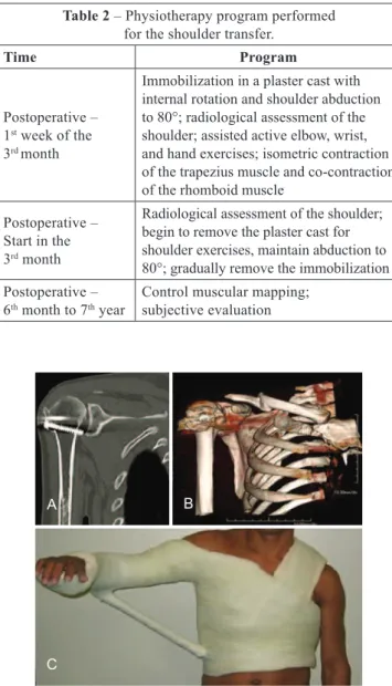
Figure 5 –In A, computed tomography examination. In B, three-dimensional reconstruction. In C, immobilization in a
plaster cast with abduction to 80° and internal rotation
of the shoulder (used for 4 months).
A B
C
with grade 4 wrist, hand, and digital extension; grade 3 thumb extension; and mobility and active elevation of the right shoulder with grade 4 muscle strength (60 degrees
of active abduction and 70 degrees of lexion) were seen.
The functional result was satisfactory, and the patient was considered rehabilitated and able to independently perform everyday activities and work activities.
DISCUSSION
Segmental injuries of the brachial plexus with shoulder paralysis and glenohumeral subluxation associated with the
paralysis of the forearm extensor muscles and the inability
to extend the wrist, hand, and ingers cause great functional
impairment of the upper extremities. According to Malessy et al.1 and Bertelli & Ghizoni2, injuries to the brachial plexus
that involve C5 and C6, such as the lesion described in this report, have a poor outcome after nerve reconstruction using grafts or nerve transfer procedures. However, some authors have reported satisfactory results after muscle tendon transfer to the shoulder16,11. Indications for surgical intervention in the
shoulder include subluxation, persistent internal contracture not resolved by physiotherapy, limited active abduction and external rotation, and a progressive deformity of the gleno humeral joint. The treatment is based on surgical contracture release, muscle balancing, and joint reduction.
Options for shoulder stabilization via muscle balancing
include transfers as described by Saha6 and Bateman7 and
shoulder arthrodesis8. Saha6 described a modiication of
Bateman’s7 technique that uses the trapezius muscle to
sta bilize and restore active function of the shoulder, and variable functional results have been reported. Rühmann et al.10 described an abduction gain of 70–90° along with
func tional recovery and subjective improvement, indings
that are in accordance with those described in the present report. The requirements for transfer are as follows: passive
shoulder abduction to 80°, patient agreement regarding the
long immobilization time, and satisfactory preoperative condition of the glenohumeral joints. If these requirements are not met, shoulder arthrodesis must be performed in the functional position, which carries a higher risk of morbidity and is considered the last resort7,8,11.
Satisfactory results have been reported with regard to
the use of the modiied Burkhalter transfer for the correction
of triple paralysis of the radial nerve13. This technique was
used in the case described in the present report with excellent functional improvement; however, the importance of preo perative evaluation and training, muscle traction line, and suture tension and immobilization should be noted. Other
options have been described, such as the transfer of the lexor carpi radialis muscle or the lexor digitorum muscles to the
extensor digitorum communis muscle1315.
In the present case report, rehabilitation of a patient with an old partial injury of the brachial plexus was achieved after performing a surgical transfer procedure to restore wrist,
hand, and digital extension using the pronator teres, lexor
carpi ulnaris, and palmaris longus muscles and to stabilize the shoulder using the trapezius muscle. The main factors that contributed to the success of the surgery included the preoperative preparation, including patient training and
participation, ixation of the trapezius muscle to the humerus,
REFERENCES
1. Malessy MJ, Ruiter GC, Boer KS, Thomeer RT. Evaluation of supras capular nerve neurotization after nerve graft or transfer in the treatment of brachial plexus traction lesions. J Neurosurg. 2004;101(3):377-89. 2. Bertelli JA, Ghizoni MF. Transfer of the accessory nerve to the su
prascapular nerve in brachial plexus reconstruction. J Hand Surg Am. 2007;32(7):989-98.
3. Birch R. Obstetric brachial plexus palsy. J Hand Surg Br. 2002;27(1):38. 4. Pearl ML. Arthroscopic release of shoulder contracture secondary to
birth palsy: an early report on indings and surgical technique. Arthros copy. 2003;19(6):577-82.
5. Waters PM, Smith GR, Jaramillo D. Glenohumeral deformity secondary to brachial plexus birth palsy. J Bone Joint Surg Am. 1998;80(5):668-77. 6. Saha AK. Surgery of the paralyzed and lail shoulder. Acta Orthop
Scand. 1967;97(Suppl):5-90.
7. Bateman JE. The shoulder and environs. St. Louis: C.V. Mosby; 1955. p. 383-93.
8. Aziz W, Singer RM, Wolff TW. Transfer of the trapezius for lail shoulder after brachial plexus injury. J Bone Joint Surg Br. 1990;72(4):701-4.
9. Mir-Bullo X, Hinarejos P, Mir-Batlle P, Busquets R, Carrera L, Navarro A. Trapezius transfer for shoulder paralysis: 6 patients with brachial ple xus injuries followed for 1 year. Acta Orthop Scand. 1998;69(1): 69-72.
10. Rühmann O, Schmolke S, Bohnsack M, Carls J, Wirth CJ. Trapezius transfer in brachial plexus palsy. Correlation of the outcome with mus cle power and operative technique. J Bone Joint Surg Br. 2005;87(2): 184-90.
11. Singh AK, Karki D. Modiied trapezius transfer technique for restoration of shoulder abduction in brachial plexus injury. Indian J Plast Surg. 2007;40(1):39-46.
12. Motta Filho GR, Mendes HM, Faria LOM. Transferências tendinosas para tratamento da paralisia radial. Rev Bras Ortop. 1990;25(10):341-8. 13. Burkhalter WE. Early tendon transfers in upper extremity peripheral
nerve injury. Clin Orthop Relat Res. 1974;104:68-79.
14. Boyes JH. Tendon transfers for radial nerve palsy. Bull Hosp Joint Dis. 1960;21:97-105.
15. Chuinard RG, Boyes JH, Stark HH, Ashworth CR. Tendon transfers for radial nerve palsy: use of supericialis tendons for digital extension. J Hand Surg Am. 1978;3(6):560-70.
Correspondence to: Kátia Torres Batista