Arq Bras Cardiol 2001; 77: 239-42.
Costa et al Laser for extraction of pacemaker
239 239 239 239 239
Instituto do Coração do Hospital das Clínicas - FMUSP
Mailing address: Roberto Costa – InCor - Av. Dr. Enéas C. Aguiar, 44 – 05403-000 – São Paulo, SP, Brazil – E-mail: rcosta@incor.usp.br
English version by Stela Maris C. e Gandour
Objective - To analyze the results of laser-assisted ex-traction of permanent pacemaker and defibrillator leads.
Methods - We operated upon 36 patients, whose me-an age was 54.2 years, me-and extracted 56 leads. The reasons for extracting the leads were as follows: infection in 19 pa-tients, elective replacement in 13, and other causes in 4 patients. The mean time of catheter placement was 7.5±5.5 years. Forty-seven leads were from pacemakers, and 9 we-re from defibrillators. Thirty-eight leads wewe-re in use, 14 had been abandoned in the pacemaker pocket, and 4 had been abandoned inside the venous system.
Results - We successfully extracted 54 catheters, ob-taining a 96.4% rate of success and an 82.1% rate for complete extraction. The 2 unsuccessful cases were due to the presence of calcium in the trajectory of the lead. The me-an duration of laser light application was 123.0±104.5 s, using 5,215.2±4,924.0 pulses, in a total of 24.4±24.2 cy-cles of application. Thirty-four leads were extracted from the myocardium with countertraction after complete pro-gression of the laser sheath, 12 leads came loose during the progression of the laser sheath, and the remaining 10 were extracted with other maneuvers. One patient experi-enced cardiac tamponade after extraction of the defibril-lator lead, requiring open emergency surgery.
Conclusion - The use of the excimer laser allowed ex-traction of the leads with a 96% rate of success; it was not effective in 2 patients who had calcification on the lead. One patient (2.8%) had a complication that required car-diac surgery on an emergency basis.
Key words: artificial cardiac pacemaker, lead extraction, la-ser light
Arq Bras Cardiol, volume 77 (nº 3), 239-42, 2001
Rober t o Cost a, Mar t i no Mar t i nel l i Fi l ho, El i zabet h Sar t or i Cr evel ar i , Noedi r Ant oni o Gr oppo St ol f , Sér gi o Al m ei da de Ol i vei r a
São Paulo, SP - Brazil
Laser Assisted Extraction of Pacemaker and Implantable
Defibrillator Leads
Original Article
The extraction of venous leads of permanent pacema-kers and defibrillators poses a great challenge to artificial cardiac stimulation, because of the high indices of failure and complications observed with the different techniques used.
The clinical conditions requiring extraction of these catheters are luckily not frequent and include infection in the stimulation system, venous thrombosis, repetitive pul-monary embolisms, or other situations associated with the presence of a catheter and that constitute a risk to the pati-ent’s life 1,2.
The routine practice of abandoning leads with no function has also become a problem with the use of multiple electrodes of the dual-chamber and multisite pacemakers. In the same way, the electrodes of implantable defibrillators, because of their large caliber and rough surface, also cause a severe problem when they require replacement or extrac-tion. This management increases the risk of pulmonary thromboembolism, syndrome of the superior vena cava, and, frequently, hinders the access route for placement of new leads.
Three major approaches for extracting venous electrodes have been used as follows: 1) direct external traction of the leads, which has a low success rate and a high risk of laceration of the cardiac and venous structures; 2) cardiotomy, usually performed with the aid of cardiopulmonary bypass; and 3) internal countertraction, which is performed with sheaths of Teflon or polypropylene with a good success rate 1.
240 240 240 240 240
Costa et al
Laser for extraction of pacemaker
Arq Bras Cardiol 2001; 77: 239-42.
Recent studies published in the literature have shown the superiority of the technique that uses the laser light, which has a higher rate of success in removing the fibrosis and a lower rate of complications.
The objective of this study is to report the results ob-tained with the use of the excimer laser for extracting perma-nent transvenous leads from 36 patients operated upon at the of the University of São Paulo. Heart Institute (Medical School)
Methods
During the period from September 1998 to June 2000, we operated upon 36 patients with ages ranging from 1 to 91 years (mean = 54.2±21.9 years). Twenty-two patients were males and 14 were females. The causes of the disorder in the cardiac conduction system were as follows: Chagas’ disea-se in 9 patients, degenerative didisea-seadisea-ses in 6 patients, ische-mic disease in 5, and other causes in 16 patients. At the mo-ment of extraction, 23 patients were bradycardic because of atrioventricular block in 17 patients and sinus dysfunction in 6 patients.
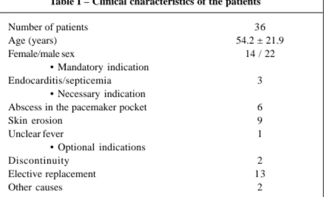
The reason for extracting the leads was infection in the stimulation system in 19 patients, elective replacement of the electrode in 13, and other causes in 4 (tab. I).
The catheters extracted had been implanted from 8 months to 21.8 years (mean = 90±66 months) previously, 47 being pacemaker leads and 9 being defibrillator leads. The leads had been in place longer in the group of patients with pa-cemakers (mean = 103.2±63.6 months). Forty-two leads had been implanted in the right ventricle and 14 in the right atrium. In the patients with defibrillators, the electrodes had been in place for a shorter period (mean = 24.0±10.8 months) (tab. II).
The access route through which the leads had been implanted was the cephalic vein for 10 leads, the subclavian vein for 40, the jugular vein for 3, and the femoral vein for 3 leads.
At the time when extraction was indicated, 38 leads were being used, 14 had been abandoned in the pacemaker pocket, and 4 had been abandoned inside the venous system.
General anesthesia was used in 34 patients, and local anesthesia associated with sedation was used in 2 patients. The CVX-300 excimer laser emits a beam of light of 308 nm, which is not visible to the human eye. It is a type of laser light that cuts using coldness: the temperature of the light emitted is approximately 50ºC. At the tissue depth of appro-ximately 0.06 mm, 64% of the energy is absorbed, and at the tissue depth of 0.18 mm, 95% of this energy is dissipated. Light is absorbed by lipids and proteins, but not by water, which is the major dissipation medium of other laser light modalities; this allows photoablation of the fibrous bands that surround the lead without damaging it, and with no harm to the venous and cardiac structures.
The beams of light are emitted by the distal extremity of flexible sheaths that are passed above the electrode to be extracted, completely encircling it after its release from the fibrous tissue, providing circumferential support to the tip of the electrode in the myocardium for the application of internal countertraction. The body of this sheath contains 82 optical fibers that conduct the laser light as far as their distal portions, each one with a diameter of 100 mm. Progres-sion of the laser sheath upon the lead is obtained as the light beam cuts the fibrous tissue, and it is followed by conti-nuous fluoroscopy. Total release of the lead is obtained when the sheath reaches the endocardium, in the cardiac chamber where the electrode is implanted.
Three different calibers of sheath exist: 12, 14, and 16 F, corresponding respectively to 2.8 mm, 3.4 mm, and 4.2 mm of internal diameter, and 4.1 mm, 4.8 mm, and 5.6 mm of exter-nal diameter. The proper choice of caliber of the sheath allows the extraction of any model of lead used in artificial venous cardiac stimulation.
Patients are preferably operated upon while they are under general anesthesia with orotracheal intubation. In the horizontal dorsal decubitus position, under continuous mo-nitoring of blood pressure and electrocardiography, the patients receive adhesive plates for cardiac stimulation and percutaneous defibrillation. Antisepsis of the skin is perfor-med with povidone iodine, and sterile fields are placed to al-low an easy access for sternal or lateral median thoracotomy. All material for thoracotomy should be easily accessible.
After opening the pacemaker pocket, and with the electrode already released from the generator of pulses, the lead is carefully dissected from the scare tissue that envelo-pes it, and the suture sleeve is extracted. A smooth manual traction is then applied to check whether the extraction is feasible without using the set of extraction sheaths, and with no damage to the lead. If the manual extraction is not possible, the extraction procedure proceeds.
Table I – Clinical characteristics of the patients
Number of patients 36
Age (years) 54.2 ± 21.9
Female/male sex 14 / 22
• Mandatory indication
Endocarditis/septicemia 3
• Necessary indication
Abscess in the pacemaker pocket 6
Skin erosion 9
Unclear fever 1
• Optional indications
Discontinuity 2
Elective replacement 13
Other causes 2
Table II - Characteristics of the extracted leads
Electrodes per patient 1.55
Atrial/ventricular 14/42
ICD/PM 9/47
Length of stay (months) 90±66
Arq Bras Cardiol 2001; 77: 239-42.
Costa et al Laser for extraction of pacemaker
241 241 241 241 241
The standard procedure for extracting the lead starts with extraction of the connector of the lead with a sterile wire cutter, and a long proximal portion of the lead is left to allow handling of the locking stylet and sheaths. As the distal part of the conductor may be deformed by the use of the wire cutter, a sharp conical dilator is used to expand the coil and allow the passage of the thickest possible locking stylet. A common guide for the pacemaker lead is passed at this time to assure that the lumen of the conductor is completely free as far as the distal portion of the lead, which is implanted in the myocardium, and also to provide an accurate measure-ment of the distance at which the locking stylet should be introduced. The internal diameter of the lumen of the con-ductor is then measured with a set of locking stylet measu-rers. The locking stylet with the greatest possible diameter is then introduced into the coil until the end of the lumen is reached and expanded, therefore, increasing the rigidity and the traction power of the lead. After this maneuver, a new traction may be applied to the lead to check whether its release from the myocardium is feasible without using coun-tertraction.
If the electrode is not freed, the set of sheaths (the in-ternal, which emits the laser light, and the exin-ternal, of poly-propylene) is applied to the lead, using it as a guide for their introduction. The applications of laser light are then started, each application lasting for 5 seconds, always accompanied by fluoroscopy. As the laser sheath progresses towards the heart, the external polypropylene sheath also advances, fre-eing the adherences of the lead to the venous and cardiac structures, until the most distal portion of the lead is reached by the laser sheath. At this point, the use of the laser light is interrupted, the laser sheath is used to provide circumferen-tial support to the myocardium, and, applying traction to the lead, it is extracted from the myocardium. Then, the entire set (external sheath, laser sheath, and lead) is extracted, and he-mostasis is performed at the site of introduction of the ca-theter by simple compression.
Results
Fifty-four catheters were extracted, resulting in a 96.4% rate of success. The extraction of 46 leads (82.1%) was complete. In 8 other leads, only the metallic tip remai-ned attached to the myocardium (14.3%), and only 2 leads could not be extracted at all (3.6%).
The duration of laser light application ranged from 20 to 540 s (mean = 123.0±104.5), and 800 to 23,380 pulses (mean = 5,215.2±4,924.0) were applied, in a total of 4 to 119 cycles of application (24.4±24.2).
The following mechanisms accounted for the release of the lead from the myocardium: 1) countertraction after complete progression of the laser sheath in 34 electrodes (60.7%); 2) traction during progression of the sheath in 12 (21.4%); 3) only traction after complete progression of the sheath due to the impossibility of countertraction (usually because of adhesion of the lead to the lumen of the sheath due to fibrosis) in 4 (7.1%); 4) only traction due to failure in
complete progression of the sheath in 2 (3.6%), and other forms in 4 (7.1%).
One patient had cardiac tamponade after extraction of the defibrillator lead, requiring open surgery on an emer-gency basis for correction of a laceration in the tip of the right ventricle.
The 2 unsuccessful cases were related to the presence of a large amount of calcium in the trajectory of the lead.
Discussion
In regard to extraction of leads, the state-of-the-art of artificial cardiac stimulation has revealed the advantages of the techniques that use internal countertraction when com-pared with other techniques.
The high rate of success obtained with the counter-traction techniques with mechanical dilators has been repor-ted in the literature. Smith et al3 have reported the following results obtained from 1988 to 1994 in the U.S. Lead Extrac-tion Database: 86.8% of complete extracExtrac-tion; 7.5% of incom-plete extraction; 5.7% of failure; 2.5% of severe complica-tions; and 0.6% of mortality. Byrd et al 4, analyzing data col-lected from January 1994 to April 1996 in the same database in a period when these techniques were considered reaso-nably stabilized, reported the results of extraction of 3,540 leads from 2,338 patients: 93% of complete extraction; 5% of partial extraction; 2% of failure in the procedure, and 1.4% of severe complications. Kantharia and Kutalek5 reported a 98% rate of success in the extraction of pacemaker and defi-brillator leads, with a 0.7% rate of severe complications, re-presented by 2 cases of cardiac tamponade.
Byrd et al 4, analyzing the results of the U.S. Lead Ex-traction Database, concluded that the risk of failure in ex-traction or of incomplete exex-traction increases as follows: the longer the time the lead is implanted (p<0.0001); the less experience the physician performing the procedure has (p<0.0001); when the electrode is implanted in the ventricle (p<0.005); and the younger the patient is (p<0.0001). On the other hand, the risk of complications increases as follows: the higher the number of leads to be extracted (p<0.005); the less experience the physician performing the procedure has (p<0.005); and in patients of the female sex (p<0.01).
According to Kantharia and Kutalek 5, the major cau-ses of failure of extraction are related to: a) the severity of the fibrous scar, which increases with the length of time that the lead is implanted; b) the experience of the physician per-forming the procedure; and c) the type of electrode to be ex-tracted. According to these authors, active fixation atrial leads coated with silicone are completely extracted more fre-quently than passive fixation ventricular leads coated with polyurethane. Fibrosis can be very intense at 4 months after implantation of electrodes coated with silicone, and at 6 months for those coated with polyurethane 5.
242 242 242 242 242
Costa et al
Laser for extraction of pacemaker
Arq Bras Cardiol 2001; 77: 239-42.
In 1999, Wilkoff et al 6 carried out a study in which 301 patients were randomized, using 2 techniques for counter-traction, as follows: in 1 group, mechanical dilators were used, and, in the other group, the excimer laser was used. They reported that the use of the laser light allowed the fol-lowing results: complete extraction of the leads in 94% of the cases; partial extraction in 2.5% of the cases; failure in 3.3% of the cases; and severe complications in 2.6% of the cases. They also reported a significant superiority (p = 0.001) in the results obtained with the use of the laser light (94% suc-cess for the laser-assisted procedure, and 64% sucsuc-cess for the conventional techniques) in a significant number of pa-tients, who, due to failure or difficulty in extracting the elec-trode by classical techniques, migrated to the group of pati-ents randomized for laser-assisted extraction (crossover in 72 electrodes) 6.
Kennergren 7, analyzing the results obtained by the European Multicenter Study on excimer laser-assisted ex-traction with 179 catheters approached in 149 patients, has reported the following: complete extraction of 89.5% of the electrodes; partial extraction of 6% of the electrodes; failure of extraction of 4.5% of the electrodes; severe complica-tions with 2.7% of the electrodes.
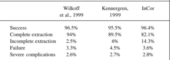
The results obtained in our study, when compared with those in the literature, show the reproducibility of the method. The 96.4% rate of success and 2.8% rate of severe complications are in accordance with the rates found in the literature, even though the incidence of incomplete extrac-tion (14.3%) was higher in our study. Using the conclusions of Byrd et al 4 and Kantharia and Kutalek 5 and analyzing data shown in tables III and IV, we can observe that the po-pulation reported in our study has a higher risk of failure and of incomplete extraction than those populations studied by Wilkoff et al 6 and Kennergren 7.
Our patients are a mean of 11 and 14 years younger than those studied by Wilkoff et al 6 and Kennergren 7; the leads in our cases had clearly been in place longer (39% and 32%, respectively), and the proportion of atrial leads as compared with the ventricular ones was lower in our study
Table III – Comparison of the results obtained with laser-assisted extraction of catheters
Wilkoff Kennergren, InCor et al., 1999 1999
Success 96.5% 95.5% 96.4%
Complete extraction 94% 89.5% 82.1% Incomplete extraction 2.5% 6% 14.3%
Failure 3.3% 4.5% 3.6%
Severe complications 2.6% 2.7% 2.8%
Table IV – Comparison of the populations undergoing laser-assisted extraction of catheters
Wilkoff Kennergren, Incor et al., 1999 1999
Number of patients 153 149 36
Mean age (years) 65±18 68.3 54.2±21.9 Permanence (months) 65±42 68.3 90±66
Leads/patient 1.59 1.2 1.55
Atrial/ventricular 125/118 104/57 14/42
Female sex 33% 35% 39%
(0.33 versus 1.06 and 1.83, respectively). All these factors suggest a higher risk of complications or of partial extrac-tion. The other 2 criteria observed, number of leads per pa-tient and proportion of female papa-tients, have similar figures for the 3 populations analyzed.
In conclusion, the use of the excimer laser has allowed extraction of catheters with a high success rate and a low level of complication. These complications, however, tend to be severe, requiring immediate action to save the pati-ent’s life, including the possibility of open thoracic surgery.
Acknowledgements
We thank Anísio Pedrosa, João Luiz Piccione, Neide Romão, Sérgio Siqueira, Silvana D’Ório Nishioka, and Wag-ner T. Tamaki.
1. Byrd CL, Shwartz SJ, Hedin N. Lead extraction: indications and techniques. Cardiol Clin 1992; 10: 735-48.
2. Klug D, Lacroix D, Savoye C, et al. Systemic infection related to endocarditis pacemaer leads. Clinical presentation and management. Circulation 1997; 95: 2098-107.
3. Smith HJ, Fearnot NE, Byrd CL, Wilkoff BL, Love CJ, Sellers TD. Five-years experience with intravascular lead extraction. U.S. Lead Extraction Database. Pacing Clin Electrophysiol 1994; 17: 2016-20.
4. Byrd CL, Wilkoff BL, Love CJ, et al. Intravascular extraction of problematic or
References
infected permanent pacemaker leads: 1994-1996. U.S. Extraction Database, MED Institute. Pacing Clin Electrophysiol. 1999; 22: 1348-57.
5. Kantharia BK, Kutalek SP. Extraction of pacemaker and implantable cardioverter defibrillator leads. Curr Opin Cardiol. 1999; 14: 44-51.
6. Wilkoff BL, Byrd CL, Love CJ, et al. Pacemaker lead extraction with the laser sheath: results of the pacing lead extraction with the excimer sheath (PLEXES) trial. J Am Coll Cardiol. 1999; 33: 1671-6.

