278 278 278 278 278
Gianisella et al
Pulmonary arteriovenous fistula in childhood
Arq Bras Cardiol 2001; 77: 278-81.
Instituto de Cardiologia do Rio Grande do Sul/Fundação Universitária de Cardiologia
Mailing address: Roberto Gianisella – Unidade de Pesquisa do IC/FUC – Av. Princesa Isabel, 395 – 90620-001 – Porto Alegre, RS, Brazil - E-mail: [email protected]
English version by Stela Maris C. e Gandour
Rober to Bar tol omeu Gi ani sel l a, Raul I vo Rossi Fi l ho, Paul o Zi el i nsk y
Porto Alegre, RS - Brazil
Diagnosis and Therapeutics of Pulmonary Arteriovenous
Fistula in Childhood. Case Report and Review of the Literature
Case Report
We report the case of a 3-year-old female patient, who, since birth, had cyanosis difficult to explain with usu-al diagnostic tests. The only findings on physicusu-al exami-nation were cyanosis and clubbing of her fingers. Chest computerized tomography showed images of excessive attenuation in the right lung, which resembled arteriove-nous fistulae that were later confirmed on cardiac cathete-rization. The fistulous trajectories were then embolized with 7 Gianturco coils, which resulted in an immediate in-crease in the arterial saturation of blood oxygen.
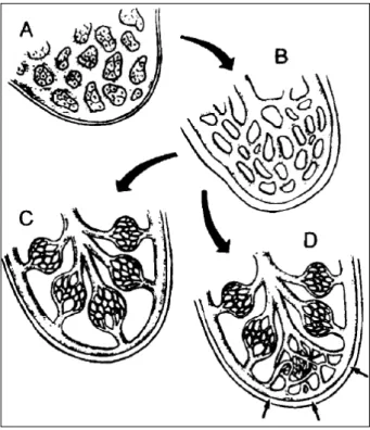
During the embryological development of the vascular system, which occurs between the 5th and 10th weeks of
in-trauterine life, a continuous differentiation of the vascular bed occurs, resulting in the creation of separate arterial and venous channels, interconnected by capillaries. When a mistake or halt occurs in this process of vascular differentia-tion, vascular malformations will appear at different anato-mical sites and with variable morphology (fig. 1), depending on the stage of differentiation 1. We report the diagnosis and
interventional therapeutics of arteriovenous fistulae in the lungs of a 3-year-old child.
Pulmonary arteriovenous fistulae are characterized by right-to-left shunt of variable magnitude, and the effect of these communications depends on the size of the vessels involved. If the anastomoses affect peripheral arterioles and venules, a small telangiectasia will result, and it usually remains small, not causing hemodynamic alterations in the pulmonary circulation. If larger veins and arteries are affected or if massive involvement of the pulmonary capillaries takes place, an increase in the size of the vessels may occur 2,
resul-ting in severe hemodynamic alterations. Arterial desatu-ration of oxygen, cyanosis, clubbing of the fingers, and
po-lycythemia may occur, secondary to the intrapulmonary shunt. Multiple neurological sequelae have been reported due to a variety of infectious, embolic, and hemorrhagic complications 3-5.
The etiology of pulmonary arteriovenous fistulae may be congenital or acquired. The congenital form has 2 varieties: a) cavernous angioma, usually fed by 1 or more tortuous and dilated branches of the pulmonary artery and b) capillary telangiectasia, which forms a net of capillaries and is usually associated with hereditary hemorrhagic telangiectasia 6 or Rendu-Osler-Weber
syndrome. This syndrome is an autosomal dominant vascular dysplasia with variable penetrance, has multiple telangiectasias, and is characterized by recurring hemorrhages. The levels of the coagulation factors are normal and the coagulation mechanism is intact. Ap-proximately 1/3 of the patients diagnosed with this syn-drome presents pulmonary vascular malformations. Some studies together with the scientific division of the Here-ditary Hemorrhagic Telangiectasia (HHT) Foundation International, Inc. reached a clinical consensus on the diagnostic criteria for this disease; 4 criteria should be sought: epistaxis, telangiectasias, visceral lesions, and a compatible familial history. The diagnosis of Rendu-Osler-Weber disease is established if 3 of the above-cited criteria are present. If less than 2 criteria are present, the diagnosis of the disease is very unlikely, even though children of parents affected should be considered at risk due to the fact that the penetrance varies with age. The clinical investigation of our patient showed only 1 of the cited criteria, which was pulmonary telangiectasia, making the diagnosis of Rendu-Osler-Weber syndrome unlikely.
Pulmonary arteriovenous fistula in its acquired form usually occurs in juvenile cirrhosis, but has also been repor-ted in patients with metastases of thyroid carcinoma and pulmonary schistosomiasis. In the latter, the pulmonary ar-teriovenous fistula occurs because of the vascular neofor-mation secondary to chronic vasculitis triggered by
Schis-tosoma eggs or their metabolic or degrading products.
Arq Bras Cardiol 2001; 77: 278-81.
Gianisella et al Pulmonary arteriovenous fistula in childhood
279 279 279 279 279
Case report
The patient is a 3-year-old white female child with pro-gressive clubbing of her fingers and cyanosis since birth. Physical examination did not disclose other abnormalities and no additional clinical symptoms were reported. Analy-sis of the arterial gases at room temperature showed pH = 7.5; PCO2 = 26 mm Hg; PO2 = 36mmHg; CO2T = 20.4 mmol/L; bi-carbonate = 19.4 mEq/L; saturation of O2 = 75%. Hematocrit = 48%; Hb = 15.5g/dL; MCV = 91. Electrocardiography, echo-cardiography, and total abdominal echography were within the normal range. The chest X-ray showed an interstitial infil-trate in the right base and perihilar regions.
Continuing with the investigation, a chest computeri-zed tomography was performed and revealed images of ex-cessive attenuation in the right lung, close to the posterior wall, with confluence of the vessels. These alterations raised the possibility of pulmonary arteriovenous fistulae (fig. 2).
Considering that pulmonary arteriovenous fistulae might be present, a cardiac catheterization was performed to elucidate the diagnosis. The angiographic study showed a vascular net with diffuse malformations in the right middle and inferior pulmonary lobes (fig. 3 and 4). Pressures in the cardiac cavities were as follows (in mmHg): RA = 2 (mean); PA = 15 (systolic), 9 (diastolic), 19 (mean); LV = 77 (systolic); Ao = 63 (systolic), 38 (diastolic), 49 (mean). Pulmonary scin-tigraphy confirmed the results of catheterization.
Because the interventional therapy was chosen, pul-monary arteriovenous fistulae were embolized with
catheteri-zation with 7 Gianturco coils, sizes 3 and 5 mm (figs. 5 and 6). The immediate result was an increase in oxygen saturation from 90% to 98%, with the patient sedated receiving oxygen by nasal catheter at the rate of 4 L/min (figs. 5 and 6). At the time of hospital discharge, oxygen saturation measured at room temperature with the pulse oximeter was 90%.
Prior to the embolization procedure of the fistulae, the right pulmonary artery was completely occluded with a bal-loon of an adequate size for investigating the existence of fistulae in the left lung. This hypothesis was discarded be-cause the hemoglobin saturation suddenly increased from 90% to 100%.
Discussion
Pulmonary arteriovenous fistula is not easily diagno-sed routinely, due to its rarity and its unspecific findings on routine examinations. However, this diagnostic hypothesis should always be considered when examining children with cyanosis, in whom the initial tests do not detect cardiac ab-normalities.
Two other alternatives in the diagnostic investiga-tion, which were not used in the present case, are the calcu-lation of the shunt through the saturations measured with catheterization in the different cardiac chambers 7 and
con-trast echocardiography 8,9, in which agitated saline solution,
for example, can be injected in a peripheral vein. The micro-bubbles resulting from this process and detected in the left atrium determine a right-to-left shunt through the lungs. The capillary alveolar filter does not allow the passage of these microbubbles.
Tc-99m MAA total body imaging 10,11 follows the same
principle, because the macroaggregates of albumin labeled with technetium-99 measure from 20 to 60 mm, but the diameter of the pulmonary capillary bed is smaller than 15 mm. Contrast medium uptake in other organs, in addition to the lungs, would show the presence of the shunt, and the pulmo-nary area with no radionuclide would delimitate its site 12.
Fig. 1 – Normal stages of vascular development according to Woolard1: a) primitive
mesenchyma; b) differentiation in plexiform structures; c) disappearance of the primitive elements and differentiation into vascular structures; d) inadequate development during the embryogenesis of the vascular system with persistence of the primitive vascular communications or arteriovenous malformations (small arrows).
280 280 280 280 280
Gianisella et al
Pulmonary arteriovenous fistula in childhood
Arq Bras Cardiol 2001; 77: 278-81.
Figs. 3 and 4 – Pulmonary arteriography showing multiple pulmonary fistulae.
Figs. 5 and 6 – Chest X-ray showing the coils (left) and one widened image of the right inferior lobe (right).
In the case we report, the diagnosis was established by chest spiral computed tomography, pulmonary scinti-graphy, and selective pulmonary angiography.
Interferon has been suggested as a less invasive the-rapeutical approach, but, until the present time, its efficacy for the treatment of pulmonary arteriovenous fistula has not been scientifically confirmed.
Another alternative would be occlusion of each pul-monary lobe with a balloon, and concomitant assessment of blood saturation, therefore checking whether selective embolizations with detachable balloon 13-15 could replace
pneumectomy 16. Embolization with coils has also been
suc-cessfully used 17-20, even in the patients with telangiectasia 21.
Chest computed tomography performed after the proce-dure could serve to confirm the success of embolization 22,
and the quantification of the percentage of the reduction in the shunt could be done with pulmonary scintigraphy with Tc-99m MAA 10,11, comparing the rate of short-circuit
es-tablished prior to the procedure.
In our case, we chose the interventional approach, wi-th multiple embolizations wiwi-th coils, because of wi-the great number of fistulous trajectories, and the result was conside-red satisfactory. Andersen et al 23 and Shioyia et al 24 had
si-milar results.
Arq Bras Cardiol 2001; 77: 278-81.
Gianisella et al Pulmonary arteriovenous fistula in childhood
281 281 281 281 281 1. Woolard HH. The development of the principal arterial stems in the forelimb of the
pig. Contemp Embryol 1992; 14: 139.
2. Moyer JH, Glantz G. Pulmonary arteriovenous fistulas, physiologic and clinical considerations. Am J Med 1962; 32: 417-35.
3. Press OW, Ramsey, PG. Central nervous system infections associated with here-ditary hemorrhagic telangiectasia. Am J Med 1984; 77: 86-92.
4. Boczko ML. Neurological implications of hereditary hemorrhagic telangiecta-sia. J Nerv Ment Dis 1964; 139: 525-36.
5. Merland JJ, Djindjian R. Manifestations cérébrales de la maladie de Rendu-Osler. J Neuroradiol 1974; 1: 257-85.
6. Batinica S, Gagro A, Bradic I, et al. Congenital pulmonary arteriovenous fistula: a rare cause of cyanosis in childhood. Thorac Cardiovasc Surg 1991; 39: 105-6. 7. Shapiro JL, Levien MG, Latson LA, et al. Diffuse pulmonary arteriovenous mal-formations (angiodysplasia) with unusual histologic features. Pediatric Pulmo-logy 1996; 21: 255-61.
8. Van Hare GF, Silverman NH. Contrast two-dimensional echocardiography in congenital heart disease: techniques, indications and clinical utility. J Am Coll Cardiol 1989; 13: 673-86.
9. Hernandez A, Strauss AW, McKnight R, et al. Diagnosis of pulmonary arteriove-nous fistula by contrast echocardiography. J Pediatrics 1978; 93: 258-61. 10. Lu G, Shih WJ, Xu SY, et al. TC-99m MAA total-body imaging to detect
intrapul-monary right-to-left shunts and to evaluate the therapeutic effect in pulintrapul-monary arteriovenous shunts. Clin Nuclear Med 1996; 21: 197-202.
11. Chilvers ER, Peters AM, George P, et al. Quantification of right to left shunt through pulmonary arteriovenous malformations using 99Tc m albumin mi-crospheres. Clinical Radiology 1988; 39: 611-4.
12. Lewis AB, Gates GF, Slanley P. Echocardiography and perfusion scintigraphy in the diagnosis of pulmonary arteriovenous fistula. Chest 1978; 73: 675-7. 13. Terry PB, White RI, Barth KH, et al. Pulmonary arteriovenous malformations.
Phy-References
siologic observations and results of therapeutic ballon embolization. N Eng J Med 1983; 308: 1197-200.
14. Goergen SK, Sacharias NR. Pulmonary arteriovenous malformations: pathology, clinical features and treatment with ballon and coil oclusion. Australas Radiol 1992; 36: 222-9.
15. Costa GPR, Cukier A. Pulmonary arteriovenous fistulae: treatment by emboli-zation. J Pneumol 1986; 12: 180-3.
16. Jatene FB, Pêgo F. Tratamento cirúrgico das fístulas arteriovenosas pulmonares. J Pneumol 1990; 16: 9-12.
17. Gianturco C, Anderson JH, Wallace S. Mechanical devices for arterial occlusion. Am J Roentgenol 1975; 124: 428-35.
18. Jackson JE, Whyte MKB, Allison DJ, et al. Coil embolization of pulmonary arte-riovenous malformations. Cor Vasa 1990; 32: 91-6.
19. Hartnell GG, Jackson JE, Allison DJ. Coil embolization of pulmonary arteriove-nous malformations. Cardiovasc Intervent Radiol 1990; 13: 347-50. 20. Hartnell GG, Allison DJ. Coil embolization in the treatment of pulmonary
arte-riovenous malformations. J Thorac Imag 1989; 4: 81-5.
21. Dutton JA, Jackson JE, Hughes JM, et al. Pulmonary arteriovenous malforma-tions: results of treatment with coil embolization in 53 patients. Am J Roentg 1995; 165: 1119-25.
22. Remy J, Remy-Jardim M, Wattinne L, Deffontaines C. Pulmonary arteriovenous malformations: evaluation with CT of the chest before and after treatment. Radio-logy 1992; 182: 809-16.
23. Andersen PE, Kjeldsen AD, Oxhj H, et al. Embolotherapy for pulmonary arterio-venous malformations in patients with hereditary hemorrhagic telangiectasia. Acta Radiol 1988; 39: 723-6.