MAGNETIC RESONANCE IMAGING IN THE STAGING OF CERVICAL
CANCER*
Claudia C. Camisão1
, Sylvia M.F. Brenna2
, Karen V.P. Lombardelli3
, Maria Célia R. Djahjah4 , Luiz Carlos Zeferino5
Cervical cancer is the worldwide leading cause of cancer-related death of women, especially in developing countries. The International Federation of Gynecology and Obstetrics recommends staging during surgery, however, surgical-pathologic staging would not be feasible in cases of more advanced cancers. Generally, in these cases, the staging is performed by means of clinical and gynecological examination and basic imag-ing studies. However, such an approach fails to demonstrate the actual extent of the disease, and does not include significant prognostic factors such as tumor volume, stromal invasion and lymph node involvement. Magnetic resonance imaging has increasingly been utilized in cervical cancer staging, since at early stages of the disease its performance may be compared to intraoperative findings and, at advanced stages, it shows to be superior to the clinical evaluation. Additionally, magnetic resonance imaging presents an excellent imaging resolution for the different densities of pelvic structures, does not require ionizing radiation, is comfortable for the patient, improves de staging, allowing the early detection of recurrence and the identification of re-liable prognostic factors which contribute to the therapeutic decision making process and results prediction with an excellent cost-effectiveness. The present article is aimed at reviewing the most significant aspects of magnetic resonance imaging in the cervical cancer staging.
Keywords: Cervical cancer; Staging; Magnetic resonance imaging.
Ressonância magnética no estadiamento dos tumores de colo uterino.
O câncer de colo uterino é a maior causa de morte entre mulheres em todo o mundo, notadamente nos países em desenvolvimento. A Federação Internacional de Ginecologia e Obstetrícia preconiza o estadiamento durante o ato operatório, porém nos casos mais avançados a abordagem terapêutica não é cirúrgica. Nestes casos, o estadiamento, em geral, é feito com o exame clínico ginecológico e exames básicos de imagem. Entretanto, essa forma de abordagem não expressa a real extensão da doença e não inclui importantes fa-tores prognósticos como volume tumoral, invasão estromal e acometimento linfonodal. A ressonância mag-nética está sendo cada vez mais utilizada para este fim, pois nos estádios iniciais seu desempenho pode ser comparado aos achados intra-operatórios e nos estádios avançados se mostra superior em relação à avaliação clínica. A ressonância magnética apresenta excelente resolução para diversas densidades das estruturas pélvicas, não utiliza radiação ionizante, é confortável, melhora o estadiamento, permite a detecção precoce de recidiva e a identificação de fatores prognósticos fidedignos que contribuem na decisão e predição dos resultados terapêuticos, com excelente custo-efetividade. Este artigo tem como objetivo revisar os aspectos da ressonância magnética mais importantes no estadiamento desta doença.
Unitermos: Câncer de colo uterino; Estadiamento; Ressonância magnética.
Abstract
Resumo
* Study developed at Hospital do Câncer (HC II) – Instituto Nacional de Câncer (INCA), Ministério da Saúde (Ministry of Health), Rio de Janeiro, RJ, Brazil.
1. MD, Radiologist at Hospital São Lucas, Head for the Service of Radiology of Hospital do Câncer (HC II) – Instituto Nacional de Câncer (INCA), Rio de Janeiro, RJ, Fellow Master Degree at Universidade Estadual de Campinas (Unicamp), Campinas, SP, Brazil.
2. PhD in Tocogynecology, Director for the Scientific Division at Hospital Maternidade Leonor Mendes de Barros, Secretaria de Estado da Saúde de São Paulo, São Paulo, SP, Brazil.
3. MD, Radiologist at Hospital do Câncer (HC II) – Instituto Nacional de Câncer (INCA), Rio de Janeiro, RJ, Fellow Master Degree at Universidade Estadual de Campinas (Unicamp), Cam-pinas, SP, Brazil.
4. PhD in Radiology, Head for the Department of Radiology at Universidade Federal do Rio de Janeiro (UFRJ), MD, Radiologist at Hospital do Câncer (HC I) – Instituto Nacional de Câncer (INCA), Rio de Janeiro, RJ, Brazil.
5. Private Docent in Gynecology, Head for the Department of Gynecology at Faculdade de Ciências Médicas da Universidade Estadual de Campinas (FCM-Unicamp), Campinas, SP, Brazil.
Mailing address: Dra. Claudia C. Camisão. Rua do Equador,
INTRODUCTION
Presently, uterine cervix carcinoma rep-resents a significant public health problem. Despite the longer survival of patients be-cause of earlier diagnoses and more effec-tive therapies, this disease still remains as the leading cause of cancer-related death of women in the majority of developing coun-tries(1). Cervical carcinoma is a
slow-grow-ing disease, usually invadslow-grow-ing the vagina and the paracervical space along the
parametrium and uterosacral ligaments. Also, bladder, rectum, pelvic and paraaortic lymph nodes may be invaded(2). The pattern
of pelvic dissemination of cervical carci-noma restricts the utilization of surgical treatment for early stages of the disease, given the lack of a safety margin in the re-section of tumors which may have already affected the paracervical space.
The staging recommended by Interna-tional Federation of Gynecology and Ob-stetrics (FIGO) is widely adopted both for therapy planning and post-therapy follow-up, however it has shown to be inaccurate in the estimation of the actual tumor extent. Additionally, the FIGO staging system does
831. Serviço de Imagem, 2º andar – Santo Cristo. Rio de Ja-neiro, RJ, Brasil, 20220-410. E-mail: [email protected]
not take into consideration relevant prog-nostic factors such as the tumor volume, vascularization, endophytic or exophytic growth, stromal invasion and lymph nodes involvement(3,4). Considering this flaw, the
FIGO Committee on Gynecologic Oncol-ogy started recommending that the definite staging is based on the surgeon’s and pathologist´s intraoperative findings (Fig-ure 1)(5).
In cases where cervical carcinoma is locally advanced (above IIb), the majority of specialized centers have opted for exclu-sive radiotherapy or radiotherapy in asso-ciation with chemotherapy. Therefore, the clinical finding that could not be intraop-eratively confirmed should be based pre-dominantly on highly accurate studies. However, in developing countries, basic imaging equipment is not always widely available in health services; so the gyneco-logical examination ends up being the main alternative for staging of cervical carcino-mas. The involvement of the parametrium evaluated by rectal examination is a param-eter which frequently characterizes a lo-cally advanced carcinoma(6). Mistakes may
occur, especially due to underestimation of the disease extent as a consequence of limi-tations of the clinical-gynecological exami-nation(7).
MAGNETIC RESONANCE IMAGING
Magnetic resonance imaging (MRI) can evaluate the actual extent of the disease because of its high spatial and contrast resolution for pelvic tissues and organs. Some advantages of MRI are short acqui-sition time with multiplanar images, com-fort for the patient, absence of ionizing ra-diation, and, mainly, the high reproducibil-ity in the evaluation of musculotendinous structures in the pelvis which are of great relevance in the parametrium evaluation(8).
T2-weighted images provide excellent details of the cervical anatomy and normal uterus, besides identifying the primary tu-mor and its extent. The normal cervical stroma presents a low density signal on this sequence, and about 95% of tumors of uter-ine cervix appear as slightly hyperintense masses in relation to the surrounding stroma(9) (Figure 2). Pre-invasive lesions of
uterine cervix carcinoma cannot be
identi-fied on T2-weighted images, but may be described as an area of marked early im-pregnation in the arterial phase of MRI dynamic studies(10).
Prognostic parameters influencing in an appropriate therapy choice, and which may be evaluated by means of gynecological examination, can be evaluated by MRI with a good cost-effectiveness ratio, considering that patients with cervical cancer submit-ted to MRI as the initial staging method, require less tests or procedures in compari-son with those submitted to a traditional staging method(11,12).
As a matter of fact, MRI has shown a better accuracy than the clinical examina-tion and computed tomography (CT) as staging methods, particularly in the para-metrial evaluation. Comparative studies of
the three methods (MRI, CT and clinical examination) have demonstrated 92% ac-curacy for MRI compared with 78% for clinical examination, and 70% for CT(13).
With the arrival of new turbo sequences and phased-array coils, sensitivity reported for parametrial invasion was 100%(14).
Other authors highlight a 98% negative predictive value for parametrial invasion in T2-weighted turbo spin-echo (TSE) and short tau inversion recovery (STIR) se-quences(15,16).
The correlation between uterine cervix carcinoma staging proposed by FIGO and MRI findings is described in Table 1(17)
.
Examination technique
MRI for staging of uterine cervix tu-mors should cover from the plane passing
Figure 1. Staging of uterine cervix carcinoma according to FIGO(3).
The staging of uterine cervix tumors requires three planes in the T2-weighted TSE sequence in high resolution, i.e., 512 matrix, small field-of-view (FOV), sections always < 5 mm (preferably 3–4 mm), with gap of zero, all of them obtained in the axial plane. Additionally, a T1-weighted TSE se-quence in the true axial plane of the pelvis with large FOV is essential for acquisition of a global pelvic view(17).
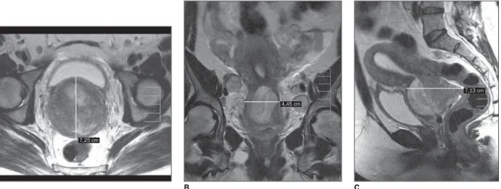
Sagittal images are useful for demon-strating the relation between tumor and cervix, uterine body, vagina and adjacent organs such as bladder and rectum. On the other hand, axial images are relevant for detection of parametrial and pelvic wall invasion, ureteral and lymph nodes in-volvement. The coronal plane, in conjunc-tion with the sagittal and axial planes, is useful in the parametrial evaluation, and particularly necessary for measurement of the tumor volume (Figure 3).
T2-weighted TSE is the sequence of choice in the evaluation of lymph nodes involvement, since in this sequence, muscles and vessels appear hyperintense, differently from lymph nodes. Fat suppres-sion improves even more the identification of structures or lesions surrounded by adi-pose tissues like parametrium and lymph nodes(15,20) (Figure 4).
Many times, the use of contrast is not necessary in the staging, since, in most of cases, precontrast sequences provide the necessary information. Besides, dynamic sequences frequently underestimate the tumor volume and the depth of the stromal invasion, and should not be utilized for these purposes(21). However, the use of
contrast agent may be useful for facilitat-ing the identification of fistulous tracts in advanced diseases or in the post-therapy follow-up(10).
TUMOR GROWTH PATTERNS
The tumor arises in the cervical canal and extends peripherally towards the cer-vical stroma, progressively replacing it. A full-thickness stromal invasion may occur, and, by contiguity, a parametrial invasion (IIb). Cervical canal obstruction is usual, and frequently causes the endometrial cav-ity to be distended with blood, serous fluid or purulent material(22) (Figure 5).
Table 1 Correlation between FIGO staging of uterine cervix cancer and MRI findings(17).
Ia Ib
Ib1 Ib2
IIa
IIb
IIIa
IIIb
IVa
IVb
Stage MRI T2-weighted sequence
Microinvasor
Invasive, confined to the cervix
Clinically visible lesion ≤ 4 cm Clinically visible lesion > 4 cm
Tumor invades the upper vaginal third, but does not affect the lower vaginal third Tumor invades the parametrium, but not the pelvic wall neither the lower vaginal third
Involvement of the lower vaginal third, without affecting the pelvic wall Pelvic wall involvement or hydronefrosis
Tumor invades the bladder or rectum mucosa
Distant metastasis
No tumor evidence
Hyperintense tumor on T2-weighted sequence in contrast with hypointense signal from cervical stro-ma
Tumor partially or completely replacing the hypo-intense cervical stroma, not surpassing the para-metrial interface represented by a hypointense halo Segmental interruption of hypointense signal on the upper third of the vaginal wall
Hyperintense tumor interrupting hypointense halo of the interface between cervical stroma and pa-rametrium
Segmental interruption of the hypointense signal of the lower vaginal third
Tumor extending to the musculature (internal ob-turator muscle, piriform muscle or levator ani mus-cle) or causing hydroureter
Loss of hypointense signal of the internal wall (mu-cosa) of the bladder or rectum
Distant metastasis
Figure 2. A: Sagittal T2-weighted TSE sequence, hyperintense uterine cervix tumor. Part of the (hypointense) cervical stroma is intact. Preserved vaginal canal (stage Ib). B: Sagittal T2-weighted TSE sequence, slightly hyperintense tumor in the posterior portion of the uterine cervix, extending to the upper vaginal third (stage IIa).
A B
〈〈〈〈〈
〈〈〈〈〈
through the inferior renal pole to the vulva, including the paraaortic and pelvic regions. The anterior saturation band should be uti-lized as a routine to reduce respiratory and peristaltic artifacts. On the other hand, the posterior saturation band is dispensable. The use of antiperistaltic agents four to six hours before the examination also is rec-ommended to reduce artifacts resulting from intestinal peristalsis(3,10). The
phased-array coil improves the signal-noise ratio, allowing the acquisition of more detailed
images than the formerly utilized body coils, and, consequently improving the imaging resolution. However, body coils may be useful for obese patients with a very protuberant abdomen, or for retroperitoneal evaluation(18). The utilization of endorectal
Figure 4. A: Axial T2-weighted TSE sequence, the same patient in Figure 3 in an uppermost plane. The arrows indicate bilateral adenomegalies, with a slightly hyperintense signal similar to the cervical tumor. B: Axial T2-weighted TSE sequence with fat suppression. The arrows indicate the same adenomegalies as a, more evident in this sequence.
A B
〈〈〈〈〈
Tumors extending into the uterine cav-ity are associated with a worst prognosis and higher prevalence of distant me-tastases(2). Clinically, tumors with
endo-phytic growth are difficult to be measured, since the largest component cannot be di-rectly visualized and evaluated in a gyne-cological examination. The clinical evalu-ation of exophytic tumors is easier, but MRI facilitates the identification of a pos-sible vaginal invasion.
Evaluation of parametrium and pelvic wall
The parametrium is the connective tis-sue between the layers of the broad
liga-ment. Medially it is contiguous with the uterus, cervix and proximal vagina; extend-ing laterally to the pelvic wall. Inferiorly, it is nearly the cardinal ligament. It is pre-dominantly consisted of fat through which run uterine vessels, nerves and lymphatic vessels(11).
Parametrial invasion (above IIb) is a significant prognostic factor influencing in the diagnosis and therapeutic choice. On T2-weighted sequences, the interface be-tween the normal cervical stroma and the parametrium appears like a hypointense ring or halo surrounding the cervix. A pre-served hypointense halo represents a high negative predictive value for parametrial
invasion(15,17). The indicator of parametrial
invasion is the segmental interruption or complete absence of this halo in the inter-face between the cervical stroma and the parametrial fat, or yet, the clear protrusion of the tumor into the parametrium(13,23).
Some authors correlate the complete re-placement of the cervical stroma and the tumor extension into the uterine body with the parametrial invasion. In these cases, 94% of the parametrium is invaded, with direct relation between the size of the tu-mor and the parametrial involvement(15,22).
Loss of parametrial fat may be an indica-tor of invasion, but this is a non-specific sign, since peritumoral inflammation also
Figure 3. A: Axial T2-weighted TSE sequence, tumor completely replacing the cervical stroma in its largest anteroposterior diameter. B: Coronal T2-weighted TSE sequence of the same patient showing the largest latero-lateral diameter of the tumor. Note the bilateral adenomegalies. C: Sagittal T2-weighted TSE sequence, largest craniocaudal diameter of the tumor. Preserved signal of the bladder mucosa.
A B C
7,25 cm
4,45 cm
Figure 5. A: Sagittal T2-weighted TSE sequence, hyperintense tu-mor completely replacing the uterine cervix stroma and ob-structing the cervical canal, caus-ing distension of the endometrial cavity filled with a highly hyper-intense fluid. B: Sagittal T2-weighted TSE sequence, tumor completely replacing the uterine cervix stroma; so there is no ac-cumulation of fluid inside the endometrial cavity.
A B
may result in loss of fat simulating inva-sion.
Contrast-enhanced T1-weighted se-quences have demonstrated higher accu-racy than the T2-weighted in parametrial evaluation(10) (Figure 6). In cases where the
tumor extends into the parametrium it may reach the ureter, causing hydronephrosis (IIIb). Hydronephrosis associated with mass in the uterine cervix are specific signs of parametrial invasion(12).
Parametrial invasion up to the pelvic wall (IIIb) is diagnosed when the tumor cannot be separated from the pelvic wall at clinical examination. At MRI, this
diagno-sis is made when the distance between the tumor and the pelvic wall is < 3 mm, or when T2-weighted sequences show partial or complete loss of a normal hyposignal of the pelvic wall musculature (piriform muscle, internal obturator muscle, levator ani muscle or coccygeal muscle) (Figure 7). In these cases, iliac vessels become com-pressed and narrowed by the tumor, and the bone destruction may occur by direct exten-sion of a diffusely infiltrated tumor(25).
Vaginal involvement
MRI is highly sensitive in the detection of vaginal invasion, with 93% accuracy(11).
The sign of vaginal involvement is better characterized on high-resolution T2-weighted sequences, showing the segmen-tal interruption of the normal hypointense signal of the vaginal wall, or yet a hyperin-tense vaginal thickening (tumor), or the mass itself in contiguity with the vaginal wall (Figure 8). Vaginal invasion corre-sponds to stage IIa; when this invasion extends up to the lower vaginal third, cor-responds to stage IIIa(25). Additionally, the
use of intravaginal ultrasonographic gel during the MRI acquisition is recom-mended to distend and fill the cavity with a highly hyperintense material on
T2-haematocolpos
cervical canal
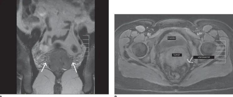
Figure 6. A: Coronal T2-weighted TSE sequence showing bilateral parametrial invasion, loss of hypointense halo separating the interface between the cervical stroma and the parametrial fat. Note the fimbrias laterally along the parametrium which also represent indirect signs of parametrial invasion (stage IIb). B: Axial T1-weighted sequence with fat suppression of the same patients, showing the bilateral involvement of uterosacral involvement.
A B
tumor bladder
Figure 7. A: Axial illustration of the pelvis showing a tumor restricted to the cervix, with no sign of parametrial invasion (stage I). B: Axial illustration of the pelvis showing a tumor invading the parametrium, without reaching the pelvic wall or right ureter (stage IIb). C: Axial illustration showing cervical tumor invading the parametrium and reaching the right ureter (stage IIIb). D: Axial illustration showing cervical tumor invading the parametrium and reaching the pelvic wall (stage IIIb). E: Axial T2-weighted TSE sequence showing a cervical tumor involving the urethra, and extending up to the pelvic wall with alteration of the ischium signal (arrow). F: An uppermost image of the same sequence clearly showing parametrial invasion (arrow).
sequences). This procedure improves the sensitivity in the evaluation of the vaginal invasion. About 20 ml of gel applied at the moment of the examination are sufficient(10).
Lymph node involvement
Several studies have demonstrated the significance of the lymph node involve-ment as a factor of worsening in the sur-vival prognosis of women affected by uter-ine cervix tumor(3,26). There are three
drain-age routes from the cervical lymph nodes through which the tumor propagates (Fig-ure 9): the lateral route, along the external iliac vessels; the hypogastric route, along the internal iliac vessels; and the presacral route, along the uterosacral ligament. All of the three routes drain into the common iliac lymph nodes, through which the tumor may reach the paraaortic lymph nodes. Gener-ally, the paracervical and parametrial lymph nodes are the first to be affected, followed by the obturator lymph nodes and, subse-quently, the external and internal iliac lymph nodes(27).
weighted sequences in order to improve the tumor contrast (slightly hyperintense on T2-weighted sequences) and the vaginal wall contrast ((hypointense on T2-weighted
Figure 8. Sagittal T2-weighted TSE sequence showing uterine cervix tumor invading the upper vaginal third. Observe that the introduction of the intravaginal ultrasonographic gel (highly hyperin-tense on T2-weighted sequences) distends the vaginal wall and improves de assessment of the invasion.
T2-weighted are the sequences of choice for evaluation of pelvic lymph nodes, since in these sequences vessels and musculature become hypointense, facilitat-ing the differentiation from lymph nodes which are slightly hyperintense on weighted sequences (Figure 10). T2-weighted TSE fat suppressed sequences allow the suppression of the adipose tissue surrounding the lymphatic vessels, improv-ing the accuracy in the detection of pelvic adenomegalies(12) (Figure 11). Up to the
present moment, the suspicion of lymph node metastasis by means of MRI is lim-ited to the increase in the size of the lymph node. Lymph nodes > 10 mm in axial di-ameter are considered as abnormal. Also, some higher limits are suggested for deter-mined specific sites, as follows: for lymph nodes in the internal iliac chain, 7 mm; for common iliac lymph nodes, 9 mm; and for external iliac lymph nodes, 10 mm. Positron emission tomography with fluoro-deoxy-D-glucose (PET-FDG) seems to of-fer higher specificity than MRI for enlarged
A B C
D E F
tumor
parametrium bladder
〈〈〈〈〈
tumor
pelvic lymph nodes(3). When lymph node
central necrosis is identified, the positive predictive value for malignancy is 100%. It has been already demonstrated that lymph nodes with necrosis or signal inten-sity similar to the tumor presented worst prognosis. The diagnosis of lymph node necrosis may improve with the use of en-dovenous contrast (25,28).
Most recently, improvement has been demonstrated in the MRI sensitivity for detection of metastatic lymph nodes in uter-ine cervix tumors, utilizing a new type of lymph node-specific contrast agent called ferumoxtran-10, with nanoparticles of iron oxide (USPIO). However the utilization of this contrast agent is not a consensus yet(29).
Considering that the FIGO staging system
does not take the lymph nodes involvement into consideration, the detection of en-larged pelvic lymph nodes on MRI corre-sponds to stage IIIb, as well as the diagno-sis of enlarged paraaortic lymph node cor-responds to stage IVb(12).
Invasion of bladder and rectum
Invasion of bladder or rectum (IVa) may be difficult to be detected only by a physi-cal examination. MRI has shown to be a reliable method for detection of bladder invasion with 83% sensitivity, specificity near 100%, and 99% accuracy. When the bladder presents invaded by the tumor, its wall, which normally is hypointense, shows a focal or diffuse area with increase in the signal intensity on T2-weighted
se-quences, or simply a vegetating mass into the lumen is observed(10,30). For defining
the bladder invasion, it is important to ob-serve that signal alteration is present both for the bladder muscle and mucosa, other-wise the tumor may be just contiguous to the bladder(4). Other indicative signs of
invasion are hyperintensity on the internal surface of the posterior wall, nodularity or irregularity in the bladder wall (Figure 12). On the other hand, the vesico-ureteral junc-tion is poorly evaluated because of the dif-ficult visualization of a non-dilated ureter on MRI.
Direct invasion of the ureter is not fre-quent, however, in the setting of ureter in-vasion, a tumor extension is observed along the uterosacral ligaments. Findings,
usu-Figure 9. Lymphatic drainage route of uterine cervix tumor. Coronal (left) and sagittal (right) illustrations showing, in black, primarily affected lymph nodes, and, in gray, those secondarily affected.
Figure 10. Coronal T2-weighted TSE sequence showing uterine cervix tumor and bilaterally en-larged lymph nodes (arrows).
Figure 11. Axial T2-weighted sequence with fat-suppression on the same plane as Figure 10, showing enlarged and hyperintense lymph nodes in this sequence.
Common iliac
Common iliac
Internal iliac
External iliac
Parametrial
Inguinal Sacral
Internal iliac
Obturator
Paracervical
Sacral
Internal iliac
Obturator Aortic
〈〈〈〈〈
〈 〈 〈 〈 〈 tumor
uterus
ally, are: focal thickening or segmental in-terruption of the hypointense signal on the anterior rectal wall(10).
FINAL CONSIDERATIONS
Even though MRI is not utilized by the majority of oncology services for staging uterine cervix tumors, and, up to this mo-ment, it has not been officially approved by FIGO yet, it is the best imaging method in terms of accuracy for assessment of tumors, and plays an essential role in the therapeu-tic planning and follow-up(31).
MRI has shown to be better than the clinical examination, and, when utilized as the initial staging method, reduces the number of invasive procedures and radio-logical studies such as urography, cytoscopy and rectosigmoidoscopy, with lower cost for the management of the disease. Addi-tionally, the correct assessment of the tu-mor extent and volume allows optimizing the planning of the fields for external pel-vic radiotherapy and brachytherapy.
A recent study has demonstrated that the MRI-aided radiotherapy planning may re-duce the possibility of geographic errors as compared with the conventional radio-therapy planning(32). It is important that the
radiologist interpreting a pelvic MRI for uterine cervix tumor, is familiarized with the findings and, mainly, provides informa-tion regarding tumor volume, invasion of parametrium, vagina and adjacent organs, besides indicating the tumor growth type and lymph nodes involvement.
Acknowledgments
The authors thank the International Fed-eration of Gynecology and Obstetrics (FIGO), for the free authorization for repro-duction of the scheme for staging of cervi-cal carcinoma; Rubens de Andrade, who has designed the figures; and the technician Marcello C. Galdino, for his dedication to the performance of the examinations.
REFERENCES
1. Ferlay F, Bray F, Pisanni P, Parkin DM. Globocan 2002: Cancer incidence, mortality and prevalence worldwide. IARC CancerBase No. 5, version 2.0. Lyon: IARC Press, 2004.
2. Eifel PJ, Berek JS, Thigpen JT. Cancer of cervix, vagina and vulva. In: DeVita VT Jr, Hellman S, Rosenberg SA, editors. Cancer: principles and practice of oncology. Philadelphia, PA: Lippin-cott, 1997;1433–1475.
3. Narayan K, Mckenzie AF, Hicks RJ, Fisher R, Bernshaw D, Bau S. Relation between FIGO stage, primary tumor volume, and presence of lymph node metastases in cervical cancer patients referred for radiotherapy. Int J Gynecol Canc-er 2003;13:657–663.
4. Soutter WP, Hanoch J, D’Arcy T, Dina R, McIndoe G.A, deSouza N.M. Pretreatment tumor volume measurement on high-resolution magnetic reso-nance imaging as a predictor of survival in cervi-cal cancer. Br J Obstet Gynaecol 2004;111:741– 747.
5. Benedet JL, Bender H, Jones H, Ngan HY, Peco-relli S. FIGO staging classifications and clinical practice guidelines in the management of gyne-cologic cancers. FIGO Committee on Gyneco-logic Oncology. Int J Gynaecol Obstet 2000;20: 209–262.
6. DiSaia PJ, Creasman WT. Clinical gynecologic oncology. 6th ed. St. Louis, MO: Mosby, 2002; 53–95.
7. Bipat S, Glas AF, van der Velden J, Zwinderman AH, Bossuyt PMM, Stoker J. Computed tomog-raphy and magnetic resonance imaging in
stag-ing of uterine cervical carcinoma: a systematic review. Gynecol Oncol 2003;91:59–66. 8. Bezerra MRL, Soares AFF, Faintuch S, et al.
Iden-tificação das estruturas músculo-ligamentares do assoalho pélvico feminino na ressonância mag-nética. Radiol Bras 2001;34:323–326. 9. Seki H, Azumi R, Kimura M, Sakai K. Stromal
invasion by carcinoma of the cervix: assessment with dynamic MR imaging. AJR Am J Roent-genol 1997;168:1579–1585.
10. Chiang SH, Quek ST. Carcinoma of the cervix: role of MR imaging. Ann Acad Med Singapore 2003;32:550–556.
11. Hricak H, Powell CB, Yu KK, et al. Invasive cer-vical carcinoma: role of MR imaging in pretreat-ment work-up-cost minimization and diagnostic efficacy analysis. Radiology 1996;198:403–409. 12. Pannu HK, Corl FM, Fishman EK. CT evaluation of cervical cancer: spectrum of disease. Radio-Graphics 2001;21:1155–1168.
13. Kim SH, Choi BI, Han JK, et al. Preoperative staging of uterine cervical carcinoma: compari-son of CT and MRI in 99 patients. J Comput Assist Tomogr 1993;17:633–640.
14. Vorgias G, Katsoulis M, Argyrou K, et al. Preop-erative imaging of primary intra-abdominal gynaecological malignancies: diagnostic accu-racy of CT-scan and MRI. A Greek cohort study. Eur J Gynaecol Oncol 2002;23:139–144. 15. Lam WW, So NM, Yang WT, Metreweli C.
De-tection of parametrial invasion in cervical carci-noma: role of short tau inversion recovery se-quence. Clin Radiol 2000;55:702–707. 16. Tanaka YO, Nishida M, Yamaguchi M, Kohno K,
Saida Y, Itai Y. MRI of gynaecological solid masses. Clin Radiol 2000;55:899–911. 17. Okamoto Y, Tanaka YO, Nishida M, Tsunoda H,
Yoshikawa H, Itai Y. MR imaging of the uterine cervix: imaging-pathologic correlation. Radio-Graphics 2003;23:425–445.
18. Gauger J, Holzknecht NG, Lackerbauer CA, et al. Breathhold imaging of the upper abdomen using a circular polarized-array coil: comparison with standard body coil imaging. MAGMA 1996;4: 93–104.
19. deSouza NM, Scoones D, Krausz T, Gilderdale DJ, Soutter WP. High-resolution MR imaging of stage I cervical neoplasia with a dedicated
trans-Figure 12. A: Sagittal T2-weighted TSE sequence show-ing uterine cervix tumor invad-ing the bladder mucosa (stage IVa). B: Axial illustration show-ing the tumor invadshow-ing the bladder and rectum mucosas. B
A
vaginal coil: MR features and correlation of im-aging and pathologic findings. AJR Am J Roent-genol 1996;166:553–559.
20. Follen M, Levenback CF, Iyer RB, et al. Imaging in cervical cancer. Cancer 2003;98(9 Suppl): 2028–2038.
21. Allen JR, Prost RW, Griffith OW, Erickson SJ, Erickson BA. In vivo proton (H1) magnetic reso-nance spectroscopy for cervical carcinoma. Am J Clin Oncol 2001;24:522–529.
22. Okuno K, Joja I, Miyagi Y, et al. Cervical carci-noma with full-thickness stromal invasion: rela-tionship between tumor size on T2-weighted images and parametrial involvement. J Comput Assist Tomogr 2002;26:119–125.
23. Stehman FB, Randall ME, Michel H, Morken JV. Uterine cervix. In: Hoskins WJ, Perez CA, Young RC, editors. Principles and practice of gyneco-logic oncology. Philadelphia, PA: Lippincott Wil-liams & Wilkins, 2005;743-822.
24. Scheidler J, Heuck AF, Steinborn M, Kimmig R, Reiser MF. Parametrial invasion in cervical
car-cinoma: evaluation of detection at MR imaging with fat suppression. Radiology 1998;206:125– 129.
25. Taylor MB, Carrington BM, Davidson SE, Swindell R, Lawrance JAL. Staging of advanced cervical carcinoma using MRI-predictors of out-come after radical radiotherapy. Clin Radiol 2003; 58:532–541.
26. Landoni F, Maneo A, Colombo A, et al. Random-ised study of radical surgery versus radiotherapy for stage Ib-IIa cervical cancer. Lancet 1997;350: 535–540.
27. Jeong YY, Kang HK, Chung TW, Seo JJ, Park JG. Uterine cervical carcinoma after therapy: CT and MR imaging findings. RadioGraphics 2003;23: 969–981.
28. Yang WT, Lam WWM, Yu MY, Cheung TH, Metreweli C. Comparison of dynamic helical CT and dynamic MR imaging in the evaluation of pelvic lymph nodes in cervical carcinoma. AJR Am J Roentgenol 2000;175:759–766. 29. Rockall AG, Sohaib SA, Harisinghani MG, et al.
Diagnostic performance of nanoparticle-en-hanced magnetic resonance imaging in the diag-nosis of lymph node metastases in patients with endometrial and cervical cancer. J Clin Oncol 2005;23:2813–2821.
30. Kim SH, Han MC. Invasion of the urinary blad-der by uterine cervical carcinoma: evaluation with MR imaging. AJR Am J Roentgenol 1997;168: 393–397.
31. Mayr NA, Taoka T, Yuh WTC, et al. Method and timing of tumor volume measurement for out-come prediction in cervical cancer using magnetic resonance imaging. Int J Radiat Oncol Biol Phys 2002;52:14–22.