Revista Brasileira de Anestesiologia 719 Received from Hospital de São João, Portugal.
1. Anesthesiology Department, Hospital de São João, Entidade Pública Empresarial (EPE), Porto, Portugal
2.General Unit of Polyvalent Intensive Care, Hospital de São João, EPE, Porto, Portugal
Submitted on August 30, 2011. Approved on September 30, 2011.
Correspondence to: Luísa Guedes, MD Hospital de São João
Alameda Professor Hernani Monteiro Porto, Portugal
E-mail: [email protected]
REVIEW ARTICLE
Regional Analgesia in Intensive Care
Luísa Guedes
1, Helena Rebelo
1, Raquel Oliveira
1, Aida Neves
2Summary: Guedes L, Rebelo H, Oliveira R, Neves A – Regional Analgesia in Intensive Care.
Justifications and objectives: regional analgesia plays an important role in multimodal pain management in critically ill patients, minimizing patient discomfort and reducing the associated physiological and psychological stress. Lower doses of systemic opioids reduce some of its side effects, such as withdrawal syndrome, possible psychological changes, and gastrointestinal dysfunction. Despite these benefits, its use is contro-versial, as patients in intensive care units often have contraindications, such as coagulopathy, hemodynamic instability, and difficulty in neurologi-cal assessment and implementation of regional technique.
Content: The authors present a review of regional analgesia in intensive care, focusing on the main advantages and limitations of its use in criti-cally ill patients, and describe the most commonly used regional techniques and its applicability.
Keywords: Analgesia; Anesthesia, Conduction; Pain; Intensive Care/complications.
©2012 Elsevier Editora Ltda. All rights reserved.
INTRODUCTION
Approximately 50% of critically ill nonsurgical patients report
pain during hospitalization in intensive care units (ICUs) 1.
There are many causes of pain, such as underlying disease at admission, trauma, surgery, nursing care (mobilization, air-way suction, physiotherapy), prolonged immobilization and invasive therapeutic procedures, diagnostic or monitoring 2,3.
The method considered more reliable for assessing pain intensity and response to analgesia is indicated by the patient with the use of objective scales (Visual Analogue Scale, Vi-sual Numeric Scale, ViVi-sual Descriptive Scale) 4,5,6. It is often
difficult or impossible to quantify the subjective experience of pain in critically ill patients due to the presence of the endo-tracheal tube, state of unconsciousness and/or administration of sedative drugs, which inhibit or preclude the application of these scales. Observation of behavioral responses (facial expression, agitation, posture) or physiological stress (blood pressure, heart rate, respiratory rate, diaphoresis, intracra-nial pressure) may be the only possible assessment of pain, but with large margin of error, as suffering and autonomic re-sponse to aggression result from activation of different zones of the central nervous system (CNS) with different sensitivity to noxious activation 7.
Effective analgesia in critically ill patients improves lung function, ventilatory weaning and early mobilization, and de-creases the plasma levels of catecholamines and myocardial
oxygen consumption 8,9. Regional analgesia is particularly
ef-fective in achieving these objectives, mainly in multimodal an-algesia in which the required doses of opioids are reduced to mitigate side effects 4,10.
However, patients in intensive care often have contraindica-tions, such as coagulation disorders, severe hypovolemia, he-modynamic instability, and difficult neurological assessment and implementation of the technique, which determines the use of regional analgesia and requires careful risk-benefit evaluation.
There are few published data on the use of regional anal-gesia in intensive care units, which hinders evidence-based decisions.
The aim of this paper is to present a review of regional anal-gesia in intensive care, focusing on the main advantages and limitations of its use in critically ill patients, and describe the most commonly used regional techniques and its applicability.
METHODS
Literature review was performed through search of Med-line articles published in the last 12 years with the following keywords: “regional analgesia”, “intensive care”, “peripheral blocks” and “critically ill patients”.
REGIONAL ANALGESIA
Peripheral nerve block
In polytraumatized patients in which orthopedic injuries of limbs are part of multiple lesions, the presence of severe pain, often accompanied by traumatic or iatrogenic altered consciousness, conditions the use of effective doses of opi-oids by fear of central depression after the use of high doses. Regional techniques for analgesia of upper and lower limbs can provide adequate pain control without central depression. Peripheral nerve blockade guided by ultrasound and/or neu-rostimulation and the use of stimulation catheters in sedated patients allow the reduction of complications associated with
the technique and increase success rate 11.
In a meta-analysis, Richman found that continuous periph-eral nerve block promoted better analgesia with fewer side
effects compared to analgesia with opioids 12.
Analgesia for both shoulder and upper limb may be ob-tained through various approaches for brachial plexus block-ade, particularly by interscalenic or axillary route. Continuous femoral nerve block is a good option in postoperative femoral
neck fractures 13,14. This technique, when combined with
sci-atic block, allows adequate analgesia for injuries affecting the entire lower limb.
In high-risk fractures, such as tibial and distal radius, epi-dural and peripheral nerve block may hinder the recognition of
compartment syndrome in intensive care 15. To minimize this
risk, the indication for peripheral nerve block analgesia should be discussed with the surgical team, and intracompartmental
pressure monitoring should be considered 16.
Diagnosis of bacteremia caused by peripheral catheter in-fection in continuous analgesia techniques may be difficult to distinguish from worsening disease. The puncture site should
be carefully inspected when administering the drug and, if infection is suspected, the catheter should be immediately removed. Microbiological analysis of the catheter-tip may be useful for guiding antibiotic therapy. To confirm suspicion as-sociated bacteremia, blood cultures should be collected prior to the administration of empiric antibiotic therapy 17.
The large volumes of local anesthetic used demands a carefully observation of the maximum recommended doses,
especially when a combination of different blocks is used 18.
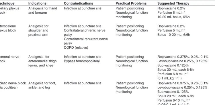
Table I shows the main practical problems, indications, contraindications, and recommended doses for upper and lower limb blockade performed in the ICU.
Epidural analgesia
Epidural analgesia is the regional technique most frequently used in critically ill patients. There is no evidence of mortal-ity reduction with its use, but several studies show that when compared to parenteral opioids it is at least equivalent regard-ing the outcome, ICU length of stay, and duration of ventilatory support. It is also reported that epidural analgesia reduces the incidence of paralytic ileus, improves analgesia and patient satisfaction in cases of thoracic trauma, abdominal surgery, vascular surgery, major orthopedic surgery, acute pancreati-tis, cardiac surgery, and intractable angina pectoris 4,19,20.
Pulmonary dysfunction after thoracic and abdominal sur-gery is mainly caused by pain that leads to diaphragmatic dys-function and hypoventilation. These factors give rise to a re-duction in functional residual capacity (FRC) and hypoxemia.
Table I – Main Indications, Contraindications, Practical Problems, and Recommended Doses for Upper and Lower Limb Blockade
Technique Indications Contraindications Practical Problems Suggested Therapy Axillary plexus
block
Analgesia for hand and forearm
Infection at puncture site Patient positioning Neurological function monitoring
Ropivacaine 0.2% Perfusion 5 mL.h-1 10-20 mL bolus, 6/6h
Interscalene plexus block
Analgesia for shoulder and proximal arm
Infection at puncture site Contralateral phrenic nerve palsy
Contralateral recurrent nerve palsy
COPD (relative)
Patient positioning Neurological function monitoring
Ropivacaine 0.2% Perfusion 5 mL.h-1 Bolus 10-20 mL, 6/6h
Femoral nerve block
Analgesia for anteromedial thigh, femur, and knee
Infection at puncture site Bypass femoropopliteal
Patient positioning Neurological function monitoring
Ropivacaine 0.375%, 0.2%, 0.1% Levobupivacaine 0.25%, 0.125% Bupivacaine 0.125%
Bolus 20 mL, each 6-8h Perfusion 6-8 mL.h-1 (0.1 mL.kg-1.h-1) Sciatic nerve block
(via popliteal)
Analgesia for foot, ankle, and leg
Infection at puncture site Patient positioning Neurological function monitoring
Ropivacaine 0.375%, 0.2%, 0.1% Levobupivacaine 0.25%, 0.125% Bupivacaine 0.125%
Revista Brasileira de Anestesiologia 721
Decreased FRC may result in atelectasis, changes in ventila-tion/perfusion, and pulmonary complications in postoperative
period, particularly pulmonary infection 21. Epidural
anesthe-sia, when compared to intravenous analgeanesthe-sia, increases FRC
by 27% and decreases the rate of pulmonary complications 22.
On the other hand, by reducing the need for mechanical ven-tilation, iatrogenic, ventilator-associated pneumonia, length of hospital stay, and hospital costs are also reduced.
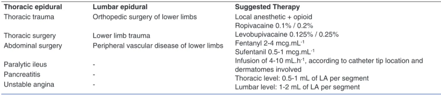
Table II shows the main indications for epidural analgesia in critically ill patients, as well as therapeutic approaches. Table III shows the absolute and relative contraindications to epidural catheter insertion.
Complications
The most common side effects of epidural analgesia are bra-dycardia and hypotension resulting from sympathetic block-ade, which may be reduced by using low concentrations or incremental doses of local anesthetics. Other side effects of opioids are pruritus, nausea, vomiting, sedation and respira-tory depression. Clonidine side effects are sedation, hypoten-sion, and bradycardia. The addition of morphine, fentanyl, sufentanil or clonidine to epidural analgesia in intensive care should be considered individually and may be an advantage in critically ill patients, as it allows lower doses of local anesthet-ics, optimal analgesia, and fewer hemodynamic changes.
Positive pressure ventilation, particularly at high pres-sures, reduces venous return, may influence local anesthetic spread in thoracic epidural blocks, and favors a dispersion trend caudally 23.
Studies have shown that the risk of infection associated with epidural anesthesia in intensive care appears to be low, and the presence of two local signs of inflammation is a strong predictor of local and epidural catheter infection 24.
Antibiotic prophylaxis for placement of epidural catheter is not routinely recommended in scenarios of intra and extra in-tensive care, although some studies show a reduction in
cath-eter colonizationrates 25. The use of maximum aseptic
mea-sures, similar to the placement of central venous catheters,
is recommended and aslo reduces the risk of infections 26.
Catheter must be removed if signs of inflammation and/or pus at the site of infection are perceived, or if there is a suspected central nervous system infection.
The use of epidural catheter in patients with clotting disor-ders may be associated with the onset of epidural or subdural hematomas, whose diagnosis is difficult in critically ill patients. It is recommended that epidural catheter should be firmly se-cured to prevent accidental removal. If this occurs, it should be given particular attention to neurological monitoring. The placement of tunneled catheters may prevent its exteriorization during maneuvers for the positioning of a critically ill patient.
The proper positioning of epidural catheter in epidural space must be assessed carefully by aspiration before drug administration. This seems to be the most simple and secure approach to prevent intravascular injection of local anesthetic in critically ill patients, as they may have an altered response to administration of test dose with adrenaline, by the use of beta blockers, calcium channel blockers, clonidine, and
en-dogenous and exogenous catecholamines 7. As an option to
this method of placement confirmation, electrical stimulation may be used via epidural catheter (Tsui test) or radiological confirmation after administration of radiopaque contrast into epidural space 27. Intravascular injection of local anesthetics is
associated with significant toxicity, which primarily affects the central nervous system (CNS) and is manifested by prodromal symptoms (dizziness, tinnitus, tongue paresthesia, perioral numbness, metallic taste, hearing and visual disorders) prior to seizures (by inhibitory pathway blockade) and central de-pression. Cardiovascular manifestations occur after the onset of signs and symptoms of CNS and are more frequent with the
Table II – Main Indications and Therapeutic Approaches for Epidural Analgesia in Critically Ill Patients
Thoracic epidural Lumbar epidural Suggested Therapy Thoracic trauma Orthopedic surgery of lower limbs Local anesthetic + opioid
Ropivacaine 0.1% / 0.2% Levobupivacaine 0.125% / 0.25% Fentanyl 2-4 mcg.mL-1
Sufentanil 0.5-1 mcg.mL-1
Infusion of 4-10 mL.h-1, according to catheter tip location and dermatomes involved
Thoracic level: 0.5-1 mL of LA per segment Lumbar level: 1-2 mL of LA per segment Thoracic surgery Lower limb trauma
Abdominal surgery Peripheral vascular disease of lower limbs
Paralytic ileus
-Pancreatitis
-Unstable angina
-Table III – Absolute and Relative Contraindications to Epidural Catheter Placement
Absolute Relative
Infection at puncture site Sepsis
Patient’s refusal Preexisting neurological deficits (Demyelinating diseases)
Coagulopathy or other bleeding disorders
Stenotic valvular disease
Intracranial hypertension Spinal deformities
Severe aortic stenosis Uncooperative patient
use of bupivacaine due to the long blockade duration of this fast-in-slow-out fashion. Systemic manifestations are related
to the patient’s acid-base status. The increase in PaCO2 and
acidosis causes a reduction in seizure threshold and an in-creased rate of systemic toxic reactions. Hypercapnia increas-es cerebral blood flow and uptake of local anincreas-esthetics 28.
Local toxicity should also be considered, for (1) Allergic re-actions, most common with amino esters and related to
pre-sensitization mechanisms of para-aminobenzoic acid (PABA).
Symptoms are predominantly urticarial skin rash, starting on the face and extending to the neck and chest, followed by in-tense itching, and anaphylactic shock. (2) Myotoxicity, related to the intramuscular administration of local anesthetic in high concentrations. (3) Neurotoxicity, common to all local anes-thetics (more frequent with lidocaine) by accumulation of high concentrations of local anesthetics near nerve trunks (Cauda Equina Syndrome).
Table IV shows a summary of the most common complica-tions associated with epidural block.
Changes in hemostasis
The techniques for regional analgesia in patients undergoing hemostasis inhibition therapy in scenarios of intra and extra intensive care should be guided according to existing
recom-Table V – Recommended Times for Suspension of Antiplatelet Agents in Neuraxial Blockade (Adapted from Fonseca C et al.30 with Author’s Permission)
COX-1 INHIBITORS NSAIDs Do not suspend Do not suspend
Aspirin Do not suspend Do not suspend. Start 6-24 hours after NAB/catheter removal
Triflusal Do not suspend Do not suspend
CAMP INHIBITORS Dypiridamole Do not suspend Do not suspend
TIENOPYRIDINE DERIVATIVES Clopidogrel 5 days Start 6-24 hours after NAB/ catheter removal
Ticlopidine 10 days
Prasugrel 7 days
GP IIB/IIIA INHIBITORS Abcximab 48 days Recommended not to start within 4 weeks after surgery/NAB
Tirofiban 8 days Neurological monitoring, if necessary
Eptifibatide 8 days
NAB: Neuraxis blockade.
Table IV – Most Frequent Complications Associated with Epidural Block
Associated with the technique
Associated with the use of LA
Associated with the use of opioids
Dura mater puncture Abscess, epidural hematoma Nerve injury
Hypotension Extended bilateral motor and sensory block
Cardiovascular and central nervous system toxicity
Respiratory Depression Sedation Nausea, vomiting Pruritus
LA: local anesthetics.
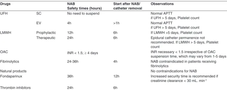
Table VI – Safety Recommendations Times for Handling Drug-treated Patients with Hemostasis Inhibitors
Drugs NAB
Safety times (hours)
Start after NAB/ catheter removal
Observations
UFH SC No need to suspend Normal APTT
if UFH > 5 days, Platelet count
EV 4h >1h Normal APTT
if UFH > 5 days, Platelet count
LMWH Prophylactic 12h 6h If LMWH >5 days, Platelet count
Therapeutic 24h 6h Epidural catheter permanence not
recommended. If LMWH > 5 days, Platelet count
OAC INR < 1.5; ≥ 4 days INR necessary < 1.5 irrespective of OAC
suspension time, which may vary from 1-5 days
Fibrinolytics 24-36h 4h NAB contraindicated in patients receiving
fibrinolytics
Natural products No contraindications for NAB
Fondaparinux 36h 12h Increased security time is recommended if
creatinine clearance < 30 mL. min-1
Thrombin inhibitors 24h 6h
Revista Brasileira de Anestesiologia 723
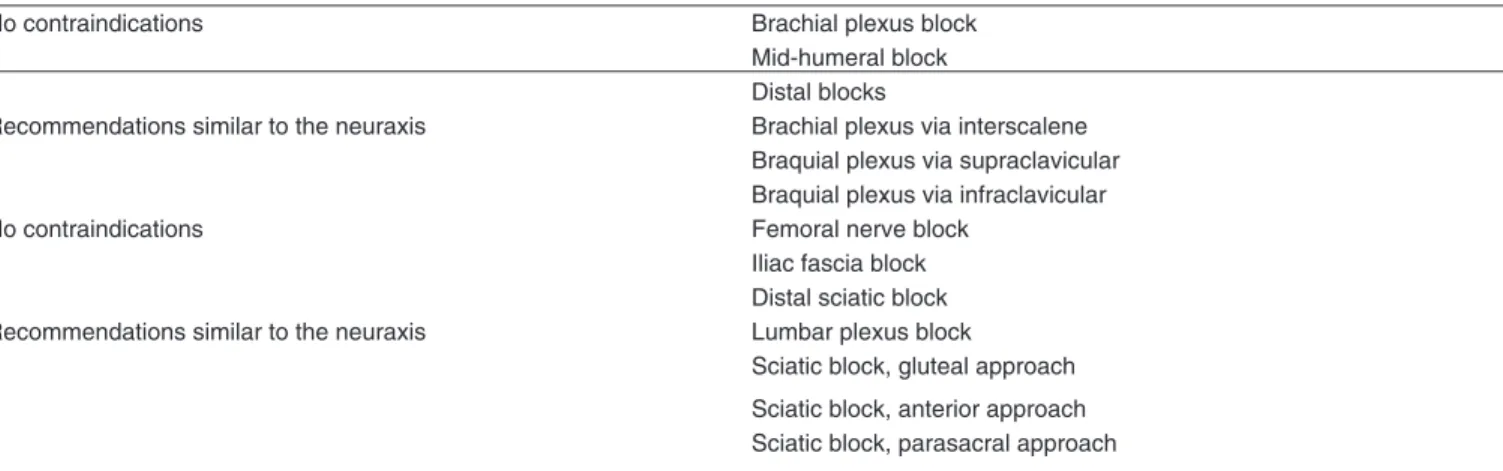
mendations to minimize risks and increase safety. Tables V, VI and VII present the current recommendations of the Por-tuguese Society of Anesthesiology validated by the College of the Anesthesiology Specialty to perform neuraxial blocks, peripheral blocks, and removal of catheters in patients under-going antiplatelet and anticoagulant therapies 29.
DISCUSSION
Regional analgesia for neuraxial or peripheral nerve block should be seen as effective and safe for critically ill patients, used alone or in multimodal context. It minimizes patient discomfort and reduces the physiological and psychological stress, as in non-critical patients. The reduced side effects of systemic opioids and sedatives allow early mobilization, reha-bilitation, and improve patient satisfaction.
The presence of coagulation disorders of iatrogenic cause or in multiple organ failure is the main limitation to its use in critically ill patients. The recommendations for patients with hypocoagulation or under antiplatelet therapy should be fol-lowed in intensive care.
The use of regional analgesia in the ICU setting should consider the risk/benefit ratio due to the limited cooperation of the patient in the placement and monitoring. The indications
for its use should be carefully and individually set according to patient’s anatomy, clinical condition, presence of contrain-dications, and logistic conditions 30.
To ensure the safety and efficacy, it is essential: a) training of health professionals (physicians, nurses, and physiothera-pists) in the prevention and early detection of complications and problems; b) creating a detailed record of the assessment of pain and complications, enabling a real time audit of the efficacy and safety of therapies used and adjusting them on time; c) development of protocols and safety rules that ex-traordinarily anticipate possible situations, allowing the opti-mization of therapeutic regimens.
Literature shows that regional analgesia in critically ill pa-tients is very little used, despite evidence of favorable out-comes. Still, the available evidence is limited to case reports, cut-off studies, or experts’ opinion and is based mainly on studies designed for intraoperative management of surgical patients in whom admission to intensive care is part of the pe-rioperative course. There are no studies specifically designed to evaluate the use of regional analgesia in this context 4,7. To
confirm the promising results found in literature, it is neces-sary to evaluate prospectively the advantages and limitations of epidural and regional blocks that allow the precise definition of its role in intensive care analgesia.
Table VII – Recommendations for Plexus/Peripheral Nerve block in Patients Treated with Hemostasis Inhibitors
No contraindications Brachial plexus block
Mid-humeral block Distal blocks
Recommendations similar to the neuraxis Brachial plexus via interscalene Braquial plexus via supraclavicular Braquial plexus via infraclavicular
No contraindications Femoral nerve block
Iliac fascia block Distal sciatic block Recommendations similar to the neuraxis Lumbar plexus block
Sciatic block, gluteal approach
Sciatic block, anterior approach Sciatic block, parasacral approach
REFERENCES
1. Desbiens NA, Wu AW, Broste SK et al. – Pain and satisfaction with pain control in seriously ill hospitalized adults: findings from the SUPPORT research investigations. For the SUPPORT investigators (Study to Understand Prognoses and Preferences for Outcomes and Risks of Treatment). Crit Care Med, 1996;24(12):1953-61.
2. Novaes MA, Knobel E, Bork AM et al. – Stressors in ICU: perception of the patient, relatives and health care team. Intensive Care Med, 1999; 25(12):1421-1426.
3. Hall LG, Oyen LJ, Murray MJ – Analgesic agents. Pharmacology and application in critical care. Crit Care Clin, 2001;17(4):899-923. 4. Schulz-Stübner S, Boezaart A, Hata JS – Regional analgesia in the
critically ill. Critical Care Med, 2005;33(6):1400-1407.
5. Fraser GL, Riker RR – Monitoring sedation, agitation, analgesia, and delirium in critically ill adult patients. Crit Care Clin, 2001;17(4):967-987.
6. Melzack R, Katz J – Pain measurement in persons in pain. In Wall PD, Maelzack R (eds): Textbook of pain, Edinburgh, Churchill Living-stone 1999, pp. 337-351.
7. Schulz-Stübner S – The critically ill patient and regional anesthesia. Curr Opin Anaesthesiol, 2006;19(5):538-44.
8. Epstein J, Breslow MJ – The stress response of critical illness. Crit Care Clin, 1999;15(1):17-33.
9. Lewis KS, Whipple JK, Michael KA, Quebbeman EJ – Effect of anal-gesic treatment on the physiological consequences of acute pain. Am J Hosp Pharm, 1994;51(12):1539-1554.
10. Clark F, Gilbert HC – Regional analgesia in the intensive care unit. Principles and practice. Crit Care Clin, 2001;17(4):943-966.
11. Marhofer P, Greher M, Kapral S – Ultrasound guidance in regional anaesthesia. Br J Anaesth, 2005;94:7-17.
12. Richman JM, Liu SS, Courpas G et al. – Does continuous peripheral nerve block provide superior pain control to opioids? A meta-analysis. Anesth Analg, 2006;102:248-257.
13. Finlayson BJ, Underhill TJ – Femoral nerve block for analgesia in frac-tures of the femoral neck. Arch Emerg Med, 1988;5:173-176. 14. Tan TT, Coleman MM – Femoral blockade for fractured neck of femur
in the emergency department. Ann Emerg Med, 2003;42: 596-597. 15. Davis ET, Harris A, Keene D et al. – The use of regional anaesthesia in
patients at risk of acute compartment syndrome. Injury, 2006;37:128-133.
16. Kostler W, Strohm PC, Sudkamp NP – Acute compartment syndrome of the limb. Injury, 2004;35:1221-1227.
17. Furuno JP, Perencevich EN, Johnson JA et al. – Methicillin-resistant Staphylococcus aureus and vancomycin-resistant enterococci co-colonization. Emerg Infect Dis, 2005;11:1539-1544.
18. Rosenberg PH, Veering BT, Urmey WF – Maximum recommended doses of local anesthetics: a multifactorial concept. Reg Anesth Pain Med, 2004;29:564-575.
19. Jorgensen H, Wetterslev J, Moiniche S, Dahl JB. Epidural local an-aesthetics versus opioid-based analgesic regimens on postoperative gastrointestinal paralysis, PONV and pain after abdominal surgery. Cochrane Database Syst Rev 2000;4:CD001893.
20. Peyton PJ, Myles PS, Silbert BS et al. – Perioperative epidural an-algesia and outcome after major abdominal surgery in high-risk pa-tients. Anesth Analg, 2003;96:548-554.
21. Ballantyne JC – Does epidural analgesia improve surgical outcome? Br J Anaesth, 2004;92:4-6.
22. Ballantyne JC, Carr DB, de Ferranti S et al. The comparative effects of postoperative analgesic therapies on pulmonary outcome. Anest Analg, 1998;86:598-612.
23. Visser WA, Gielen MJ, Giele JL – Continuous positive airway pressure breathing increases the spread of sensory blockade after low-thoracic epidural injection of lidocaine. Anesth Analg, 2006;102:268-271. 24. Darchy B, Forceville X, Bavoux E, Soriot F, Domart Y – Clinical and
bacteriologic survey of epidural analgesia in patients in the intensive care unit. Anesthesiology, 1996;85(5):988-998.
25. Morin AM, Kerwat KM, Klotz M et al. – Risk factors for bacterial cathe-ter colonization in regional anaesthesia. BMC Anesthesiol, 2005;5:1. 26. Herwaldt LA, Coffin SA, Schulz-Stübner S – Nosocomial infections
associated with anesthesia. Em: Hospital Epidemiology and Infection Control. Third Edition. Mayhall CG, ed. Philadelphia, Lippincott, Wil-liams & Wilkins, 2004, pp 1073-1117.
27. Tsui BC, Guenther C, Emery D, Finucane B – Determining epidural catheter location using nerve stimulation with radiological confirma-tion. Reg Anesth Pain Med, 2000;25:306-309.
28. Borges S, Azevedo A, Bezerra M, Canas M – Efeitos adversos dos anestésicos locais. CAR, 2001;28-32.
29. Fonseca C, Lages N, Correia C et al. – II Reunião de Consenso de Doentes medicados com fármacos inibidores da hemostase propos-tos para anestesia locorregional. Rev SPA, 2010;19(2):12-29. 30. Burton AW, Eappen S – Regional anesthesia techniques for pain