Rev Bras Anestesiol CLINICAL INFORMATION 2012; 62: 5: 736-740
736 Revista Brasileira de Anestesiologia
Vol. 62, No 5, September-October, 2012
Received from Jawaharlal, Nehru Medical College, AMU, Aligarh, UP, India.
1. Associate Professor, Department of Anesthesiology, Jawaharlal, Nehru Medical College, AMU, Aligarh, UP, India.
2. Assistant Professor, Department of Anesthesiology, Jawaharlal, Nehru Medical College, AMU, Aligarh, UP, India.
3. Resident, Department of Anesthesiology, Jawaharlal, Nehru Medical College, AMU, Aligarh, UP, India.
Submitted on October 8, 2011. Approved on November 19, 2011. Correspondence to:
Qazi Ehsan Ali, Associate Professor Dept of Anesthesiology
AMU, Aligarh, UP, India. E-mail: qaziehsanali@gmail.com
CLINICAL INFORMATION
Airtraq
®
Optical Laryngoscope for Tracheal Intubation in a
Patient with Giant Lipoma at the Nape: a Case Report
Qazi Ehsan Ali
1, Obaid Ahmed Siddiqui
2, Syed Hussain Amir
2, Abdulla Zoheb Azhar
3, Kashif Ali
3Summary: Ali QE, Siddiqui OA, Amir SH, Azhar AZ, Ali K –Airtraq® Optical Laryngoscope for Tracheal Intubation in a Patient with Giant Lipoma at the Nape: a Case Report.
Background and objectives: Lipoma is a progressively increasing disease which may appear anywhere in the body. Its appearance at the back of the neck, especially when it is large enough to cause restriction of neck extension, poses challenges to anesthesiologists in airway management whenever needed. This paper evaluates the role of Airtraq® in restricted neck movement.
Case Report: Case with a huge lipoma of 14 x 12 cm at the nape, and its surgical removal during an elective operation theatre posed difficulty in securing the airway by conventional laryngoscopy. To overcome the problem we successfully used a newly developed device, the Airtraq®, which is an optical laryngoscope for securing the airway.
Conclusion: Airtraq® can be used for elective intubation in patients with restricted neck movements.
Keywords: Intubation, Intracheal; Lipoma; Laryngoscopes; Neck Injuries.
©2012 Elsevier Editora Ltda. All rights reserved.
INTRODUCTION
Conventional laryngoscopy and tracheal intubation is consid-ered to be the gold standard of airway management 1.
Howev-er, this may prove to be difficult in situations where achieving an optimal sniffing position may not be possible or difficult and restricted neck movements. The airway management in such patients presents a unique challenge to anesthesiologists, and failure to secure airway in a timely and effective manner may lead to catastrophe.
Certain newer airway devices are presently available and have been used to facilitate airway management in patients with restricted neck movements.The Airtraq® (Prodol Meditec
S.A., Vizcaya, Spain) is a recently introduced airway device to facilitate tracheal intubation in patients with both normal and difficult airways. The device provides a high quality view of the glottis without the need to align the oral, pharyngeal and tra-cheal axes. The blade of the Airtraq® consists of two channels.
One channel acts as a conduit for passing the tracheal tube (ETT) while the other channel consists of an optical system, that transfers the image from the illuminated tip to a proximal
viewfinder. The Airtraq® is anatomically shaped and standard
ETTs of all sizes can be used (Figure 1). We describe a case
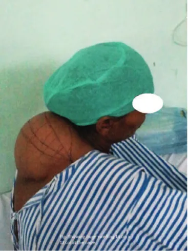
of a massive swelling (lipoma) on the posterior aspect of neck that renders restricted movements in a patient who was suc-cessfully intubated using Airtraq® laryngoscope (Prodol Ltd.
Vizcaya, Spain).
Case Report
A female, 50 years old, weighing 57 kg, American Society of Anesthesiologists (ASA) Class I, presented to the surgical outpatient department with a history of gradually progressive swelling on the posterior (nape) aspect of neck for the last 10 years. On clinical examination and investigations of the swell-ing the diagnosis of lipoma was made and the patient was planned for excision under general anesthesia. On preopera-tive airway assessment the thyromental distance was 5 cm and the interincisor distance was 5.5 cm. Neck movements, especially head extension, were severely restricted. The pa-tient had a normal mouth opening and was classified as Mal-lampati II. Routine preanesthetic investigations were normal. X-Ray cervical spine showed no bony or articular abnormality and the joint spaces were maintained. A preoprative assess-ment of difficult intubation was made and all the preparations for managing a difficult airway were kept ready. She did not give consent for awake intubation, so a general anesthesia was planned. Patient was premedicated with i.v. midazolam and i.m. glycopyrrolate. The patient was made to lie supine with the head supported by pillows in a way to avoid com-pression of the swelling and all the standard monitors were applied. After preoxygenation, anesthesia was induced with i.v. fentanyl 1 µg.kg-1 and i.v. propofol 2 mg.kg-1. After
AIRTRAQ® OPTICAL LARYNGOSCOPE FOR TRACHEAL INTUBATION IN A PATIENT WITH GIANT LIPOMA AT THE NAPE: A CASE REPORT
Revista Brasileira de Anestesiologia 737
Vol. 62, No 5, September-October, 2012
relaxation was achieved with 1.5 mg.kg-1 of succinylcholine.
The table was adjusted to the head down position with flexion of both the knees. The blade of the laryngoscope was intro-duced into the oral cavity in the midline, over the base of the tongue and the tip positioned in the vallecula. Trachea was intubated with size 7.0 mm endotracheal tube (PVC) in the first attempt after adequate visualization of the vocal cords, which required minor adjustments of Airtraq® and wrist
move-ments. Anesthesia was maintained with nitrous oxide (66%) and sevoflurane (1-2%) in oxygen. The introperative course was uneventful and the patient was extubated after reversal of neuromuscular blockade.
DISCUSSION
Difficult airway increases the risk associated with anesthesia and leads to higher chances of mortality and morbidity. Con-ventional laryngoscopy is performed in an optimal sniffing
po-sition, which requires an extension at the atlanto-occipital joint (80°-85°) and flexion at the lower cervical joint (25°-30°). Head extension is an important movement during laryngoscopy and an adequate extension of the atlanto-occipital joint is important to align the three axes i.e. oral, pharyngeal and laryngeal 2-5.
Patients with restricted neck movements therefore present a difficult airway situation because of improper positioning and non alignment of the three axes. El-Ganzouri 6 and colleagues
demonstrated restricted head and neck movements as one of the variables to have a significant association with difficult intubation. Awake fiberoptic intubation is considered to be the gold standard and the safest option in patients of difficult air-way. However, awake intubation is technically more difficult and a relatively painful procedure. Moreover, some patients remain apprehensive about the procedure and refuse to re-main awake. Supraglottic airway devices i.e. LMA/ILMA are of proven value in difficult airway situation, but present limited value in patients with limited head extension and airway pat-ency in these situations cannot be guaranteed. Ishimura 7 et
al described that success of LMA/ILMA insertion for airway man agement is determined by angle between oral, pharyn-geal and larynpharyn-geal axis. An angle greater than 90 degree is required for in sertion of LMA/ILMA. Any condition where angle is smaller than 90 degree, the LMA has a tendency to kink at the corner leading to airway obstruction 7. Our patient had
an adequate mouth opening, but the difficulty in conventional laryngoscopy was because of a huge lipoma at the posterior aspect of neck causing extremely restricted neck movements leading to improper positioning of the patient. Awake intuba-tion was not planned because of the refusal of the patient to remain awake during the procedure. Intubation with Airtraq@
laryngoscope was therefore planned because of number of advantages it offers in these situations. Airtraq® laryngoscope
(Prodol Ltd. Vizcaya, Spain) is a newly introduced intubation aid. The extreme curvature of the blade and the optical com-ponents help to visualize the glottis without the need for align-ing the three airway axes, i.e. oral, pharyngeal and laryngeal. It also does not obstruct the endoscopic view of the vocal cord during laryngoscopy because of its inbuilt conduit for endo-tracheal tube 8. Studies have reported the effectiveness and
utility of the Airtraq® for tracheal intubation in patients with
cer-vical spine immobilization and in morbidly obese patients 9,10.
Dimitriou et al. 11 reported a case series of four patients with
difficult airway with successful awake intubation using Airtraq®
laryngoscope 11. Basaranoglu et al. 12 also used Airtraq®
suc-cessfully as rescue device following failed awake fibreoptic intubation in a patient with severe ankylosing spondylitis 12.
The conclusion was that Airtraq® can be used for elective
intubation in patients of restricted neck movements and many other situations where conventional laryngoscopy fails.
740 Revista Brasileira de Anestesiologia Vol. 62, No 5, Setembro-Outubro, 2012 ALI, SIDDIQUI, AMIR E COL.
REFERENCES
1. Gupta AK, Ommid M, Nengroo S, Naqash I, Mehta A – Predictors of dif-ficult intubation: Study in Kashmiri population. BJMP, 2010;3(1):307. 2. Horton WA, Fahy L, Charters P – Defining a standard intubating
posi-tion using “angle finder”. Br J Anaesth, 1989;62:6-12.
3. Benumof JL – Difficult laryngoscopy: obtaining the best view. (Edito-rial) Can J Anaesth, 1994;41:361-365.
4. Benumof JL – Conventional (laryngoscopic) orotracheal and nasotra-cheal intubation (single-lumen tube). Em: Benumof JL (Ed.). Airway Management: Principles and Practice, 1st ed. St. Louis: Mosby; 1996: 261-276.
5. Gal TJ – Airway management. Em: Miller RD (Ed.). Anesthesia, 6th ed. Philadelphia: Elsevier Churchill Livingstone; 2005: 1637–5. 6. El-Ganzouri AR, McCarthy RJ, Tuman KJ, Tanck EN, IvanKovich AD
– Preoperative airway assessment: Predictive value of a multivariate risk index. Anesth Analg, 1996;82:1197-1204.
7. Ishimura H, Minami K, Sata T – Impossible insertion of laryngeal mask airway and orophrayngeal axes. Anes thesiology, 1995;83:867-869. 8. Martin F, Buggy DJ – New airway equipment: opportunities for
en-hanced safety. Br J Anaesth, 2006;102(6):734-738.
9. Maharaj CH, Buckley E, Harte BH, Laffey LG – Endotracheal intuba-tion in patients with cervical spine immobilizaintuba-tion. A comparison of Ma-cintosh and Airtraq laryngoscopes. Anesthesiology, 2007;107:53-59. 10. Ndoko SK, Amathieu R, Tual L et al. – Tracheal intubation of morbidly
obese patients: a randomized trial comparing performance of Macin-tosh and AirtraqTM laryngoscopes. Br J Anaesth, 2008;100:734-738.
11. Dimitriou VK, Zogogiannis ID, Liotiri DG – Awake tracheal intuba-tion using the Airtraq laryngoscope: a case series. Acta Anaesthesiol Scand, 2009;53(7):964-967.