Pelvic Floor 3D Ultrasound of Women
with a TVT, TVT-O, or TVT-S for Stress
Urinary Incontinence at the Three-year
Follow-up
Ultrassonogra
fi
a tridimensional do assoalho pélvico após 3
anos de correção cirúrgica de incontinência urinária de
esforço por sling retropúbico, transobturador, ou de incisão
única
Claudinei A. Rodrigues
1Ana Maria H. M. Bianchi-Ferraro
1Eliana Viana Monteiro Zucchi
1Marair G. F. Sartori
1Manoel J. B. C. Girão
1Zsuzsanna I. K. Jarmy-Di Bella
11Sector of Urogynecology and Vaginal Surgery, Department of Gynecology, Universidade Federal de São Paulo, São Paulo, Brazil
Rev Bras Ginecol Obstet 2017;39:471–479.
Address for correspondence Claudinei Alves Rodrigues, MD, Setor de Uroginecologia e Cirurgia Vaginal, Departmento de Ginecologia, Universidade Federal de São Paulo, Alameda Joaquim Eugênio de Lima, 913, Apt. 91, São Paulo, SP, 01403-001, Brazil
(e-mail: [email protected]).
Keywords
►
urinary incontinence
►
ultrasonography
►
pelvic
fl
oor
Abstract
Objective
Using three-dimensional ultrasound (3D-US), we aimed to compare the
tape position and the angle formed by the sling arms in different techniques of
mid-urethral sling insertion for the surgical treatment of stress urinary incontinence, three
years after surgery. In addition, we examined the correlations between the US
fi
ndings
and the clinical late postoperative results.
Methods
A prospective cross-sectional cohort study of 170 patients who underwent
a sling procedure between May 2009 and December 2011 was performed. The
fi
nal
sample, with US images of suf
fi
cient quality, included 26 retropubic slings (tension-free
vaginal tape, TVT), 42 transobturator slings (tension-free vaginal tape-obturator,
TVT-O), and 37 single-incision slings (tension-free vaginal tape-Secur, TVT-S). The images (at
rest, during the Valsalva maneuver, and during pelvic
fl
oor contraction) were analyzed
of
fl
ine by 2 different observers blinded against the surgical and urinary continence
status. Group comparisons were performed using the Student
t-test, the chi-squared
and the Kruskal-Wallis tests, and analyses of variance with Tukey multiple comparisons.
Results
Differences among the groups were found in the mean angle of the tape arms
(TVT
¼
119.94°, TVT-O
¼
141.93°, TVT-S
¼
121.06°;
p
<
0.001) and in the distance
between the bladder neck and the tape at rest (TVT
¼
1.65 cm, TVT-O
¼
1.93 cm,
TVT-S
¼
1.95 cm;
p
¼
0.010). The global objective cure rate was of 87.8% (TVT
¼
88.5%,
TVT-O
¼
90.5%, TVT-S
¼
83.8%;
p
¼
0.701). The overall subjective cure rate was of
received
November 5, 2016 accepted June 12, 2017
DOIhttps://doi.org/ 10.1055/s-0037-1606125. ISSN 0100-7203.
Introduction
The current standard surgical treatment for stress urinary incontinence (SUI) involves tension-free mid-urethral sling (MUS) placement, either using the retropubic or transobtu-rator approach, which has a reported success rate of up to
80% at the long-term follow-up.1More recently,
single-inci-sion slings were developed to minimize some of the risks related to MUSs, such as infection and chronic pain. However, this technique has not been well-accepted by the medical community due to the variability in insertion techniques and
the diversity of materials used to ensure thefixation of the
83.8% (TVT
¼
88.5%, TVT-O
¼
88.5% and TVT-S
¼
78.4%;
p
¼
0.514). The slings were
located in the mid-urethra in 85.7% of the patients (TVT
¼
100%, TVT-O
¼
73.8%,
TVT-S
¼
89.2%;
p
¼
0.001), with a more distal location associated with obesity (distal:
66.7% obese; mid-urethra: 34% obese;
p
¼
0.003). Urgency-related symptoms were
observed in 23.8% of the patients (TVT
¼
30.8%, TVT-O
¼
21.4%, TVT-S
¼
21.6%;
p
¼
0.630).
Conclusions
The angle formed by the arms of the sling tape was more obtuse for the
transobturator slings compared with the angles for the retropubic or single-incision
slings. Retropubic slings were more frequently located in the mid-urethra compared
with the other slings, regardless of obesity. However, the analyzed sonographic
measures did not correlate with the urinary symptoms three years after the surgery.
Resumo
Objetivo
Comparar por meio de ultrassom tridimensional (US-3D) a posição e o
ângulo entre os braços da faixa, em diferentes técnicas de inserção de
sling
de uretra
média, para tratamento de incontinência urinária de esforço, 3 anos após a cirurgia,
correlacionando os achados ultrassonográ
fi
cos aos resultados clínicos pós-operatórios.
Métodos
Este é um estudo de coorte transversal prospectivo de 170 pacientes que se
submeteram a um procedimento de
sling
entre maio de 2009 e dezembro de 2011. Foi
possível avaliar as imagens de US em 105 pacientes: 26 com
tension-free vaginal tape
(TVT), 42 com
tension-free vaginal tape-obturator
(TVT-O) e 37 com
tension-free vaginal
tape-Secur
(TVT-S). As imagens (em repouso, em manobra de Valsalva e em contração
perineal) foram analisadas por dois observadores diferentes, que desconheciam o tipo
de
sling
utilizado na cirurgia, assim como as queixas da paciente. A análise estatística foi
realizada por meio dos testes
t
de Student, qui-quadrado, Kruskal-Wallis, e análise de
variância com comparações múltiplas de Tukey.
Resultados
As médias dos ângulos entre os braços da faixa foram: TVT
¼
119,94°,
TVT-O
¼
141,93°, TVT-S
¼
121,06° (p
<
0,001). As médias das distâncias entre o colo
vesical e a faixa, em repouso, foram: TVT
¼
1,65 cm, TVT-O
¼
1,93 cm,
TVT-S
¼
1,95 cm (p
¼
0,010). A taxa de cura objetiva dos
slings
foi de 87,8% (TVT
¼
88,5%,
TVT-O
¼
90,5% e TVT-S
¼
83,8%;
p
¼
0,701). A taxa de cura subjetiva foi de 83,8%
(TVT
¼
88,5%, TVT-O
¼
88,5% e TVT-S
¼
78,4%;
p
¼
0,514). Os
slings
estavam na
uretra média em 85,7% (TVT
¼
100%, TVT-O
¼
73,8% e TVT-S
¼
89,2%;
p
¼
0,001)
dos pacientes, e a localização mais distal foi associada a obesidade (distal: 66,7%
obesas; uretra média: 34% obesas;
p
¼
0.003). Os sintomas de urgência foram
observados em 23,8% das pacientes (TVT
¼
30,8%, TVT-O
¼
21,4%, TVT-S
¼
21,6%;
p
¼
0,630). Não houve diferenças signi
fi
cativas quando se comparam os achados
ultrassonográ
fi
cos e os grupos de pacientes com sintomas de urgência, cura subjetiva e
objetiva.
Conclusão
O ângulo formado pelos braços da faixa foi mais obtuso no TVT-O quando
comparado com o TVT ou o TVT-S. Os TVTs foram localizados mais frequentemente na
uretra média quando comparados com os outros dois grupos, mesmo em pacientes
obesas. Entretanto, as medidas ultrassonográ
fi
cas não tiveram correlação com os
sintomas urinários três anos após a cirurgia.
Palavras-Chave
►
incontinência
urinária
device, which has interfered with continence success. Thus,
traditional MUSs are considered significantly superior to
mini-slings in terms of cure outcomes.2–4
Although MUS operations are considered safe, complica-tions such as urinary obstruction and postoperative urgency may occur. Imaging techniques can provide assistance in the
diagnosis of these complications.5Ultrasonography (US) is a
widespread tool used to assess the anatomy and function of the
pelvicfloor structures. It is a non-invasive, reproducible, and
technically simple method to visualize the lower urinary tract, particularly the urethra and the bladder, and an endovaginal or
translabial convex probe can be used.6Moreover, translabial
three-dimensional ultrasound (3D-US) allows good visualiza-tion of the suburethral polypropylene sling tape in orthogonal planes (axial, coronal, mid-sagittal), and can be used to explain the pathogenesis of voiding dysfunction following synthetic
sling procedures.7Thus, US during the postoperative period
has shown an increasing role in the monitoring of surgically treated patients, especially for complications such as urinary retention and urinary disorders. However, there are few studies that correlate the information obtained by 3D-US and the prognostic and predictive markers of SUI treatment
with synthetic slings.7
Therefore, the present study utilized translabial 3D-US 3 years after surgeries for the cure of SUI to evaluate and compare the spatial position of the polypropylene sling tape for 3 different SUI correction techniques: MUS using the retropubic approach (tension-free vaginal tape, TVT), the transobturator approach (tension-free vaginal tape-obturator, TVT-O), and the single-incision sling (tension-free vaginal tape-Secur, TVT-S). In addition, the relationship between the
3D-US findings and the objective/subjective cure rates and
urgency-related symptoms was examined. Our hypothesis was that the angle formed by the arms of the tape following TVT insertion is the most acute, causing more post-operative urgencies, whereas the angle following TVT-O insertion is more obtuse and, consequently, results in more failures of SUI correction. In addition, we expected the TVT-S, which is not currently on the market, to act similarly to the TVT. Moreover, we hypothesized that if the tape is located in the mid-urethra, the better the long-term clinical postoperative results.
Methods
Subjects
A prospective cross-sectional cohort study of patients who underwent a sling procedure at the Urogynecology and
Vaginal Surgery Sector of our institution between
May 2009 and December 2011 was performed. All patients who underwent the surgery during the study period were invited (via phone) to participate. The inclusion criteria were: women over 18 years-old who had undergone SUI treatment with MUS (using the retropubic or transobturator routes) or a single-incision sling without any concomitant
pelvic floor surgical procedures. In addition, the patient
should have had a preoperative diagnosis of SUI without detrusor overactivity. The exclusion criteria were as follows:
incomplete medical records relating to surgical hospitaliza-tion, urinary incontinence previously treated by surgery with a polypropylene implant, or undergoing new treat-ments for urinary incontinence during the follow-up period. Total of 170 patients were eligible to participate in the study (48 with TVTs, 56 with TVT-Os, and 66 with TVT-Ss). Among these, 115 accepted the invitation, being thus selected and placed in each group according to the surgery performed (TVT: 26; TVT-O: 46; TVT-S: 43). However, 10 evaluated patients were excluded from the analysis (2 women pre-sented with 2 synthetic slings at US, and 3D-US images did not have perfect technical quality in 8 cases), resulting in a
final sample of 105 patients (TVT: 26; TVT-O: 42; TVT-S: 37).
The patient selection process is depicted in►Fig. 1.
An urodynamic study was performed before the surgery, and the results of the Valsalva leak point pressure (VLPP,
mean and standard deviation) were 67.6419.20 cmH2O,
87.5732.14 cmH2O and 85.1427.05cmH2O, for the
TVT, TVT-O and TVT-S groups respectively.
A sample power calculation was performed using the
results of a previous publication,7which involved the
evalu-ation of the angles formed by the arms of the polypropylene tape in sonographic images at rest and during the Valsalva maneuver. At rest, the mean values considered in the sample calculation were 116° and 137°, respectively for the TVT and TVT-O groups, and the overall standard deviation considered was 7. During the Valsalva maneuver, the mean values were 130° (TVT) and 140° (TVT-O), with a standard deviation of 10. Considering the power of the sample of 80%, 4 patients per group at rest and 17 patients per group during the Valsalva
maneuver would be necessary. Ap-value<0.05 was
consid-ered statistically significant.
The study was approved by the National Ethics in Research Committee, and the trial was appropriately registered on www.clinicaltrials.gov (NCT 02406638). All participants pro-vided written informed consent, and the research was performed according to the Declaration of Helsinki, as revised in 2008.
Procedures
The TVT surgery was performed according to the classical
technique,8 using Gynecare TVT (Ethicon Inc., Somerville,
New Jersey, US). The TVT-O procedure was performed
according to the inside-out technique proposed by de Leval,9
using the Gynecare TVT Obturator System (Ethicon Inc., Somerville, New Jersey, US). The single-incision sling (TVT-Secur, Gynecare TVT Secur System, Ethicon Inc., Somerville,
New Jersey, US) was inserted using the“U”insertion
tech-nique.10The manufacturer discontinued the
commercializa-tion of the TVT-S in 2012.11
Three-dimensional US imaging and a clinical evaluation were performed between April 2013 and June 2014. The physical examination involved stress tests, including the
250-ml bladder volume and the 20-minute pad tests.12 In
addition, the quality of life was assessed using the King Health Questionnaire (KHQ), which had been previously validated for
the Portuguese language.13Objective cure was defined as the
cure was defined as the absence of self-reported urinary leakage as indicated by a KHQ symptoms scale score of 0. The presence of urgency-related symptoms was also evaluated using the KHQ symptoms scale.
Three-dimensional US was performed after voiding, with the
patient in the lithotomy position, with the hips flexed and
abducted; it was performed at rest, during the Valsalva maneuver, and during perineal contraction. The 3D-US equip-ment (Voluson 730 Expert, General Electric [GE] Healthcare, Zipf, Austria) included a convex volumetric transducer covered by a
plastic transducer (4–8 Mhz) with an acquisition angle of 85°.
The obtained images of the pelvicfloor were later evaluated
using the 4D View (version 14, ext 0; GE Kretz Ultrasound, GE Healthcare, Zipf, Austria) software on a computer with the Windows system. For the analysis of the images, the sagittal plane (two-dimensional [2D] image) was selected with the sight line through the pubis, the urethra, and the polypropylene
sling tape.14The assessors of the ultrasound data were blinded
against all clinical data. The measurements of the urethral length (UL) and the distance between the bladder neck and the central point of the tape (BT) were performed using a 2D-US
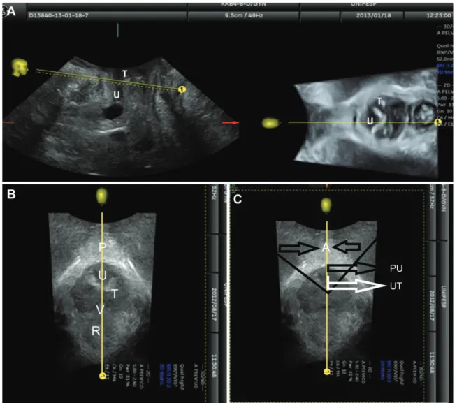
image in the mid-sagittal plane (►Fig. 2). The tape
displace-ment in relation to the UL was measured as the difference between the ratio of the BT and UL at rest and during the
Valsalva maneuver or pelvicfloor contraction. For an evaluation
of the relative position of the tape, we divided the BT by the UL, and the resulting number was used to classify the position of the tape within the urethra. The tape was considered to be: in the proximal-third (closest to the bladder neck) if the result was between 0 and 0.33; in the middle-third if the result was between 0.34 and 0.66; and in the distal-third (closest to the external urethral ostium) if the result was between 0.67 and 1.0.
The OmniView volume contrast imaging (VCI) mode (GE Healthcare, Zipf, Austria) was used during the image analysis, sliding digitally with a straight line passing through the lower edge of the pubic symphysis, the urethra, and the lower tape. In order to improve visibility, VCI was selected, with a thickness of 3.0 mm, which allowed the visualization of the pubis, the urethra, the vagina, the tape, and the rectum (►Fig. 2).15The OmniView-VCI mode was used to evaluate the
angle formed by the tape arms (Ao), as well as the distance
between the posterior inferior symphysis margin and the echogenic center of the urethra (PU), and the distance between the echogenic center of the urethra and the tape (UT) (►Fig. 2). The mobility of the urethra during movement was calculated as the difference between the PU at rest and the PU
during the Valsalva maneuver or pelvicfloor contraction.
Statistical analyses were performed using the Minitab software (Minitab, Inc., State College, PA, US), version 16. The Mann-Whitney test was used for the continuous
non-parametric variables, and the Studentt-test was used for the
continuous parametric variables. The Chi-squared, Fisher, Kruskal-Wallis, and Pearson correlation tests were used for the nominal variables. Analyses of variance (ANOVAs) for continuous variables were performed using the Tukey
multiple comparison procedure. Ap-value<0.05 was
con-sidered statistically significant.16,17
Results
The three surgical groups (26 TVT, 42 TVT-O, and 37 Minis-ling TVT-S) were similar in terms of age, body mass index (BMI), the number of vaginal or caesarean deliveries, and
hormonal status, as shown in►Table 1. At the time of the
Fig. 1 Patient selectionflowchart: the patient selection process is depicted. Abbreviations: TVT, tension-free vaginal tape (retropubic sling);
evaluation, the postoperative time ranged between 36 and 40 months.
The 2D-US measurements obtained in the mid-sagittal
plane are presented in►Table 2. The UL at rest, during the
Valsalva maneuver, and during pelvicfloor contraction did
not significantly differ among the three groups. In contrast,
there were differences among the groups regarding the BT.
Specifically for the TVT group, the tape was significantly
Fig. 2 Example images: (A) a 2D-US mid-sagittal plane (left) and 3D OmniView axial image (right) image of the pelvicfloor are shown. Note the sling position
(white hyperechoic structure–T) and urethra (hypoechoic structure–U). Two images below - OmniView mode: The axial plane on the OmniView mode with
pelvicfloor organs, at the left (B), and measurements, at the right (C), is shown. Abbreviations: Ao, angle between the two arms of the sling; P, symphysis pubis; PU, distance between the symphysis pubis and the urethra; R, rectum; T, polypropylene tape (sling); U, urethra; UT, distance between the tape and the urethra; V, vagina.
Table 1 Patient characteristics
TVT (n¼26) Mean (SD)
TVT-O (n¼42) Mean (SD)
TVT-S (n¼37) Mean (SD)
p
Age (Years) 58.35 (9.79) 55.00 (10.77) 54.51 (13.2) 0.384a
BMI (Kg/m2) 27.87 (3.66) 30.41 (5.16) 29.50 (5.35) 0.121a
Caesarean delivery 0.42 (0.86) 0.52 (0.89) 0.62 (0.83) 0.372b
Vaginal delivery 1.89 (1.11) 2.71 (2.11) 3.05 (2.73) 0.130b
Menopause (%) 22 (84.60%) 26 (61.90%) 24 (64.86%) 0.122c
Abbreviations: BMI, body mass index; SD, standard deviation; TVT, tension-free vaginal tape (retropubic sling); TVT-O, tension-free vaginal tape-obturator (transobturator sling); TVT-S, tension-free vaginal tape-Secur (single-incision sling).
Notes:aANOVA;bKruskal-Wallis test;cChi
–squared test. The values are represented as mean and SD (range) and n (%), and are attributed to patients who
closer to the bladder neck compared with the other groups
during rest (p¼0.001), but not during the Valsalva
maneu-ver (p¼0.08) or pelvicfloor contraction (p¼0.11). There
was no significant difference in the displacement of the tape
among the three groups.
The location of the sling relative to the urethra was different among the groups at rest and during movement
(rest:p<0.001; Valsalva: p¼0.008; pelvic floor
contrac-tion: p<0.001). Specifically in the TVT group, all tapes
(100%) were located in the mid-urethra during rest and
during movement (Valsalva maneuver and pelvicfloor
con-traction). However, for the TVT-O group, only 31 out of 42 tapes (73.8%) were located in the mid-urethra at rest, and for the TVT-S group, 33 out of 37 tapes (89.2%) were located in the mid-urethra at rest. Furthermore, during the Valsalva maneuver, 78.6% of tapes in the TVT-O group and 78.4% in the TVT-S group were located in the mid-urethra. During
pelvicfloor contraction, only 73.8% of the tapes in the TVT-0
group and 86.5% in the TVT-S group were located in the mid-urethra.
Of the 15 patients with the tape located in the distal
urethra, 66.7% had a BMI>30 kg/m2 (obese status). In
contrast, of the 90 patients with the tape located in the mid-urethra, only 34% were obese as assessed by BMI
(mid-urethra versus distal (mid-urethra:p¼0.003). The total sample
had 40/105 (38%) obese patients.
The measurements taken using the OmniView-VCI mode
are shown in►Table 2. The angle formed by the arms of the
sling was not significantly different between the TVT and
TVT-S groups, but was more obtuse in the TVT-O group
compared with the other groups during rest (p<0.001),
during the Valsalva maneuver (p<0.001), and during pelvic
floor contraction (p<0.001). There were no significant
group differences regarding the PU, at rest or during
move-ment. While the UT at rest and during pelvicfloor
contrac-tion did not show differences among the groups, during the
Valsalva maneuver, the UT significantly differed among the
groups (p¼0.01), with a smaller UT in the TVT group
compared with the TVT-S group. No significant differences
among the groups regarding urethral mobility during the
Valsalva maneuver and during pelvicfloor contraction were
found (p¼0.78 andp¼0.51 respectively).
Information on subjective and objective cure is presented in►Table 3. A subjective cure was achieved by 88 of the 105 patients (83.8%), with no difference among the groups
(p¼0.701). An objective cure was verified in 92/105
(87.8%) patients, with no difference among the groups
(p¼0.514). The presence of urgency-related symptoms
Table 2 Midsagittal sonographic and OmniView-VCI mode measurements
TVT (n¼26) Mean (SD)
TVT-O (n¼42) Mean (SD)
TVT-S (n¼37) Mean (SD)
p
Urethral length: rest (cm) 3.34 (0.37) 3.24 (0.40) 3.36 (0.45) 0.38
Urethral length: Valsalva (cm) 3.26 (0.30) 3.18 (0.40) 3.25 (0.42) 0.63
Urethral length: contraction (cm) 3.56 (0.42) 3.34 (0.39) 3.53 (0.49) 0.07
BT: rest (cm) 1.65(0.26) 1.93 (0.38) 1.95 (0.51) 0.01
BT: Valsalva (cm) 1.67 (0.24) 1.85 (0.37) 1.89 (0.50) 0.08
BT: contraction (cm) 1.81 (0.38) 2.03 (0.39) 2.03 (0.57) 0.11
Mid-urethra tape n (%): rest 26 (100%) 31 (73.8%) 33 (89.2%) 0.008
Mid-urethra tape n (%):Valsalva 26 (100%) 33 (78.6%) 29 (78.4%) 0.036
Mid-urethra tape n (%): contraction 26 (100%) 31 (73.8%) 32 (86.5%) 0.013
A°: rest 119.94 (19.66) 141.93 (14. 25) 121.06 (14. 24) <0.001
A°: Valsalva 130.79 (21.78) 14.,48 (13. 23) 124.40 (24. 79) <0.001
A°: contraction 120.94 (16.63) 144.77 (16. 51) 126.53 (18. 20) <0.001
PU: rest (cm) 1.68 (0.28) 1.53 (.,25) 1.64 (0.29) 0.08
PU: Valsalva (cm) 1.72 (0.29) 1.59 (0.28) 1.64 (0.14) 0.19
PU: contraction (cm) 1.65 (0.27) 1.49 (0.29) 1.66 (0.31) 0.02
UT: rest (cm) 0.67 (0.14) 0.73 (0.15) 0.71 (0.11) 0.21
UT: Valsalva (cm) 0.65 (0.13) 0.68 (0.14) 0.75 (0.14) 0.01
UT: contraction (cm) 0.60 (0.01) 0.65 (0.15) 0.66 (0.10) 0.16
Abbreviations: Ao, angle of the two arms of the sling; BT, distance between the bladder neck and the tape; PU, distance between the symphysis pubis and the urethra; SD, standard deviation; TVT, tension-free vaginal tape (retropubic sling); TVT-O, tension-free vaginal tape-obturator (transobturator sling); TVT-S, tension-free vaginal tape-secure (single-incision sling); UT, distance between the tape and the urethra.
Table 3 Urinary symptoms
TVT TVT-O TVT-S TOTAL p
Subjective cure 23 (88.5%) 36 (85.7%) 29 (78.4%) 88 (83.8%) 0.514a
Objective cure 23 (88.5%) 38 (90.5%) 31 (83.8%) 92 (87.8%) 0.701b
Urgency symptoms 8 (30.8%) 9 (21.4%) 8 (21.6%) 25 (23.8%) 0.630a
TOTAL 26 (100%) 42 (100%) 37 (100%) 105 (100%)
Abbreviations: TVT, tension-free vaginal tape (retropubic sling); TVT-O, tension-free vaginal tape-obturator (transobturator sling); TVT-S, tension-free vaginal tape-secure (single-incision sling).
Notes:aChi-squared test;bFisher exact test. The subjective and objective cure rates and the rate of urgency-related symptoms for patients who underwent surgical treatment for stress urinary incontinence using a retropubic sling (TVT), a transobturator sling (TVT-O), or a single-incision sling (TVT-S) are provided.
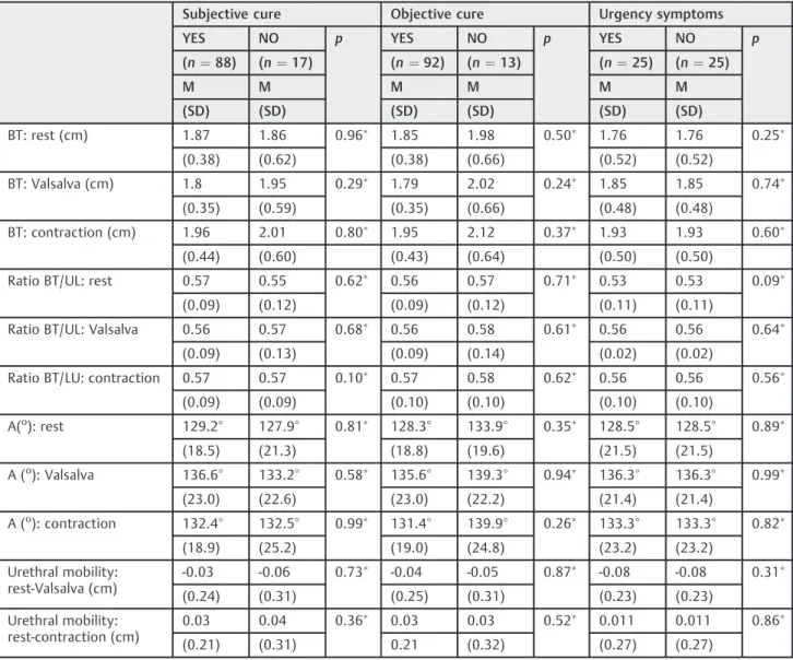
Table 4 Associations between ultrasound measurements and urinary symptoms
Subjective cure Objective cure Urgency symptoms
YES NO p YES NO p YES NO p
(n¼88) (n¼17) (n¼92) (n¼13) (n¼25) (n¼25)
M M M M M M
(SD) (SD) (SD) (SD) (SD) (SD)
BT: rest (cm) 1.87 1.86 0.96 1.85 1.98 0.50 1.76 1.76 0.25
(0.38) (0.62) (0.38) (0.66) (0.52) (0.52)
BT: Valsalva (cm) 1.8 1.95 0.29 1.79 2.02 0.24 1.85 1.85 0.74
(0.35) (0.59) (0.35) (0.66) (0.48) (0.48)
BT: contraction (cm) 1.96 2.01 0.80 1.95 2.12 0.37 1.93 1.93 0.60
(0.44) (0.60) (0.43) (0.64) (0.50) (0.50)
Ratio BT/UL: rest 0.57 0.55 0.62 0.56 0.57 0.71 0.53 0.53 0.09
(0.09) (0.12) (0.09) (0.12) (0.11) (0.11)
Ratio BT/UL: Valsalva 0.56 0.57 0.68 0.56 0.58 0.61 0.56 0.56 0.64
(0.09) (0.13) (0.09) (0.14) (0.02) (0.02)
Ratio BT/LU: contraction 0.57 0.57 0.10 0.57 0.58 0.62 0.56 0.56 0.56
(0.09) (0.09) (0.10) (0.10) (0.10) (0.10)
A(o): rest 129.2° 127.9° 0.81 128.3° 133.9° 0.35 128.5° 128.5° 0.89
(18.5) (21.3) (18.8) (19.6) (21.5) (21.5)
A (o): Valsalva 136.6° 133.2° 0.58 135.6° 139.3° 0.94 136.3° 136.3° 0.99
(23.0) (22.6) (23.0) (22.2) (21.4) (21.4)
A (o): contraction 132.4° 132.5° 0.99 131.4° 139.9° 0.26 133.3° 133.3° 0.82
(18.9) (25.2) (19.0) (24.8) (23.2) (23.2)
Urethral mobility: rest-Valsalva (cm)
-0.03 -0.06 0.73 -0.04 -0.05 0.87 -0.08 -0.08 0.31
(0.24) (0.31) (0.25) (0.31) (0.23) (0.23)
Urethral mobility: rest-contraction (cm)
0.03 0.04 0.36 0.03 0.03 0.52 0.011 0.011 0.86
(0.21) (0.31) 0.21 (0.32) (0.27) (0.27)
was observed in 25/105 (23.8%) patients, with no difference
among the groups (p¼0.630).
Correlations between the tape’s spatial position and the
clinical results are shown in►Table 4. No significant
rela-tionships were found, except for the displacement of the tape in relation to the UL between rest and contraction, which was slightly greater in the direction of the bladder neck in patients with urgency-related symptoms compared with
those without symptoms (p¼0.03). No significant
differ-ences among the groups were found for the domains of the
KHQ (►Table 5).
Discussion
Three-dimensional US imaging of the pelvicfloor is a
non-invasive and reproducible technique for the evaluation of postoperative MUS, and it enables a dynamic assessment of the polypropylene sling tape, with good visibility during rest,
pelvicfloor contraction, and the Valsalva maneuver.7
The BT found in the present study is consistent with
previously published data.18,19In addition, for the majority
of the patients (85.7%), the sling was located in the mid-urethra, and all other patients had slings located in the distal urethra (14.3%). These results are also similar to those
obtained by others researchers.19–21 We observed that
66.7% of the patients with the tape located in the distal
urethra had a BMI>30 kg/m2(obese status), while among
those with the tape located in the mid-urethra, only 34% were obese. Thus, obesity appears to be a factor favoring a more distal position of the sling. However, the tape remained located in the mid-urethra in all individuals in the TVT group,
thus showing a better location in obese patients as well, three years after the surgery.
The angle formed by the tape arms was less obtuse in the TVT and TVT-S groups compared with the TVT-O group (at rest,
during the Valsalva maneuver, and during pelvicfloor
contrac-tion), with no differences between the TVT and TVT-S groups.
Thisfinding was as we expected, and it may be explained by
features of the sling insertion technique.7,22For the TVT-O, the
insertion was in a“hammock”position, while for the TVT and
TVT-S, the insertion was in a“U”position. The“U”position was
considered the best way to insert the TVT-S before its com-mercialization was discontinued.
It is interesting to note that no differences among the
surgical techniques were identified with regard to tape
displacement or urethra mobility, in rest or during move-ment, which is as expected, given that the three surgical procedures are meant to stabilize urethral mobility.
The distance between the urethra and the tape was smaller in the TVT group compared with the TVT-S group
during the Valsalva maneuver only. Thisfinding could
sug-gest a higher frequency of postoperative urgency-related symptoms with the TVT. However, similarly to previous studies, we did not observe any differences between TVT
and TVT-O groups in terms of this sonographic measure.22
Even though no associations between the position of the MUS tape and the clinical results were found, it was noted that, in patients with urgency-related symptoms, the tape tended to move more toward the bladder neck during the
Valsalva maneuver. On the other hand, during pelvicfloor
contraction, there was a greater displacement of the tape toward the bladder neck in patients who reported urgency-related symptoms compared with patients without these symptoms. This may be a mechanism that explains the onset of de novo urgency in the postoperative period.
A lack of correlation between the tape position and the
clinical outcomes has been previously reported6,20,23 and
was confirmed in the present study, suggesting that factors
other than tape position could influence the results of the
insertion of the MUS.23,24Although some sonographic
meas-urements reached statistical significance in the group
com-parisons, the differences are on the order of millimeters, and
are not clinically significant.
The limitations of the present study include the cross-sectional design, with only a midterm postoperative follow-up. Therefore, we were not able to compare the preoperative and postoperative results or evaluate the earlier failures requiring surgical re-intervention, which could be related
to tape position, as suggested by other studies.24,25However,
our study sample does reproduce the results from the literature regarding the objective and subjective cure rates and the frequency of urinary urgency symptoms, according
to the different surgical techniques.1,2
Conclusion
In conclusion, we observed differences in the synthetic sling position three years after surgery when comparing different routes of insertion. In the retropubic approach, the tape was Table 5 Quality of life assessments (King Health Questionnaire)
TVT-R Mean TVT-O Mean TVT-S Mean p
General health 29.81 24.40 25.68 0.28
Incontinence impact 12.82 13.49 13.51 0.92
Limitations to daily activities
10.26 8.33 13.06 0.67
Limitations to physical activities
11.54 7.14 11.56 0.56
Social limitations 10.26 3.70 6.01 0.62
Impact on personal relationships
3.92 3.09 5.36 0.86
Impact on emotions 6.41 9.52 9.31 0.73
Impact on sleep arrangement
8.97 6.75 5.41 0.58
Severity measure 13.14 10.52 9.01 0.69
Abbreviations: TVT, tension-free vaginal tape (retropubic sling); TVT-O, tension-free vaginal tape-obturator (transobturator sling); TVT-S, tension-free vaginal tape-secur (single-incision sling).
more frequently located in the mid-urethra compared with the transobturator route and the single-incision sling. Regarding the angle of the sling arms, it was more obtuse with the transobturator route, and it was, in a similar manner, more acute with the retropubic route and the single-incision sling. Although a relationship between the position of the MUS tape and the subjective/objective cure three years after surgery was not demonstrated, there was a correlation between the movement of the tape during
pelvic floor contraction and the presence of symptoms of
urgency.
Funding
This work was supported by Universidade Federal de São Paulo.
Conflicts of Interest
Authors have no conflicts of interest to disclose.
Registration
Clinical Trials.gov Protocol Registration System, http://www. clinicaltrials.gov, NCT 02406638, Pelvic Floor 3D USG Three Years After Mid-urethral Slings (TVT-R, TVT-O, TVT-S).
References
1 Ogah J, Cody DJ, Rogerson L. Minimally invasive synthetic sub-urethral sling operations for stress urinary incontinence in women: a short version Cochrane review. Neurourol Urodyn 2011;30(03):284–291
2 Mostafa A, Lim CP, Hopper L, Madhuvrata P, Abdel-Fattah M. Single-incision mini-slings versus standard midurethral slings in surgical management of female stress urinary incontinence: an updated systematic review and meta-analysis of effectiveness and complications. Eur Urol 2014;65(02):402–427
3 Bianchi-Ferraro AM, Jarmy-DiBella ZI, de Aquino Castro R, Borto-lini MA, Sartori MG, Girão MJ. Randomized controlled trial comparing TVT-O and TVT-S for the treatment of stress urinary incontinence: 2-year results. Int Urogynecol J Pelvic Floor Dys-funct 2014;25(10):1343–1348
4 Schimpf MO, Rahn DD, Wheeler TL, et al; Society of Gynecologic Surgeons Systematic Review Group. Sling surgery for stress urinary incontinence in women: a systematic review and metaa-nalysis. Am J Obstet Gynecol 2014;211(01):71.e1–71.e27 5 Chantarasorn V, Shek KL, Dietz HP. Sonographic appearance of
transobturator slings: implications for function and dysfunction. Int Urogynecol J Pelvic Floor Dysfunct 2011;22(04):493–498 6 Dietz HP. Ultrasound imaging of the pelvicfloor. Part II:
three-dimensional or volume imaging. Ultrasound Obstet Gynecol 2004;23(06):615–625
7 Chene G, Cotte B, Tardieu AS, Savary D, Mansoor A. Clinical and ultrasonographic correlations following three surgical anti-in-continence procedures (TOT, TVT and TVT-O). Int Urogynecol J Pelvic Floor Dysfunct 2008;19(08):1125–1131
8 Ulmsten U, Henriksson L, Johnson P, Varhos G. An ambulatory surgical procedure under local anesthesia for treatment of female
urinary incontinence. Int Urogynecol J Pelvic Floor Dysfunct 1996;7(02):81–85, discussion 85–86
9 de Leval J. Novel surgical technique for the treatment of female stress urinary incontinence: transobturator vaginal tape inside-out. Eur Urol 2003;44(06):724–730
10 Bianchi-Ferraro AM, Jarmy-Di Bella ZI, Castro RdeA, Bortolini MA, Sartori MG, Girão MJ. Single-incision sling compared with trans-obturator sling for treating stress urinary incontinence: a rando-mized controlled trial. Int Urogynecol J Pelvic Floor Dysfunct 2013;24(09):1459–1465
11 Ethicon, a Johnson & Johnson Company, makes allegedly danger-ous transvaginal mesh and hernia mesh products [Internet]. 2012 [cited 2016 Jan 12]. Available from: http://www.yourlawyer.com/ topics/overview/johnson-johnson-ethicon-gynecare-transvagi-nal-mesh-complications-side-effects-lawsuits
12 Hahn I, Fall M. Objective quantification of stress urinary incon-tinence: a short, reproducible, provocative pad-test. Neurourol Urodyn 1991;10(05):475–481
13 Fonseca ESM, Camargo ALM, Castro RA, et al. Validation of a quality of life questionnaire (King’s Health Questionnaire) in Brazilian women with urinary incontinence. Rev Bras Ginecol Obstet 2005;27(05):235–242
14 Dietz HP. Pelvicfloor ultrasound in incontinence: what’s in it for the surgeon? Int Urogynecol J Pelvic Floor Dysfunct 2011;22(09): 1085–1097
15 Tonni G, Lituania M. OmniView algorithm: a novel 3-dimensional sonographic technique in the study of the fetal hard and soft palates. J Ultrasound Med 2012;31(02):313–318
16 Agrest A. Categorical data analysis. New York: Wiley Interscience; 1990
17 Neter J, Kutner MH, Nachtsheim CJ, Wasserman W. Applied linear statistical models. 4th ed. Boston: Irwin; 1996
18 deTayrac R, Deffieux X, Droupy S, Chauveaud-Lambling A, Calva-nèse-Benamour L, Fernandez H. A prospective randomized trial comparing tension-free vaginal tape and transobturator subure-thral tape for surgical treatment of stress urinary incontinence. Am J Obstet Gynecol 2004;190(03):602–608
19 Foulot H, Uzan I, Chopin N, Borghese B, Chapron C. Monarc trans-obturator sling system for the treatment of female urinary stress incontinence: results of a post-operative transvaginal ultrasonogra-phy. Int Urogynecol J Pelvic Floor Dysfunct 2007;18(08):857–861 20 Duckett J, Aggarwal I, Patil A, Vella M. Effect of tension-free
vaginal tape position on the resolution of irritative bladder symptoms in women with mixed incontinence. Int Urogynecol J Pelvic Floor Dysfunct 2008;19(02):237–239
21 Dietz HP, Mouritsen L, Ellis G, Wilson PD. Does the tension-free vaginal tape stay where you put it? Am J Obstet Gynecol 2003;188 (04):950–953
22 Lin KL, Juan YS, Lo TS, Liu CM, Tsai EM, Long CY. Three-dimensional ultrasonographic assessment of compression effect on urethra following tension-free vaginal tape and transobturator tape procedures. Ultrasound Obstet Gynecol 2012;39(04):452–457 23 Dietz HP, Mouritsen L, Ellis G, Wilson PD. How important is TVT
location? Acta Obstet Gynecol Scand 2004;83(10):904–908 24 Kociszewski J, Rautenberg O, Kolben S, Eberhard J, Hilgers R,
Viereck V. Tape functionality: position, change in shape, and outcome after TVT procedure–mid-term results. Int Urogynecol J Pelvic Floor Dysfunct 2010;21(07):795–800