59
Silveira RB et al. Dysplasia epiphysealis hemimelica
Radiol Bras. 2013 Jan/Fev;46(1):59–60
Dysplasia epiphysealis hemimelica (Trevor-Fairbank disease):
case report
*
Displasia epifisária hemimélica (doença de Trevor-Fairbank): relato de caso
Regina Bosenbecker da Silveira1, Felipe Augusto Rozales Lopes2, Ana Luiza Behrensdorf Reis3, Enrico Granzotto4, André Guimarães de Oliveira2
Dysplasia epiphysealis hemimelica is a rare benign disease (incidence 1:1,000,000), characterized by an osteochondral overgrowth affecting one or more epiphyses. Generally, the age of onset is between two and 14 years. The characteristic imaging findings are sufficient for the diagnosis. Surgical excision of the lesion is only indicated in cases where a functional limitation is present.
Keywords: Epiphysis; Osteochondrodysplasia; Dysplasia epiphysealis hemimelica; Trevor disease.
A displasia epifisária hemimélica é uma doença benigna rara (incidência de 1:1.000.000), caracterizada por um cres-cimento osteocondral decorrente de uma ou mais epífises. Em geral a idade de início é entre 2 e 14 anos. Os achados característicos dos exames de imagem são suficientes para o diagnóstico. A excisão cirúrgica da lesão só é indicada caso haja limitação funcional.
Unitermos: Epífise; Osteocondrodisplasia; Displasia epifisária hemimélica; Doença de Trevor.
Abstract
Resumo
* Study developed at Hospital Universitário São Francisco de Paula, Pelotas, RS, Brazil.
1. Master of Health and Behavior, Professor of Pediatrics, Universidade Católica de Pelotas (UCPel), MD, Pediatric Physi-cian at Hospital Universitário São Francisco de Paula, Pelotas, RS, Brazil.
2. Graduate Students of Medicine, School of Medicine, Uni-versidade Católica de Pelotas (UCPel), Pelotas, RS, Brazil.
3. MD, Pediatric Physician, Hospital Universitário São Fran-cisco de Paula, Pelotas, RS, Brazil.
4. Titular Member of Colégio Brasileiro de Radiologia e Diag-nóstico por Imagem (CBR), MD, Radiologist, Hospital Universi-tário São Francisco de Paula and Hospital Santa Casa de Mise-ricórdia de Pelotas, Pelotas, RS, Brazil.
Mailing Address: Felipe Augusto Rozales Lopes. Rua Emílio Jorge dos Reis, 512, Bairro Três Vendas. Pelotas, RS, Brazil, 96020-440. E-mail: [email protected].
Received March 30, 2012. Accepted after revision August 20, 2012.
Silveira RB, Lopes FAR, Reis ALB, Granzotto E, Oliveira AG. Dysplasia epiphysealis hemimelica (Trevor-Fairbank disease): case report. Radiol Bras. 2013 Jan/Fev;46(1):59–60.
0100-3984 © Colégio Brasileiro de Radiologia e Diagnóstico por Imagem CASE REPORT
significantly when running. At physical examination, a moderate swelling and lo-cal warmth were observed, with pain and flexion limitation.
At radiography, irregularities on the ar-ticular surfaces with exuberant calcifica-tions were observed in the articular space and adjacent to the distal femoral and proximal tibial epiphyses (Figure 1).
Computed tomography (CT) revealed the presence of moderate joint effusion with irregularity in the contours of the Because of such disease rarity, there is
a scarcity of cases described in the litera-ture, hence the authors’ objective of pre-senting the case of a three-year-old boy with DEH in his right knee.
CASE REPORT
A male, three-year-old patient presented pain and edema in his right knee for three days. Along the previous two months the patient presented abnormality in gait, more INTRODUCTION
Dysplasia epiphysealis hemimelica (DEH) is defined as a localized osteochon-dral overgrowth arising from half of an epi-physis and affecting either one or several epiphyses or ossification centers(1–3). It is
an abnormal and asymmetrical prolifera-tion of cartilage with osteochondral ossi-fication which ceases once the growth pro-cess is completed(3).
It is a rare disease, with a reported inci-dence of 1:1.000.000, whose etiology still remains unknown. However, it is known that such condition is connected with the group of osteochondromatoses(1,2).
Figure 1.A: Anteroposterior radiograph demonstrating irregularities on articular surfaces with calcifica-tions adjacent to the distal femoral (large arrow) and proximal tibial (small arrow) epiphyses. B: Lateral radiograph of knee identifying exuberant calcifications in the articular space (arrow).
60
Silveira RB et al. Dysplasia epiphysealis hemimelica
Radiol Bras. 2013 Jan/Fev;46(1):59–60 femoral condyle and tibial plateau in the
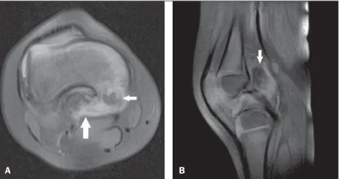
medial compartment, and multiple, irregu-lar calcifications within the joint, particu-larly on the posterior aspect of the femur. Magnetic resonance imaging (MRI) demonstrated irregularities and dysplastic alterations on articular surfaces of the me-dial femoral condyle and internal tibial pla-teau, with calcifications and adjacent intrasynovial ossifications (Figure 2).
Surgical excision of the lesion was per-formed because of the significant func-tional limitation and pain. Anatomopatho-logical analysis revealed osteochondroma.
DISCUSSION
DEH – also known as Trevor-Fairbank disease – generally affects children in the age range between 2 and 14 years and is most frequent in boys. Generally with uni-lateral involvement, the lesion originates a tone of the epiphyseal aspects, preferen-tially affecting the lower limbs, in decreas-ing order of occurrence: knee, ankle and foot(4,5).
Initially the clinical presentation corre-sponds to local painless swelling deter-mined by the cartilage overgrowth, which may become painful and generate deformi-ties, limited range of motion and limb length discrepancy(1,6).
According to Peduto et al., the diagno-sis is based on radiographic findings(1), and
biopsy is not required, except in case of sur-gical excision of the lesion or in the pres-ence of atypical radiological findings(5).
Histologically, the lesion characteristics are identical to those of osteochondroma(2).
According to Carlson et al., the osteo-cartilaginous overgrowth of DEH, origi-nated from the epiphysis, must not be con-fused with exostosis originated from the metaphysis. Such authors describe the fol-lowing differential diagnoses: chondrodys-plasia punctata and multiple epiphyseal dysplasia, which occur bilaterally, and ar-eas of asceptic necrosis presenting irregu-larities, but without any characteristic of osteochondral overgrowth(6).
Radiography shows one or more irregu-lar masses with focal ossification adjacent to the border of one of the halves of the epi-physis (with overgrowth), with irregular enlargement of epiphyseal centers and
ad-jacent metaphysis, like in the present case, where the lesion was adjacent to the distal femoral and proximal tibial epiphyses. Such mass may be located on the underly-ing bone in cases where bone maturation is present(1,2,6).
CT can more accurately define the ana-tomical relationship between the mass and the bone, as well as the continuity of the bone cortical and bone marrow, but it is inferior to MRI in the evaluation of changes in cartilage and soft tissues(1,2).
With MRI, one can better evaluate the epiphyseal osteochondral growth and its probable effects on adjacent structures, al-lowing a better definition of bone and car-tilage structures at multiple planes, but scarce reports describing MRI findings of DEH are found in the literature(1,2,7).
MRI is useful at early phases of the dis-ease, when a small mass with calcifications inside is observed, likewise in the present case(1,2). At the T1-weighted sequence, the
signal intensity is the same for both the healthy epiphyseal cartilage and the carti-lage affected by disease, but the latter pre-sents small spots of low intensity corre-sponding to calcifications. On the other hand, at T2-weighted sequences, the higher intensity is observed in the cartilaginous cap as compared with the osteocartilagi-nous junction(8).
As regards treatment, radiographic fol-low-up is performed to evaluate the lesion progression. In cases of pain, skeletal de-formities or functional limitations, surgical
excision is indicated(2). Follow-up is
re-quired to evaluate lesion recidivation after surgery(2,3).
Finally, DEH is a rare disease character-ized by asymmetrical epiphyseal over-growth, whose diagnosis is made by means of radiography, supplemented by MRI which can better evaluate the osteochon-dral overgrowth as well as adjacent struc-tures.
REFERENCES
1. Peduto AJ, Frawley KJ, Bellemore MC, et al. MR imaging of dysplasia epiphysealis hemimelica: bony and soft-tissue abnormalities. AJR Am J Roentgenol. 1999;172:819–23.
2. Araujo CR Jr, Montandon S, Montandon C, et al. Best cases of the AFIP: dysplasia epiphysealis hemimelica of the patella. Radiographics. 2006; 26:581–6.
3. Bansal P, Khare R, Lal H, et al. Dysplasia epiphy-sealis hemimelica or Trevor’s disease of proximal tibia mimicking loose body. J Clin Orthop Trauma. 2010;1:105–6.
4. Wiart E, Budzik JF, Fron D, et al. Bilateral dyspla-sia epiphysealis hemimelica of the talus associated with a lower leg intramuscular cartilaginous mass. Pediatr Radiol. 2012;42:503–7.
5. Rosero VM, Kiss S, Terebessy T, et al. Dysplasia epiphysealis hemimelica (Trevor’s disease): 7 of our own cases and a review of the literature. Acta Orthop. 2007;78:856–61.
6. Carlson DH, Wilkinson RH. Variability of unilat-eral epiphyseal dysplasia (dysplasia epiphysealis hemimelica). Radiology. 1979;133:369–73. 7. Vogel T, Skuban T, Kirchhoff C, et al. Dysplasia
epiphysealis hemimelica of the distal ulna: a case report and review of the literature. Eur J Med Res. 2009;14:272–6.
8. Iwasawa T, Aida N, Kobayashi N, et al. MRI find-ings of dysplasia epiphysealis hemimelica. Pediatr Radiol. 1996;26:65–7.
Figure 2.A: Axial MRI proton-density-weighted fat sat image demonstrating involvement of the distal femur by the disease, with irregularities, dysplastic alterations (small arrow), calcifications and intra-ar-ticular ossification (large arrow). B: Sagittal MRI proton-density-weighted fat sat image showing the pres-ence of calcifications and intrasynovial ossification (arrow). Soft tissue preservation is also observed.