Camilla Regina Galvão Bengtson(a) Antonio Lucindo Bengtson(b) Nadya Galvão Bengtson(c) Miriam Lacalle Turbino(d)
(a) PhD Student; (d)PhD, Professor – Department of Restorative Dentistry, School of Dentistry, University of São Paulo, São Paulo, SP, Brazil.
(b) PhD, Professor; (c)MSc, Professor
– Department of Pediatric Dentistry, School of Dentistry, Universidade Metropolitana de Santos, Santos, SP, Brazil.
Corresponding author:
Camilla Regina Galvão Bengtson Rua Itararé, 177/76 - Bela Vista São Paulo - SP - Brazil CEP: 01308-030 E-mail: [email protected]
Received for publication on Dec 01, 2009 Accepted for publication on Jun 06, 2010
Do the origins of primary teeth affect the
bond strength of a self-etching adhesive
system to dentin?
Abstract: The aim of this in vitro study was to evaluate the tensile bond strength of a self-etching adhesive system to three different dentinal sub-strates. Primary molar teeth that had been recently exfoliated (RE), with unknown time of exfoliation (UT), and extracted due to prolonged reten-tion (PR) were used for this investigareten-tion. Ten primary molar teeth of each group were cut in the middle following the mesio-distal direction, creat-ing a total of twenty specimens per group. The specimens were included in acrylic resin and had a lat dentin surface exposed. The self-etching adhesive system was applied to this surface and a 3-millimeter high cone with diameter of 2 mm in the adhesion area was constructed using com-posite resin. The specimens were stored in distilled water at 37°C for 24 hours. Fifteen specimens of each substrate were used for the tensile bond test (n = 15) and 5 had the interface analyzed by scanning electron mi-croscopy (SEM). The data was examined by one-way ANOVA and pre-sented no signiicant differences between groups (p = 0.5787). The mean values obtained for RE, UT and PR were 18.39 ± 9.70, 19.41 ± 7.80, and 23.30 ± 9.37 MPa, respectively. Any dentinal substrates of primary teeth studied are safe for tensile bond strength tests with adhesive systems.
Descriptors: Adhesives; Tensile strength; Dental materials; Dentin; Tooth, deciduous.
Introduction
In vitro tests, such as bond strength measurement, microleakage eval-uation and marginal gap calculation are vital screening tests that serve to predict the clinical behavior of new bonding systems.1 In any attempt
to have more accurate knowledge of the retention capability of bonding systems in the clinical situation, the evaluation of bond strength to hard dental tissue is usually required.2,3,4
Another condition that can cause alterations in dentin substrate is related to age change. Older teeth experience gradual enlargement of peritubular den-tin and intratubular mineral deposits, which often result in narrowed or completely occluded tubules. In human primary teeth, root resorption is a physi-ological process which occurs at the end of that life span. During this phase, some morphological and functional modiications occur to the pulp cells and consequently to the tissues involved in the process.7
These modiications may occur because, during this period, dentin and pulp are eliminated by odonto-clasts. This condition may suggest that primary teeth in different phases of their vital cycle could have different dentin characteristics.8,9,10
The formation of the hybrid layer, by the impreg-nation of resin ingredients into the demineralized intertubular dentin with subsequent polymeriza-tion, is thought to create an adequate bond between resin and dentin. Resin iniltration into the dentinal tubules or resin tags also contribute to bonding.11
Thus, bonding mechanism of resin to dentin is de-pendent on the microstructure of the substrate at the point of bonding.12,13
Although the number of studies regarding labo-ratory tests of the adhesive force of restorative mate-rials (tensile, microtensile, shear, and microshear) in primary teeth has increased,3,14,15,16,17,18 only a few of
these report the quality of dentin used in the tests. The substrate has an important role in hybrid layer formation. Due to this, substrate standardization of dentin which is used in bond strength is necessary. The purpose of this study was to investigate the in-luence of storage and exfoliation modes of primary teeth, on the tensile bond strength of a self etch hesive system to primary dentin. In addition, the ad-hesive interfaces were analyzed by SEM to observe differences in hybrid layer formation among groups.
Material and Methods
Thirty sound primary extracted molars were used. The teeth were obtained according to proto-cols (82/06) approved by the appropriate institution-al review board of the School of Dentistry – Univer-sity of São Paulo, and with the formal consent of the Human Tooth Bank of the School of Dentistry
– University of São Paulo. The teeth were cleaned of debris with pumice paste via a slow-speed handpiece and stored in distilled water until they were used. The teeth were manipulated with individual protec-tion equipment throughout the experiment and only one operator carried out all procedures.
The teeth were divided into three groups accord-ing to the exfoliation and storaccord-ing characteristics. Group 1: recently exfoliated primary molar teeth (RE), obtained and used on the same day of exfolia-tion. Group 2: primary molar teeth, which were sur-gically extracted due to prolonged retention (RP), used on the same day of extraction. Group 3: pri-mary molar teeth that were collected with unknown time of exfoliation. This inal group of teeth were kept dry for 2 months in an ambient temperature, and then rehydrated in distilled water over a period of 7 days at 4°C (UT).
The crowns of the thirty teeth were sectioned parallel to the long axis of the tooth, using a low-speed diamond saw under water cooling condi-tions (Buehler Ltd., Lake Bluff, IL, USA). As a re-sult, twenty specimens per group were obtained. Each dental fragment was included in acrylic resin. Enamel surfaces were lattened with 120-, 240-, 400-, and 600- grit silicon carbide papers on a pol-isher machine (Ecomet 3, Buehler Ltd., Lake Bluff, IL, USA), in order to expose the surface dentin, and standardize the smear layer.
The dentin surfaces were bonded with the self-etching adhesive system, AdheSE (Ivoclar Viva-dent AG, Schaan, the Principality of Liechtenstein) according to the manufacturer’s instructions. A silicone mold a with cone-shaped perforation mea-suring 2 mm in diameter at the base, and 3 mm in diameter at the top (3 mm in height), adhesion area = 0.0314 cm², was adjusted on this surface. A composite resin was used to ill the mold. Two in-crements were applied, each one photoactivated for 40 s using a light unit with 450 mW/cm² (3M Curing Light, 3M ESPE, St. Paul, MN, USA). All specimens were stored in distilled water at 37°C for 24 hours.
ten-sile tests. The failure load divided by the cross-sec-tional area at the point of fracture for each specimen was used to calculate the tensile bond strength in MPa. After conirmation that the values had normal and homogeneous distribution, the data was statisti-cally analyzed by one-way ANOVA. The SPSS Sta-tistics for the Windows program, version 17.0 (SPSS Inc., Chicago, IL, USA) was utilized and statistical signiicance was established at α = 0.05.
After testing, the fracture modes of each speci-men were determined by examination in a dissect-ing microscope at 25 x magniication (Olympus Co., Tokyo, Kanto, Japan). The fracture mode was classiied as an adhesive/mixed failure if debond-ing occurred between the resin and dentin, and as a cohesive failure if it occurred in composite resin or dentin.
The other ive specimens of each group were se-lected for SEM analysis of adhesive interface (model Jeol 2800, Jeol Co., Tokyo, Kanto, Japan). They were partly decalciied in phosphoric acid (36% solu-tion, 10 s), deproteinized with sodium hypochlorite (2% solution, 60 s), and washed in tap water. There-after, the specimens were dehydrated in increasing concentrations of ethanol, immersed (10 min) in hexamethyldisilazane (HMDS), and completely air-dried. These specimens were covered with a gold-palladium layer (20 nm) for visualization in SEM. Complete extension of the union zone between dentin and resin was analyzed. Electromicrographs (1,500 x and 3,000 x magniications) were obtained for descriptive analysis of the region.
Results
The one-way ANOVA revealed no statistically
signiicant difference in the tensile bond strength to dentin among the three groups (p = 0.5787 and f = 0.8895). The mean tensile bond strength values and standard deviations for each experimental con-dition are shown in Table 1.
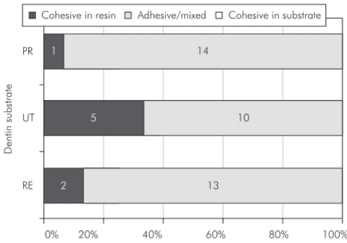
The failure modes of tested specimens are resented in Graph 1. Adhesive/mixed failure was pre-dominant in all groups, independent of the exfolia-tion mode or storing condiexfolia-tions.
The SEM observation of adhesive interfaces in-dicated that the hybrid layer was not uniform in complete extension of the union zone, with values in the range of 1-3 µm. In addition, a high number of resinous tags, homogeneously distributed, were observed in this area. Differences between the three groups were not detected (Figure 1).
Discussion
The microstructure of dentin at the site of bond-ing is extremely important in the formation of the bonding mechanism of resin to dentin.19 Since
den-tin is a dynamic substrate,20 this research proposed
analyzing the quality of bonding in three differ-ent situations. Studies have shown that the tensile bond strength of adhesive systems can be inluenced by dentin age and time of storage in permanent teeth.21,22
Data from literature demonstrates that adhesion force of composite resin systems, as applied to pri-mary dentin, ranges from 5.53 to 70.1 MPa.3,23 This
Table 1 - Mean bond strength (MPa) and standard devia-tion of the adhesive system to the different dentin substrates (n = 15).
Groups Bond Strength (MPa) ANOVA
Recently exfoliated 18.39 ± 9.70 A
Prolonged retention 23.30 ± 9.37 A
Unknown time 19.41 ± 7.80 A
Mean values followed by the same letter were not significantly different (p > 0.05).
RE UT PR
0%
1
5
2
14
10
13
20%
D
e
ntin
su
bs
trate
40% 60% 80% 100%
Cohesive in resin Adhesive/mixed Cohesive in substrate
wide variation is explained by differences between the methods employed, as well as factors related to the tooth and material used. In a small number of such studies, the origin of the primary teeth was re-ported. Due to the lack of studies in the literature concerning different conditions with primary den-tin, results from studies with permanent teeth were taken as reference, and our results are in agreement with them.
Considering time and storage conditions of the teeth after the extraction, some questions concern how changes within dentin after extraction may in-luence adhesion in vitro dentin bond studies. Titley et al.5 stated that post-mortem changes could occur
in dentin, with the potential to affect the outcome of bond strength tests. However, in another study
that compares shear bond strength of dentin to re-storative material, no signiicant differences were observed between the group stored in distilled water for eight days and the other stored for six months.22
Research about utilization of dry and rehydrated tooth in tensile bond strength showed that the dif-ferences between the dentin substrates do not ap-pear to be critical for the tensile bond strength test, observing that the dentinal substrates showed simi-lar performances with three adhesive systems.24 In
this study, these observations may be illustrated by electromicrographs of hybrid layers formed in the groups of recently-extracted teeth and those of un-known time of extraction, which present long resin-ous extensions and similar thickness (Figure 1).
A reduction in the tensile bond strength values
Figure 1 - Hybrid layer and tags in (A) recently exfoliated dentin substrate, (B) dentin substrate with unknown exfoliation time, and (C) dentin substrate with prolonged retention (1,500 x magnification); (D) higher tag magnification (3,000 x).
A B
could be expected for the group of teeth with pro-longed retention, because they present substrate alterations, such as sclerotic dentin and dentinal tubules with smaller diameter, density, and perme-ability due to the aging process and enlarged vital cycle.9,10 Since this physiological process occurs at
different speeds and in different ways in teeth with prolonged retention, some differences in dentin mi-crostructure, when compared to exfoliated primary teeth, are expected to be found.
Some studies that used self-etching adhesive sys-tems such as the ones employed in this study ob-served smaller adhesion, formation of hybrid layer, and tags in sclerotic dentin.25,26 However, the
self-conditioning adhesive utilized was able to eficient-ly improve the mineralized parts in that surface,27
forming a hybrid layer similar to that observed in the group of recently-extracted teeth (Figure 1), with similar tensile bond strength results (Table 1).
In this study, a group formed by rehydrated primary molar teeth, with unknown time of exfo-liation, was tested. Although this group can cause some doubt about the variability of what is being analyzed, that is, storage or exfoliation mode, it did not demonstrate any statistical difference compared to the other groups. Additionally, because of the dif-iculty in the collection of specimens, most studies on bond strength using primary teeth are formed by teeth with unknown time of exfoliation and storing, representative of this group.
There is a tendency for large bonded surface ar-eas to produce cohesive failures in dentin at relative-ly low bond strength.17 In this study, primary
mo-lar teeth were used, which have a small lat dentin surface area. The manipulation of thin specimens in conventional microtensile bond strength tests is ex-tremely critical. A smaller diameter matrix (2.0 mm diameter hole) for tensile bond strength was used to prepare the specimens with reduced bonding area (0.0314 cm²).28 The tensile bond strength test
ap-plied with a reduced bonding area permitted high bond values and eliminated the occurrence of cohe-sive failures (Graph 1) in dentin without dificulties in specimen manipulation.
Scanning electron microscopy (SEM) is a
meth-od that allows eficient evaluation of adhesive in-terfaces. In addition, it permits careful observation and analysis of structural characteristics (thickness, porosity, length, penetration and interaction) of the dental adhesive system set, namely composite resin, adhesive system, hybrid layer, tags, gaps, and dentin tubules.1,2,3,4
Presence of a small number of structural defects (microcracks and gaps) was observed in electromi-crographs of all groups studied. However, in the majority of occasions such defects were located in peripheral parts of the specimens, suggesting that they could have been inserted during preparation. Soares et al.3 and Uekusa et al.4 studied adhesion
of self-conditioning systems in primary dentin by the microtensile bond strength test and SEM, and observed that the hybrid layer formed had optimal, uniform, and continuous adaptation. In our indings with the self-etching adhesive system, ADheSe, the hybrid layer observed in the three substrates were not uniform, with differences in thickness being detected throughout the analyzed areas. However, they are well adapted, continuous, with long res-inous tags, and distributed homogeneously, result-ing in a good-quality adhesive layer (Figure 1). It is known that the variations observed in the thickness of the hybrid layer of primary teeth has not been as-sociated with adhesion values.29
Many factors can interfere in the bonding qual-ity of resin to dentin substrate. The results of this study can contribute to the reduction in preoccupa-tion with dentin substrates in future studies using deciduous tooth dentin for bonding tests with self etch adhesive systems.
Conclusion
References
1. Puppin-Rontani RM, De Góes MF, Voelske CE, García-Go-doy F. Clinical performance and SEM evaluation of direct composite restorations in primary molars. Am J Dent. 2006 Oct;19(5):255-61.
2. Nör JE, Feigal RJ, Dennison JB, Edwards CA. Dentin bond-ing: SEM comparison of the resin-dentin interface in primary and permanent teeth. J Dent Res. 1996 Jun;75(6):1396-403. 3. Soares FZ, Rocha R de O, Raggio DP, Sadek FT, Cardoso
PE. Microtensile bond strength of different adhesive systems to primary and permanent dentin. Pediatr Dent. 2005 Nov-Dec;27(6):457-62.
4. Uekusa S, Yamaguchi K, Miyazaki M, Tsubota K, Kurokawa H, Hosoya Y. Bonding efficacy of single-step self-etch systems to sound primary and permanent tooth dentin. Oper Dent. 2006 Sep-Oct;31(5):569-76.
5. Titley KC, Chernecky R, Rossouw PE, Kulkarni GV. The effect of various storage methods and media on shear-bond strengths of dental composite resin to bovine dentine. Arch Oral Biol. 1998 Apr;43(4):305-11
6. Susin AH, Vasconcellos WA, Saad JRC, Oliveira-Junior OB. Tensile bond strength of self-etching versus total-etching ad-hesive systems under different dentinal substrate conditions. Braz Oral Res. 2007 Jan-Mar;21(1):81-6.
7. Rodrigues LV, Vasconcelos AC, Campos PA, Brant JMC. Apoptosis in pulp elimination during physiological root re-sorption in human primary teeth. Braz Dent J. 2009;20(3):179-85.
8. Watanabe LG, Marshall Jr GW, Marshall SJ. Dentin shear strength: effects of tubule orientation and intratooth location. Dent Mater. 1996 Mar;12(2):109-15.
9. Klinge RF. Further observations on tertiary dentin in human primary teeth. Adv Dent Res. 2001 Aug;15:76-9.
10. Nalla RK, Porter AE, Daraio C, Minor AM, Radmilovic V, Stach EA, et al. Ultrastructural examination of dentin using focused ion-beam cross-sectioning and transmission electron microscopy. Micron. 2005 Oct-Dec;36(7-8):672-80. 11. Nakabayashi N, Ashizawa M, Nakamura M. Identification
of a resin-dentin hybrid layer in vital human dentin created in vivo: durable bonding to vital dentin. Quintessence Int. 1992
Feb;23(2):135-41.
12. Rocha PI, Borges AB, Rodrigues JR, Arrais CAG, Giannini M. Effect of dentinal surface preparation on bond strength of self-etching adhesive systems. Braz Oral Res. 2006 Jan-Mar;20(1):52-8.
13. Tay FR, Sano H, Carvalho RM, Pashley EL, Pashley DH. An ultrastructural study of the influence of acidity of self-etching primers and smear layer thickness on bonding to intact dentin. 2000 Summer;2(2):83-98.
14. Araújo FB, García-Godoy F, Issáo M. A comparison of three resin bonding agents to primary tooth dentin. Pediatr Dent. 1997 May-Jun;19(4):253-7.
15. Bolaños-Carmona V, González-López S, Briones-Luján T, De Haro-Muñoz C, de la Macorra JC. Effects of etching time of primary dentin on interface morphology and microtensile bond strength. Dent Mater. 2006 Dec;22(12):1121-9. 16. Cehreli ZC, Akça T. Effect of dentinal tubule orientation on
the microtensile bond strength to primary dentin. J Dent Child (Chic). 2003 May-Aug;70(2):139-44.
17. Pashley DH, Sano H, Ciucchi B, Yoshiyama M, Carvalho RM. Adhesion testing of dentin bonding agents: a review. Dent Mat. 1995 Mar;11(2):117-25.
18. Santana FR, Pereira JC, Pereira CA, Fernandes-Neto AJ, Soares CJ. Influence of method and period of storage on the microtensile bond strength of indirect composite resin restora-tions to dentine. Braz Oral Res. 2008 Oct-Dec;22(4):352-7. 19. Marshall GW, Marshall SJ, Kinney JH, Balooch M. The
den-tin substrate: structure and properties related to bonding. J Dent. 1997 Nov;25(6):441-58.
20. Pashley DH, Carvalho RM. Dentine permeability and dentine adhesion. J Dent. 1997 Sep;25(5):355-72
21. Giannini M, Chaves P, Oliveira MT. Effect of tooth age on bond strength to dentin. J Appl Oral Sci. 2003 Oct-Dec;11(4):342-7.
22. Goodis HE, Marshall GW Jr, White JM, Gee L, Hornberger B, Marshall SJ. Storage effects on dentin permeability and shear bond strengths. Dent Mater. 1993 Mar;9(2):79-84. 23. Salama FS. Gluma bond strength to the dentin of primary
molars. J Clin Pediatr Dent. 1994 Fall;19(1):35-40. 24. Muench A, Silva EM, Ballester RI. Influence of different
den-tinal substrates on the tensile bond strength of three adhesive systems. J Adhes Dent. 2000 Autumn;2(3):209-12.
25. Kwong SM, Tay FR, Yip HK, Kei LH, Pashley DH. An ultra-structural study of the application of dentine adhesives to acid-conditioned sclerotic dentine. J Dent. 2000 Sep;28(7):515-28.
26. Tay FR, Kwong SM, Itthagarun A, King NM, Yip HK, Mould-ing KM, et al. Bonding of a self-etching primer to non-cari-ous cervical sclerotic dentin: interfacial ultrastructure and microtensile bond strength evaluation. J Adhes Dent. 2000 Spring;2(1):9-28.
27. Ferrari M, Mannocci F, Vichi A, Davidson CL. Effect of two etching times on the sealing ability of Clearfil Liner Bond 2 in class V restorations. Am J Dent. 1997 Apr;10(2):66-70. 28. Stalin A, Varma BR, Jayanthi. Comparative evaluation of
ten-sile-bond strength, fracture mode and microleakage of fifth, and sixth generation adhesive systems in primary dentition. J Indian Soc Pedod Prev Dent. 2005 Jun;23(2):83-8. 29. Nakornchai S, Harnirattisai C, Surarit R, Thiradilok S.