ABSTRACT
Cleft lip and palate: recommendations for dental
anesthetic procedure based on anatomic evidences
1, Bruno Felipe GAIA22! 3, José
"#$%&4%'"() "&5
1- DDS, MSc, PhD, Assistant Professor, Department of Biological Sciences, Bauru School of Dentistry; Laboratory of Physiology, Hospital for Rehabilitation of Craniofacial Anomalies, University of São Paulo, Bauru, SP, Brazil.
2- DDS, Oral and Maxillofacial Surgeon, Private Practice, São Paulo, SP, Brazil.
3- DDS, Oral and Maxillofacial Surgery Resident, Department of Oral and Maxillofacial Surgery and Periodontology, Dental School, University of São Paulo, Ribeirão Preto, SP, Brazil.
4- DDS, Oral Maxillofacial Surgeon, Hospital for Rehabilitation of Craniofacial Anomalies, University of São Paulo, Bauru, SP, Brazil.
5- DDS, MS, PhD, Associate Professor, Department of Oral and Maxillofacial Surgery, Prosthodontics and Traumatology, Dental School, University of São Paulo, São Paulo, SP, Brazil.
""#*$$$"##+ Ivy Kiemle Trindade-Suedam - Disciplina de Fisiologia. Al. Dr. Octávio Pinheiro Brisolla, 9-75, V. Universitária - Bauru - SP - Brasil - 17012-901 - Phone: 55(0xx14)3235-8282 - Fax: 55(0xx14)3234-1590 - e-mail: [email protected]
Received: October 30, 2009 - Accepted: April 27, 2010
P
atients with cleft lip and palate usually present dental anomalies of number, shape, structure and position in the cleft area and the general dentist is frequently asked to restore or extract those teeth. Considering that several anatomic variations are expected in teeth adjacent to cleft areas and that knowledge of these variations by general dentists is required for optimal treatment, the objectives of this paper are: 1) to describe changes in the innervation pattern of anterior teeth and soft tissue caused by the presence of a cleft, 2) to describe a local anesthetic procedure in unilateral and bilateral clefts, and 3) to provide recommendations to improve anesthetic procedures in patients with cleft lip and palate. The cases of 2 patients are presented: one with complete unilateral cleft lip and palate, and the other with complete bilateral cleft lip and palate. The patients underwent local anesthesia in the cleft area in order to extract teeth with poor bone support. The and soft tissue alterations at the cleft site, was accomplished successfully and the tooth of the anatomic variations in nerve courses in the cleft area to offer high quality treatment to patients with cleft lip and palate.Key words: Cleft palate. Anesthesia. Innervation.
INTRODUCTION
Cleft lip and palate (CLP) comprises the most prevalent congenital malformation in the maxillofacial region. Epidemiological data shows that 1:611 children are born with CLP in South America7. The embryological mechanism that causes CLP in the premaxillary region occurs between the 5th and 8th4,12 week of embryologic development, when fusion of the lateral and medial nasal processes with the anterior extension of maxillary processes on either side fails. A change in nasal process growth direction may be one explanation for developing CLP2. On the other hand, development of a cleft in
the secondary palate occurs between the 7th and 12th4,12 weeks of intrauterine life and can result from: 1) failure of palatal ridges to contact because elevation mechanism, 2) failure of the ridges to merge after contact has been established because the epithelium lining does not reabsorb, 3) an unexpected rupture after the palatine ridges merge, or, 4) defective consolidation of mesenchymal palatine ridges12.
of expertise. The rehabilitation protocol requires continuous monitoring throughout the growing phase of patients with CLP, so that the patient is aesthetically and functionally rehabilitated to reach adulthood13. However, it is very important that general dentists, although not directly tied to a team, have basic knowledge of the anatomic changes in the CLP area and the possible functional disturbances arising from these changes.
Because patients with CLP have a high prevalence of dental anomalies in number, shape and position, especially in teeth adjacent to the alveolar cleft, rehabilitation frequently involves dental procedures in the anterior maxilla that require anesthesia5,6, such as restorations, scaling and root planning or even extractions. Knowledge of normal anatomy3 favors the understanding of changes determined by a cleft, which may impair clinical procedures, especially anesthesia. Thus, general dentists must adapt their procedures to the abnormal morphology of the cleft maxilla.
This paper discusses the innervation pattern seen in the cleft area and presents the management of 2 cases to demonstrate that the general dentist can carry out the anesthetic procedure effectively in patients with CLP, without causing discomfort.
CASE REPORT
Case 1
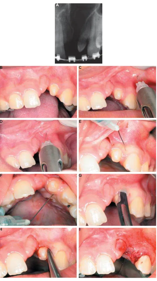
A 20-year-old male patient with a complete unilateral CLP was seen at the Orthognathic Surgery Clinic of the Hospital for Rehabilitation of Craniofacial Anomalies, University of São Paulo, Brazil, for extraction of the pre-canine supernumerary tooth adjacent to the cleft for prosthetic rehabilitation purposes (Figures 1A and 1B).
After skin and oral antisepsis with 0.12% ®, Colgate-Palmolive, São
Bernardo do Campo, SP, Brazil), a topical anesthetic gel with 20% benzocaine (Benzotop, DFL, Rio de Janeiro, RJ, Brazil) was applied at the bottom of the vestibule to reduce pain during the regional injectable anesthetic procedure (mepivacaine with !"""""#$& '*+/
A first puncture was performed in the first premolar region (Figure 1C), away from the area to be handled in order to avoid a puncture near usually very painful. Then, after approximately 5 min, when the area was partially anesthetized, a second puncture was made at the apex of the tooth to be extracted (Figure 1D), in a slow and gradual manner, with the needle inclined 45° to the apex of the pre-canine tooth. Injections throughout the marginal buccal mucosa (Figure 1E) and palate (Figure 1F) were also performed. After the anesthetic procedure, the tooth was extracted
following the steps: intra-sulcular incision (Figure / *9/ *;/
Case 2
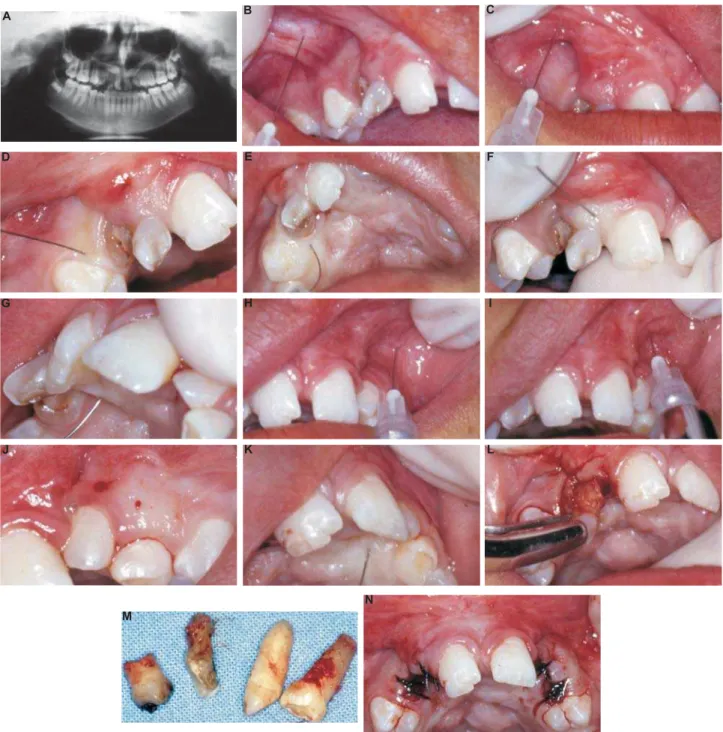
A 13-year-old female patient with a complete bilateral CLP was seen at the Outpatient Clinic of the Department of Oral and Maxillofacial Surgery, Prosthodontics and Traumatology of the Dental School, University of São Paulo, after referral from an orthodontist for extraction of residual roots located in the margins of the right alveolar cleft and premaxilla, and over-retained primary canine and supernumerary tooth located in the left maxillary segment. All teeth had poor bone insertion or root malformation (Figure 2A).
For anesthesia of teeth located on the margin of the right cleft, topical anesthetic gel with 20% benzocaine (Benzotop, DFL) was applied at the bottom of the vestibule, in the right maxillary < using 2% lidocaine with epinephrine 1:100.000 (Alphacaine, DFL), was made in the premolar region, avoiding the introduction of the needle in the area near the nose, which is often very painful (Figure 2B). A second puncture was made in the right maxillary segment close to the buccal aspect of the cleft and, in the third puncture, the tooth located on the right margin of the premaxilla was anesthetized, with the needle inserted at 45° * =>/ ; along the mucosa in the buccal (Figure 2D) and palatal aspects (Figure 2E) of the right maxillary segment and premaxillary region (Figures 2F and = / local anesthesia and hemostasia during the surgical procedure.
On the left side, the anesthetic technique was very similar to the procedure performed on the * =9/ the cleft area inclined 45o towards the tooth apex (Figure 2I) and anesthesia of the buccal (Figure 2J) and palatal mucosa (Figure 2K). After anesthesia, tooth extraction was conducted in a conventional manner (Figures 2L, 2M and 2N).
DISCUSSION
Although several studies have been conducted regarding the complex treatment procedures in patients with CLP, only a few have focused on the course and anatomy of vessels and nerves in ? in dental procedures resulting from this anatomic variation3,10,11.
Figure 1- Patient with a complete cleft lip and palate:A: Periapical radiograph of the cleft area showing the supernumerary ! " " # H:extraction procedure; 1I: suture
A
B
D E
F G
and vascular supply in the cleft maxillary complex, based on an in situ assessment. That study, which became a reference for further research, described innervation of the maxillofacial complex of stillborn children suffering from CLP by means of histological
analysis.
According to Bohn1 (1963), in unilateral CLP, the nasopalatine nerve descends along the vomer, on both sides, and reaches the pre-maxilla. From there, both nerves converge and form the median nerve
Figure 2- Patient with a complete bilateral cleft lip and palate:A: Panoramic radiograph showing residual teeth in the right "$ % & '" * +! % & '" * + % &'"+# %&'"+ / 6 %& '" * +< of the marginal gengiva adjacent to the cleft (the posterior maxillary segment - left side); L:extraction procedure; M: teeth extracted; N:suture
A B
D E F
G I
J L
that runs through the cleft, innervating the soft tissue in the cleft area. A major nerve branch stems from the nasopalatine nerve located on the cleft side and is responsible for innervating the palatal mucosa. This branch penetrates the premaxilla reaching primary and permanent anterior teeth, and the buccal mucosa. On the non-cleft side, the nasopalatine nerve runs toward the palatal mucosa conferring sensitivity. In this segment of the jaw, the upper alveolar nerve innervates the teeth and buccal mucosa. The anastomosis between right, left and median nerves that take place in the cleft region are responsible for sensitivity in this area (Figure 3).
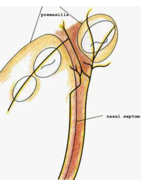
On the other hand, in bilateral CLP, nerve supply of the premaxilla and its teeth is derived from bilateral bundles of nasopalatine nerve that follow a downward course along each side of the vomer (Figure 4A). Upon reaching the premaxilla, the nasopalatine branches unite, forming the median nerve which runs close to the median suture of the premaxilla in the palatal region and, at this point, penetrates the premaxilla and sends terminal branches to the buccal mucosa and labial frenum. The main branch of median nerve also sends and incisive papilla (Figure 4B)1.
Patients with CLP have a higher prevalence of anomalies in number or shape of teeth adjacent to the alveolar cleft9. In some cases, these teeth need to be extracted due to mispositioning or malformation and local anesthesia is an important step for a successful extraction procedure, avoiding distress to patients and professional anxiety. In face of anatomic variations in the cleft maxilla, it seems obvious that adjustments to conventional anesthetic techniques should be considered to perform surgical, endodontic or restorative procedures properly in teeth located along the cleft.
* a correct anesthetic technique in the cleft area: 1)
use of topical anesthetics: it is important that the
professional always use a topical anesthetic in the < surgical reconstruction of the lip performed in early childhood, around 3 months of age, can leave scars that stretch during the anesthetic procedure, promoting excessive pain, ¿UVW SXQFWXUH IDU
from the cleft area: it is recommended to make
avoiding needle introduction in the area near the cleft or nose which is always painful. When the area of interest is partially anesthetized, a second puncture can be performed in the bottom of the vestibule, near the tooth to be treated or extracted,
3) slow administration of anesthetic: injection
of anesthetic liquid must be as slow as possible avoiding the painful sensation caused by distension Figure 3- Diagram of nerves and vessels of a unilateral
=>?1 (1963)]
?
Figure 4- Diagram of nerves and vessels of a bilateral cleft lip and palate. 4A: axial view; 4B: sagittal view =>?1 (1963)]
?
A
LQ¿OWUDWLRQRIORFDODQHVWKHWLFLQFOHIWPDUJLQV
?G gingiva serve as a complement to anesthesia and promote increased local vasoconstriction, allowing a procedure free of pain and bleeding.
Special attention should be given to premaxillary anesthesia in bilateral CLP. As previously described, innervation of this area stems from the median nerve, which penetrates premaxillary bone, innervates anterior teeth and sends branches to the buccal mucosa. Therefore, it is extremely important that, in addition to anesthesia at the bottom of the vestibule, completion of anesthesia be performed in the palatal aspect of the premaxilla, providing complete anesthesia of this area.
Finally, it should be considered that, although the dental arch of patients with CLP may appear normal after reconstructive surgery, the nerve course was embryologically determined by the presence of the cleft, and is not corrected by plastic surgery. Therefore, it is very important for the general dentist and for the oral surgeon to be aware of the anatomic course of nerves in both cleft types – unilateral and bilateral - and to use ? order to ensure safety for the clinical procedure and comfort for the patient, minimizing trauma arising from inappropriate anesthetic procedures.
ACKNOWLEDGEMENT
We are thankful to CAPES for the financial support.
REFERENCES
1- Bonh A. The course of the premaxillary and maxillary vessels and nerves in cleft jaw. Acta Odontol Scand. 1963;21:463-513. 2- Cohen MM Jr. Etiology and pathogenesis of orofacial clefting. Oral Maxillofac Surg Clin North Am. 2000;12(3):379-97. VX' Y$Z> [\+<' clinical technique for tooth extraction in the cleft lip and palate area. Int J Paediatr Dent. 2001;11(2):143-6.
4- Diewert VM. Development of human craniofacial morphology during the late embryonic and early fetal periods. Am J Orthod. 1985;88(1):64-76.
^X_>\+<$Z` central incisors of infants with unilateral cleft lip. Cleft Palate Craniofac J. 2009;46(4):420-4.
{X \Z > [ $Z ' permanent lateral incisor in patients with incomplete and complete unilateral cleft lip. Cleft Palate Craniofac J. 2005;42(5):517-20. 7- Poletta FA, Castilla EE, Orioli IM, Lopez-Camelo JS. Regional analysis on the occurence of oral clefts in South America. Am J $_=""|#}V_=}/!V={X=|
~XY >Y\[ $ X _ [ < ` {X="""! overview. J Craniomaxillofac Surg. 2001;29(3):131-40.
9- Silva AP, Costa B, Carvalho Carrara CF. Dental anomalies of number in the permanent dentition of patients with bilateral cleft lip and palate: radiographic study. Cleft Palate Craniofac J. 2008;45(5):473-6.
"X Y ' > [ $Z \ +< ' anesthetic procedures for cleft lip and palate patients. J Clin Pediatr Dent. 2000;24(3):153-8.
11- Slaughter WB, Henry JW, Berger JC. Changes in blood vessel patterns in bilateral cleft lip. Plast Reconstr Surg Transplant Bull. 1960;26:166-79.
12- Ten Cate AR. Embryology of the head, face, and oral cavity. In: Ten Cate AR. Oral histology: development, structure, and function. 7th edition: St. Louis: Elsevier; 2008. p. 32-56.