INTRODUCTION
The development of HIV infection in children has different characteristics to those noted in adults, due mainly to the earlier acquisition of the virus, combined with the immaturity of the immunologic system and other body structures8. It is estimated that in Brazil there are approximately 597,000 people infected by HIV, of which 12,800 are children aged between 0 and 14 years20. It rep-resents the number of vertically infected children, which is the principal way of HIV transmission in children.
The clinical feature of pediatric HIV infection includes the appearance of various oral lesions, some of which are considered AIDS diagnosis markers, such as recurrent oral candidiasis and chronic enlargement of the parotid5. The most fre-quently associated oral lesions are: candidiasis, herpes simplex infection, linear gingival erythema (LGE), parotid enlargement and recurrent aph-thous stomatitis. Other viral and bacterial infec-tions, including periodontal infections are less commonly associated, while hairy leukoplakia and
Pediatric HIV-related oral manifestations – a five-year
retrospective study
Manifestações bucais associadas à infecção pelo HIV em crianças
– estudo retrospectivo de cinco anos
Lívia Ferreira Soares*
Glória Fernanda Barbosa de Araújo Castro** Ivete Pomarico Ribeiro de Souza***
Melissa Pinheiro****
* Master’s Degree Student; ***Chairman, Professor; ****Scientific Initiation Student – Discipline of Pediatric Dentistry, School of Dentistry, Federal University of Rio de Janeiro.
** Professor, Discipline of Pediatric Dentistry, School of Dentistry, University Center of Volta Redonda.
ABSTRACT: The purpose of this study was to carry out a five-year retrospective descriptive follow-up of the oral manifestation frequency, systemic condition and type of medication used in HIV-infected children and adolescents after the introduction of combined antiretroviral therapy. Fifty-eight patients were examined in 2001/2002, and their previous medical and dental records (1997 to 2000) were researched from files. There was an occurrence of 7 new cases of AIDS in a sample of 19 children, while 46.5% of the entire sample (n = 58) progressed as to clas-sification of HIV infection. No difference was noted among the frequencies of oral manifestations, categories of the immunosuppression and viral load categories. The oral manifestations in the group of children and adolescents followed up in this study remained stable, even after treatment with combined antiretroviral therapy. However, a downward trend in the frequency of oral candidiasis and parotid enlargement was noted.
DESCRIPTORS: HIV; Oral manifestations; Child; Immunosuppression; Combined modality therapy; Protease in-hibitors.
RESUMO: O objetivo deste estudo foi realizar um acompanhamento descritivo retrospectivo da freqüência de mani-festações bucais, da condição sistêmica e do tipo de medicação utilizada em um grupo de crianças e adolescentes infectados pelo HIV após a introdução da terapia anti-retroviral combinada. Cinqüenta e oito pacientes foram examinados em 2001/2002, enquanto seus exames médicos e odontológicos retrospectivos (1997 a 2000) foram pesquisados em prontuário. Foram observados 7 novos casos de AIDS em uma amostra de 19 pacientes, enquanto 46,5% da amostra total (n = 58) progrediram quanto à classificação da infecção pelo HIV. Não foram observadas diferenças entre as freqüências de manifestações bucais, das categorias de imunossupressão e da carga viral. O quadro de manifestações bucais no grupo de crianças e adolescentes acompanhados neste estudo manteve-se estável, mesmo após a introdução da terapia anti-retroviral combinada. Contudo, foi observada uma tendência de diminuição da freqüência de candidíase bucal e hipertrofia de parótidas.
Kaposi’s sarcoma are rarely seen in HIV-infected children18.
The development of antiretroviral drugs for treating HIV infection began with the introduc-tion of zidovudine (AZT) in 1987. Protease inhibi-tors became available in 1997, and they led to the preparation of therapeutic schemes that provided greater control of viral replication, improving the immunologic response of HIV-infected patients. Broad access to antiretroviral therapy, principally to the combined therapy (two or more drugs), re-sulted in a better quality of life for such patients4 and, according to some studies, was responsible for a drop in the prevalence of oral manifesta-tions1,6,17.
This study sought to report those observa-tions through a five-year retrospective descriptive follow-up of oral manifestations and systemic con-ditions (laboratory data and use of antiretroviral therapy) in HIV-infected children and adolescents, after they were treated with combined antiretro-viral therapy.
MATERIAL AND METHODS
SubjectsThe population studied consisted entirely of children treated at the pediatric AIDS clinic of a public university institution of the city of Rio de Janeiro, from March 2001 to February 2002 (n = 307). The oral health (oral manifestations and dental caries) of this population is routinely followed up by a dental team through periodical clinical examinations (usually quarterly), at the same time as medical consultations.
In that period (2001-2002), the dental team examined 160 children and/or adolescents. Their previous medical and dental records were re-searched in files, and those that had at least 3 complete annual examinations in the period be-tween 1997 to 2002 were included in the research. Consent was obtained from the person responsible for each patient and the study was approved by the local ethics committee.
The study period was divided into five groups of annual intervals, according to the period of the examination:
• Interval 1 (I1) - March 1997 to February 1998.
• Interval 2 (I2) - March 1998 to February 1999.
• Interval 3 (I3) - March 1999 to February 2000.
• Interval 4 (I4) - March 2000 to February
2001.
• Interval 5 (I5) - March 2001 to February 2002.
Data collection procedures
The patients of the I5 Group (2001/2002) were examined by a trained professional and the criteria for diagnosing the principal lesions associated to HIV infection in children were those described by EC-Clearinghouse (1993)10 and Ramos-Gomez et al.18 (1999). The extrabuccal examination consisted of a visual inspection of the face and palpation of the cervical, submandibular and submentonian lymph nodes and parotid glands; the intrabuccal examination was performed using an oral mirror, wooden spatula and flashlight to improve illumina-tion. Soft tissues were examined in the following sequence: lips, buccal mucosa, tongue, floor of the mouth, hard and soft palate and gums18.
Information on previous records of oral mani-festations of the sample of Groups I1 to I4 (1997 to 2000) was researched in the dental files, while the medical files of all the patients included in the study were researched to find out what type of medication was used, data on CD4+ cell count and viral load, as well as the classification of the clinical stage of the disease8.
The resulting data was stored in a databank created by the SPSS Program, version 11.0 (SPSS Inc., Chicago, USA). Descriptive statistics was used to analyze the data.
RESULTS AND DISCUSSION
Description of sampleIt was discovered that not all of the 58 chil-dren chosen for I5 (2001/2002) had the complete examination (doctor and dentist) for all intervals of the study. In this case, only 33 patients qualified (56.9%). However, all the patients chosen had at least 3 complete examinations from I1 to I5. Ad-ditionally, of those 58 patients, 46 (79.3%) began the follow-up in this study in I1, and 12 (20.7%) in I2. There was no difference as to sex and as to age, the latter increased with time because the same children were followed up for 5 years, as seen in Table 1. Regarding the distribution of the catego-ries of exposure of the patients to the HIV virus, 55 patients (94.9%) had been vertically contaminated, while only two cases (3.4%) resulted from blood contamination (transfusion) and one case (1.7%) had unknown way of contamination.
symptoms (N - no symptoms, A - mild symptoms, B - moderate symptoms, C - serious symptoms) and the degree of immunosuppression (1 - none, 2 - moderate, 3 - serious). Fifty-four patients (93.1%) already showed, at the start of follow-up, some symptom of the disease (classification A, B or C), and this number rose to 56 (96.5%) patients in I5.
The proportion of the number of patients with AIDS (classification: N3, A3, B3, C1, C2 or C3) at the start (I1/I2) and at the end of the follow-up (I5), was 39 and 46 children, respectively. The progress of HIV infection in children is different to that in adults, because they can develop the HIV infection as fast progressors (they evolve to AIDS in the first two years of life), intermediate progressors (they show only mild symptoms dur-ing the first five years of life) or non-progressors (they did not develop the disease until eight years of age). However, the majority of children develop the disease as an intermediate progressor, similar to the findings of this study4.
The evolution of HIV infection to the AIDS
stage or death can be influenced by various fac-tors, among which are the early appearance of clinical symptoms and the presence of generalized lymphadenopathy19. It could be suggested that the occurrence of 7 cases of AIDS in the sample, in the five years of follow-up, may be due to the fact that the majority (93.1%) already showed some clinical symptoms of the infection at the beginning of the study, as well as a high frequency of lymphade-nopathy, as Table 2 shows.
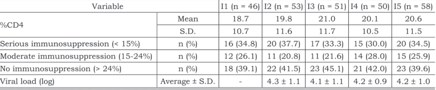
Laboratory data
The values of CD4+ cells, viral load, in ad-dition to the classification of the sample of each interval of the study into immunosuppression groups, according to criteria established by the Centers for Disease Control and Prevention (1994)8 can be seen in Table 3.
The results of the viral load levels are described from I2 to I5, because in I1 the examination for qualifying the viral load for routine clinical use was not yet available. The results are shown in log of number of copies of viral particles per ml of
TABLE 1 - Description of the sample - sex and age. Rio de Janeiro, 2002.
Interval n n (%)Sex Age
Male Female Interval Mean S.D.
I1 46 22 (47.8) 24 (52.2) 1.3 - 13 5.3 2.9
I2 53 24 (45.3) 29 (54.7) 0.6 - 13.8 6.1 3.0
I3 51 23 (45.1) 28 (54.9) 1.5 - 14.3 7.2 3.0
I4 50 24 (48.0) 26 (52.0) 2.4 - 14.6 7.8 2.7
I5 58 28 (48.3) 30 (51.7) 3.6 - 16 9.2 2.8
S.D.: standard deviation.
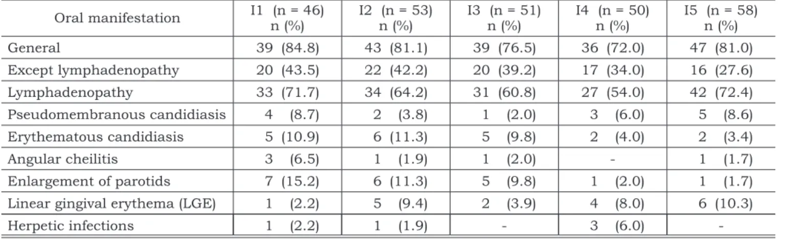
TABLE 2 - Frequency of the principal oral manifestations associated with HIV infection in children. Rio de Janeiro, 2002.
Oral manifestation I1 (n = 46) n (%) I2 (n = 53) n (%) I3 (n = 51) n (%) I4 (n = 50) n (%) I5 (n = 58) n (%)
General 39 (84.8) 43 (81.1) 39 (76.5) 36 (72.0) 47 (81.0)
Except lymphadenopathy 20 (43.5) 22 (42.2) 20 (39.2) 17 (34.0) 16 (27.6) Lymphadenopathy 33 (71.7) 34 (64.2) 31 (60.8) 27 (54.0) 42 (72.4)
Pseudomembranous candidiasis 4 (8.7) 2 (3.8) 1 (2.0) 3 (6.0) 5 (8.6)
Erythematous candidiasis 5 (10.9) 6 (11.3) 5 (9.8) 2 (4.0) 2 (3.4)
Angular cheilitis 3 (6.5) 1 (1.9) 1 (2.0) - 1 (1.7)
Enlargement of parotids 7 (15.2) 6 (11.3) 5 (9.8) 1 (2.0) 1 (1.7)
Linear gingival erythema (LGE) 1 (2.2) 5 (9.4) 2 (3.9) 4 (8.0) 6 (10.3)
-the plasma sample. The CD4+ cell count and -the viral load are parameters used for evaluating the progression of the disease and for knowing when to begin the antiretroviral treatment4.
Even though the independent predictor of pro-gression of the disease seemed more effective in adults16, the evaluation of the viral load in verti-cally infected children takes into account that the patient is already born with high levels of RNA-HIV, which tend to decline slowly during the first years of life, sometimes even without any antiretroviral medication. However, it is believed that in children older than 30 months, viral load levels higher than 100,000 copies/ml and a CD4+ lymphocytes count below 15% indicate increased risk of progression of the disease to the AIDS stage or death4. In this study, the frequency of serious immunosuppres-sion (%CD4 < 15%) varied from 30.0 to 37.7% of the sample, and although the viral load levels an-alyzed in log form did not show a difference from interval to interval, these were very high (averaging above 100,000 copies/ml after I3), and may be related to the progression of the disease observed in the sample.
Antiretroviral therapy
The type of antiretroviral therapy is described
in Table 4. The use of combined antiretroviral ther-apy increased with time, and in the case of the patients shown in Table 4 as not using any medica-tion, this means that up to that time there was no clinical and/or laboratory indication for beginning therapy4. The results show that monotherapy is no longer suitable as a therapeutic scheme for treat-ing HIV disease, and the combination of drugs may vary specifically from patient to patient, according to the infection markers4,9. The time of use and the type of therapy can influence the evaluation of its impact on the patient’s oral and systemic condi-tion (which is why more studies are necessary with more follow-up) and/or of the case-control type for clarifying that relation.
Oral manifestations
Table 2 shows the frequency of the principal oral lesions during the follow-up period (1997-2002).
Oral manifestations are frequently found in HIV-infected children, and in some cases are the first clinical sign of the disease14. In this study, no difference was noted among the frequencies of lesions between the consecutive intervals of the study, because in all the intervals, more than 70% of the patients showed some type of manifestation.
TABLE 3 - Percentage of CD4+ cells, frequency of the degree of immunosuppression and viral load levels, of each interval of the study (I1-I5). Rio de Janeiro, 2002.
Variable I1 (n = 46) I2 (n = 53) I3 (n = 51) I4 (n = 50) I5 (n = 58)
%CD4 Mean 18.7 19.8 21.0 20.1 20.6
S.D. 10.7 11.6 11.7 10.5 11.5
Serious immunosuppression (< 15%) n (%) 16 (34.8) 20 (37.7) 17 (33.3) 15 (30.0) 20 (34.5) Moderate immunosuppression (15-24%) n (%) 12 (26.1) 11 (20.8) 11 (21.6) 14 (28.0) 15 (25.9) No immunosuppression (> 24%) n (%) 18 (39.1) 22 (41.5) 23 (45.1) 21 (42.0) 23 (39.6) Viral load (log) Average ± S.D. - 4.3 ± 1.1 4.1 ± 1.1 4.2 ± 0.9 4.2 ± 1.0
S.D.: standard deviation.
TABLE 4 - Type of antiretroviral therapy, adhesion and time of use (I1-I5). Rio de Janeiro, 2002.
Antiretroviral therapy I1 (n = 46) I2 (n = 53) I3 (n = 51) I4 (n = 50) I5 (n = 58) % (n)
None 23.9 (11) 11.3 (6) 9.8 (5) 4.0 (2) 15.5 (9)
Monotherapy 10.9 (5) 1.9 (1) - -
-Double therapy 65.2 (30) 64.2 (34) 52.9 (27) 46.0 (23) 36.2 (21)
Triple therapy with protease inhibitor - 22.6 (12) 37.3 (19) 48.0 (24) 43.1 (25)
That fact was also observed by various studies, where oral manifestations occurred, regardless of constant medical treatment3,7,11,13,14.
However, when lymphadenopathy was exclud-ed, a downward trend in the frequency of lesions could be seen (Table 2). Lymphadenopathy was diagnosed in the majority of the children in all the intervals (Table 2), and this frequency can be ex-pected in the case of immunodeficient patients3.
Candidiasis is one of the most frequent oral manifestations in HIV-infected patients and is con-sidered a sign of a poor prognosis of the disease15. The study of Aguirre et al.1 (1999) proved the de-cline of the occurrence of this lesion in adults af-ter the introduction of antiretroviral therapy with protease inhibitors, due mainly to the patient’s improved immunological condition. In the study of Flanagan et al.12 (2000), no difference was not-ed between the prevalence of oral manifestations among the groups of children treated with double therapy and with triple therapy with protease in-hibitors. There was just a tendency for candidiasis to be observed more frequently in the group treated without a protease inhibitor. In this study, the fre-quency of candidiasis observed in I1 (17.4%) was already lower than that of other studies (22%10; 24%12), while a high frequency of the use of double therapy was noted. These two factors may have
contributed to the observation of a decline in the observation of the lesion over time6.
Linear gingival erythema was seen more fre-quently in the final intervals of the study (Table 2), although evidence has already been found that this lesion is more common in children over 12 years2, like in the majority of the children of I4-I5 of this study.
Enlargement of the parotids tended to drop with time, although some authors relate it to a good prognosis of the disease15 and with an in-crease in the lymphocytary infiltrate17. Among the less frequent lesions noted were herpetic infections (prevalence varying from 0 to 6.0%), and this fig-ure agrees with the majority of the reports in the literature3,18.
CONCLUSIONS
The frequency of oral manifestations in the group of children and adolescents followed up in this study remained stable, even after the intro-duction of combined antiretroviral therapy. How-ever, a downward trend in the frequency of oral candidiasis and parotid enlargement was noted. There was no difference in the frequency of the immunosuppression groups, while there was an increase in the use of combined antiretroviral ther-apy, principally with protease inhibitors.
REFERENCES
1. Aguirre JM, Echebarria MA, Ocina E, Ribacoba L, Montejo M. Reduction of HIV-associated oral lesions after highly active antiretroviral therapy. Oral Surg Oral Med Oral Pathol 1999;88:114-5.
2. Barasch A, Safford MM, Catalanotto FA, Fine DH, Katz RV. Oral soft tissue manifestations in HIV-positive vs.
HIV-negative children from an inner city population: a two year observational study. Pediatr Dent 2000;22:215-20. 3. Bosco VL, Birman EG. Oral manifestations in chil-dren with AIDS and in controls. Pesqui Odontol Bras 2002;16:7-11.
4. Brasil. Ministério da Saúde. Coordenação nacional de DST e AIDS. Guia de tratamento clínico da infecção pelo HIV em crianças. Brasília; 2001. 30 p.
5. Brasil. Ministério da Saúde. Definição nacional de caso de AIDS em indivíduos menores de 13 anos, para fins de vigilância epidemiológica. Brasília; 2000.
6. Castro GF. Correlação entre manifestações bucais e clas-sificação clínica e imunológica em crianças infectadas pelo HIV [Dissertação de Mestrado]. Rio de Janeiro: Faculdade de Odontologia da UFRJ; 1998.
7. Castro GF, Portela MB, Souza IP. Frequency of oral mani-festations and antiretroviral drugs in HIV+ children [ab-stract]. J Dent Res 1999;78(Spec Issue):181.
8. Centers for Disease Control and Prevention. 1994 revised classification system for HIV infection in children less
than 13 years of age. MMWR 1994;43(RR-12):1-10. 9. Centers for Disease Control and Prevention. Guidelines for
the use of antiretroviral agents in pediatric HIV infection. MMWR 1998;47(RR-4):1-31.
10. Classification and diagnostic criteria for oral lesions in HIV infection. EC-Clearinghouse on Oral Problems to HIV Infection and WHO Collaborating Centre on Oral Mani-festations of the Immunodeficiency Virus. J Oral Pathol Med 1993;22:289-91.
11. Costa LR, Villena RS, Sucasas PS, Birman EG. Oral find-ings in pediatric AIDS: a case control study in Brazilian children. ASDC J Dent Child 1998;65:186-90.
12. Flanagan MA, Barasch A, Koenisgsberg SR, Fine D, Houpt M. Prevalence of oral soft tissue lesions in HIV-infected minority children treated with highly active antiretroviral therapy. Pediatr Dent 2000;22:287-91.
13. Grando LI, Yurgel LS, Machado DC, Silva CL, Menezes M, Picolli C. Manifestações estomatológicas, contagem de linfócitos T-CD4+ e carga viral de crianças brasileiras e norte-americanas infectadas pelo HIV. Pesqui Odontol Bras 2002;16:18-25.
14. Howell BR, Jandinski J, Palumbo P, Shey Z, Houpt MI. Oral soft tissue manifestations and CD4 lymphocyte counts in HIV-infected children. Pediatr Dent 1996;18:117-20. 15. Katz MH, Mastrucci MT, Leggott P, Westenhouse J,
oral lesions in children with perinatally acquired hu-man immunodeficiency virus infection. Am J Dis Child 1993;147:45-8.
16. Mellors JW, Muñoz A, Giorgi JV, Margolick JB, Tassoni CJ, Gupta P, et al. Plasma viral load and CD4+ lym-phocytes as prognostic markers of HIV-1 infection. Ann Intern Med 1997;126:946-54.
17. Patton L, McKaig R, Strauss R, Rogers D, Eron JJ Jr. Changing prevalence of oral manifestations of human immunodeficiency virus in the era of protease inhibitor therapy. Oral Surg Oral Med Oral Pathol 2000;89:299-304.
18. Ramos-Gomez FJ, Flaitz C, Catapano P, Murray P, Milnes
AR, Dorenbaum A, et al. Classification, diagnostic criteria, and treatment recommendations for orofacial manifesta-tions in HIV-infected pediatric patients. J Clin Pediatr Dent 1999;23:85-96.
19. Spira R, Lepage P, Msellati P, van de Perre P, Leroy V, Simonon A, et al. Natural history of human immuno-deficiency virus type 1 infection in children: a five-year prospective study in Rwanda. Pediatrics 1999;104:1-9. 20. Szwarcwald C, Barbosa Jr A, Fonseca MGP. Estimativa
do número de crianças (0 a 14 anos) infectadas pelo HIV. Brasil. 2000. [citado 2002 jun 30]. Disponível em: URL:
http://www.aids.gov.br/final/biblioteca/bol_set_2001/ artigo.htm.