Journal of Environmental
Analysis and Progress
Journal homepage: www.jeap.ufrpe.br/ 10.24221/JEAP.4.3.2019.2523.216-222 ISSN: 2525-815XGreen synthesis of silver nanoparticles using leaf extract from
Tabebuia roseoalba and T. pentaphylla
Laureen Michelle Houlloua, Robson Antonio de Souzaa, Carolina Barbosa Malafaiaa, Débora Lorrane Montenegro da Paixãob, Alisson Tito Bezerra de Araújob, Mariana Gomes da Silvab, Gian Carlos
Silva Duartea
a Centro de Tecnologias Estratégicas do Nordeste-CETENE, Av. Prof. Luís Freire, n. 01, Cidade Universitária, Recife, PE, Brasil. CEP: 50.740-540. Corresponding author: l.houllou@gmail.com.
b Instituto Federal de Pernambuco-IFPE, Av. Prof. Luís Freire, n. 500, Cidade Universitária, Recife, PE, Brasil. CEP: 50.740-540. A R T I C L E I N F O Received 03 Jun 2019 Accepted 29 Jul 2019 Published 31 Jul 2019 A B S T R A C T
Metal nanoparticles are nanostructures that can be applied to biotechnology because they present different biological activities. Among them, the silver nanoparticles (AgNPs) are known to present antimicrobial activity allowing their application in several areas such as medicine and industry. The biological synthesis of AgNPs is ecologically correct and advantageous techniques. The objective of this study was to evaluate the synthesis of AgNps through the green synthesis using extracts of leaves of Tabebuia roseoalba, and T. pentaphylla grew in vivo and in vitro. The nanoparticle synthesis solution was colorimetrically evaluated, and the nanoparticles were physically characterized. The results obtained demonstrate that both extracts of both Tabebuia species tested can synthesize AgNPs, however only when cultured under in vivo conditions. These data suggest that photosynthesis under natural conditions promotes the production of metabolites that are essential to green synthesis.
Keywords: Nanobiotechnology, AgNPs, Nanosilver, leaves extract. Introduction
Nanobiotechnology is an area of
nanotechnology-related to the creation, use, and enhancement of nanostructures in biotechnological processes. It can be said in a simplified form that this is the manipulation of matter at the atomic and molecular levels. In this context, the synthesis of metal nanoparticles (MNPs) is inserted (Tarafdar et al., 2013). Notable metal nanoparticles such as silver, gold, platinum, and palladium have aroused considerable interest because of their small size and large surface area about their volume (Geonmonond et al., 2018). At this nanometric scale, these particles present very different characteristics of larger particles present in the same material on a larger scale.
Noble metal nanoparticles can be applied in the field of medicine, biology, physics, chemistry, and materials science (Chandra et al., 2014). Gold nanoparticles, for example, are present in combined imaging techniques and photothermal tumor therapy (Khoshgard et al., 2014). Platinum nanoparticles can be found in the manufacture of electrochemical sensors (Yang et al., 2006). In
contrast, a study shows that nanoparticles of palladium have the electrocatalytic capacity (Chen & Ostrom, 2015). Among the MNPs, silver nanoparticles (AgNPs) can also be applied in various areas such as integrated circuits, sensors, filters, fibers, cell electrodes, and antimicrobial agents. Thus, AgNPs has captured the interest of
many researchers (Iravani, 2011). The
antimicrobial property of AgNPs makes these nanostructures applicable in different areas of medicine, industry, livestock, and other areas where it is necessary to combat the disordered proliferation of microorganisms. Also, studies have shown that surface-immobilized AgNPs can be used as support for the immobilization of enzymes, as well as assigning an antimicrobial property to a particular surface (Khan et al., 2011).
For the synthesis of nanoparticles, physical, chemical, or biological methods are used. Physical and chemical methods consume more energy and can also be toxic, yet the biological method is characterized by being an ecologically correct technique and more advantageous (Iravani, 2011). Also, nanoparticles synthesized through the
biological path most often have biocompatibility, biodegradability, and lower production costs (Jiang et al., 2018).
Previously, the production of nanoparticles through the biological pathway has been carried out using fungi, bacteria, cyanobacteria, and plants. The green synthesis is one of the routes used for the biological synthesis of MNPs in vitro, which consists in the use of plant extracts to produce MNPs by the process of bioreduction, in this process the silver ions are reduced in metallic silver, which in turn succeed in the production of nanoparticles (Kuppusamy et al., 2016).
Considering the importance of AgNPs, the objective of this study is to evaluate the potential of the foliar extract of arboreal species of the Atlantic Forest of the genus Tabebuia as catalysts of the synthesis and characterization of AgNPs through the green synthesis, using extracts of the plants maintained in vivo and in vitro.
Material and Methods Plant material
Leaves of Tabebuia roseoalba (Tr) and T. pentaphylla (Tp) were obtained from two forms of plant growth: I - young leaves collected from adult plants located at the Campus of the Centro de Tecnologias Estratégicas do Nordeste (CETENE), II - leaves collected from plants grown in tissue culture (under heterotrophic conditions).
Vegetable extract
Two different types of extracts were performed: in vivo and in vitro according to the collections I and II above mentioned. The leaves fresh were properly washed, and 2.5g of the plant samples were macerated in 20 mL of deionized water using mortar and pistil and later filtered on filter paper. The solutions were titrated to 100 mL and stored at 4 °C.
Synthesis of AgNps
The synthesis of nanoparticles was based on the Okafor et al. (2013) methodology. Initially, was prepared a silver nitrate solution in 1 mM concentration in deionized water. The solution was stored in an opaque container to avoid contact with light. The solutions, for the synthesis of AgNps, were prepared using 9.75 mL of silver nitrate solution and 250 μL of each of the plant extracts, obtaining the final volume of 10 mL. The tubes containing the synthetic solutions were maintained at 37.1°C in a water bath for 1 h, protected from
nitrate and in vitro extract of material and Tr and Tp according to each of the species used.
Antioxidant PVP
The antioxidant power of
polyvinylpyrrolidone (PVP) was evaluated in order to prevent aggregation of AgNPs formed. Three concentrations of PVP were tested. In this, the extracts were mixed, in the ratio of 1:9 (v/v), with
AgNO3 solution (1mM) and added with 0, 34, 67
and 100 mg.mL-1 of PVP. The blends were held at
37°C for 1h protected from light. After the synthesis was stored for 30 days at 25 ± 2°, C protected from light, and the process of formation of the AgNPs was evaluated at one, eight, and 15 days.
Photostability
After the synthesis, the solutions
containing the nanoparticles were maintained for 15h at 25 ± 2°C in the presence of light for evaluation of Photostability of the AgNPs observing the alteration of the coloration visually. Physical characterization
UV-Vis
UV−vis Diffuse Reflectance spectra of the powders were obtained using a LAMBDA 650 UV−vis spectrometer (PerkinElmer) equipped with an integrating sphere.
X-ray diffraction
A thin film of the AgNPs was made by overlapping 20 layers each of 100 μL solution of AgNPs formed on a heated plate (100°C) of silicon. The test was performed with the tube fixed at an angle of incidence of 2°. The samples were analyzed in X-ray Diffractometer Bruker D8 Advance Davinci under radiation γ in copper tubes. The scans were made with angulation: 10°-70° (Nickel filter) voltage: 40 KV current: 40 mA and a time constant was 5s.
Results
The formation of AgNps promoted a change in the coloration of the synthetic solutions that initially presented orange coloration and after
the incubation time presented as dark
yellow/brown (Figure 1). The UV-Visible spectrophotometry analysis, shown in Figure 2, indicates that the AgNPs were formed since the synthesis is characterized as having the absorbance peak at the wavelength of 400-500 nm. Comparing
defining the formation of nanoparticles using the green synthesis. The extract from in vivo grown plants of T. roseoalba produced a higher number of nanoparticles when compared to T. pentaphylla. However, the formation of AgNPs from in vitro culture extracts was not evidenced by the results obtained, as observed in Figure 2CD.
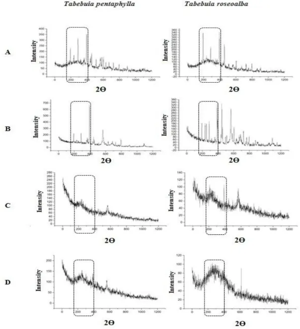
The results of X-ray diffraction analyses are presented in Figure 3. According to the graphs, the control treatment of the two plant species presented peaks in the expected range for the formation of silver nanoparticles (200-380 nm). It is possible to determine the synthesis of silver nanoparticles with leaf extracts from both tested species. However, the best concentration of PVP
for the formation of AgNPs was 34 mg.mL-1
(Figure 3B). Higher concentrations of PVP negatively affected the synthesis of AgNPs as can be observed by the high level of noise in the obtained spectra. From the results obtained, the X-ray diffraction pattern is clear that the AgNPs formed using the Tabebuia sheet were essentially crystalline.
Figure 1. Synthesis of silver nanoparticles using silver nitrate solution added with plant extracts: Negative control, silver nitrate added (a), Tabebuia roseoalba (b) and T. pentaphylla (c) leaf extract in
vivo, T. roseoalba (d) and T. pentaphylla (e) in
vivo.
Figure 2. UV-Vis spectrophotometry analyzes: A. Tabebuia roseoalba in vivo; B. T. pentaphylla in vivo; C. T. roseoalba in vitro; D. T. pentaphylla in vitro.
Figure 3. DR-X results of AgNPs green synthesis. A. Synthesis without PVP – control; B. adding 1 g; C. adding 2 g; D. adding 3 g of PVP.
Discussion
Due to its possible applications in different productive chains (pharmacological, energy, bioremediation process, etc.), the study of the synthesis of AgNPs has been gaining interest in recent years. As the chemical synthesis has the disadvantage of a large number of toxic residues generating, the study of metallic nanoparticles green synthesis, using natural compounds or plant components, has emerged as an alternative environmentally friendly methodology (Ahmed et
al., 2016; Taheriniya & Azad, 2016). Using this
strategic process, the toxicity of the chemicals
been the most used to synthesize NPs without the need for additional reducing agents and stabilizers (Ahmed et al., 2016; Moghaddam et al., 2015). According to Prasad (2014), plants are better synthesizers for nanoparticles probable because plants can actively uptake and bioreduce metal ions from soils and solutions during the detoxification process. As the extracts of plants contain functional substances with proven action in bioreduction, these substances may be responsible for the
synthesis and stabilization of metallic
nanoparticles (Rajan et al., 2015). In the present study, it was verified that extract from Tabebuia
environmentally friendly process. As so, these processes have a potential advantage of being employed and are suitable in the large-scale production of nanoparticles (Olad et al., 2018). Therefore, this study focused on the biosynthesis of AgNPs with plant extracts of Tabebuia leaves (Tabebuia spp.) from different species. It has been previously described that the size and shape of the nanoparticles could influence by varying the pH, temperature, and ratio between the leaf extract and the silver nitrate solution (Kredy, 2018). However, it was observed in the present study that the condition of plant culture (in vivo or in vitro) was also an essential factor that can affect the synthesis of silver particles since the extracts made with leaves of plants grown in vitro were ineffective to produce AgNPs
The colorimetry observed in the formation of the nanoparticles indicated that the original format of the synthesis would be spherical nanoparticles. It is believed that the weak binding of proteins in the solution with silver ions, leading to the isotropic growth of AgNPs (Tippayawat et al., 2016), may have generated the spherical form of AgNPs. The change in coloration in silver nanoparticle synthesis solutions is due to excitation of surface vibration of silver nanoparticles (Khalil et al., 2014; Rao & Tang, 2017). A color variation from reddish-brown to dark brown can occurs according to the size of the nanoparticles produced (Albernaz, 2014). Differences between ions solutions before Tabebuia leaves extract additions were observed. However, the color of the solution obtained with in vitro Tabebeuia leaves was lighter than the color observed in the solution of in vivo leaves. These results indicate that physiological differences between plants from the same species could interfere in the process reproducibility.
Although high temperatures and pressure facilitate the reduction processes (Liu et al., 2013), the synthesis of AgNPs from Tabebuia (in vivo) leaves extracts occurred rapidly and proved to be stable under ambient temperature and pressure conditions.
It is well described that the use of capping agents, such as Polyvinylpyrrolidone (PVP) can positively interfere in the nanoparticles synthesis, favoring anisotropic crystalline growth. According to (Koczkur et al., 2015), well-dispersed silver nanoparticles can be obtained with PVP as a reaction dispersant between silver ions and glucose. According to these authors, PVP may have protected AgNPs from excessive ion accumulation, preventing weak growth and agglomeration. In the case of Aloe vera, a plan of AgNPs arose predominantly as the main peak using the PVP as a capping agent (Tippayawat et al.,
2016). The same result was observed in the formation of AgNPs with Tabebuia spp. extract (in vivo cultured plants). This result corroborates the relevance of using capping agents to improve nanoparticle synthesis.
However, as the plant condition of culture interfere drastically on leaves extract potential use to nanoparticle synthesis, and the most relevant physiological differences between in vitro plants compared to in vivo plants have low photosynthetic rates (Arigita et al., 2002). It was suggested that during the photosynthesis, essential molecules are produced for the process of AgNP synthesis.
Some other studies suggest that binding proteins can be involved in AgNPs formation and stabilization. Under suitable conditions, some substances found in leaf extracts (triterpene acids,
flavonoids, sesquiterpene glycosides,
polysaccharides, and proteins) may act as silver nitrate reducers for the formation of AgNPs (Rao & Tang, 2017). However, the precise class of substance that mediates AgNPs synthesis using
Tabebuia leaves extract must be further
investigated.
The use of X-ray diffraction analysis could be helpful to indicate AgNPs formation. In the present study, X-ray diffraction showed that the peak pattern observed in the silver nitrate sample differed from the peaks observed in the nanoparticle samples. The free silver ions may have been complexed with the organic substances present in the plant extract, thus providing the formation of nanoparticles. This organization of silver ions in the form of nanoparticles may interfere with the X-ray diffraction pattern, as seen in the spectra. The typical X-ray spectrum of nanomaterials consists of crystalline component peaks. The identification of the structure, composition, and degree of the texture of the crystalline phases can be inferred by the relative positions and intensities of the peaks. Peak width is related to the crystallinity of the material, i.e., grain size and grid distortion. According to Khalil et al. (2014), this result may suggest that the crystallization of the bioorganic phase occurs on the surface of silver nanoparticles.
The temperature and time of reaction are the main parameters in order to establish a nanoparticle synthesis routine process (Liu et al., 2014). The nanoparticle particle shape can interfere with antibacterial agent potential (Feng et al., 2000). Determining favorable synthesis conditions are essential to control morphology, size, and
purity of nanoparticle formation. Another
important characteristic, previously reported in the
literature, is that the inorganic (metal)
improved its bactericidal effect (Yousefzadi et al., 2014). According to Tippayawat et al. (2016), the increase in bactericidal activity was possibly caused by synergistic antibacterial effects of AgNPs and naturally occurring chemicals in Aloe
vera. As various natural ligands may interact with
the microbial membrane, such as saponin, tannin, terpenoids, and flavonoids (Griffin et al., 1999; Sahu et al., 2013), it is possible that silver nanoparticles produced by green synthesis are potentially more efficient in the control of microbial contamination if combined with some of these compounds during nanoparticle formation. As so, the AgNPs produced by Tabebuia leaves extracts could be potentially used to control microorganism contamination (ex. During plant in vitro culture). However, this antimicrobial effect must be later further investigated.
Conclusion
The results obtained in this study show that organic compounds which originate from the Tabebuia leaf extract probable function as reductants and stabilizers for AgNPs green synthesis. A wide range of substances found in leaves extracts, such as triterpenic acids,
flavonoids, sesquiterpenes glycosides,
polysaccharides, and proteins can be act as silver nitrate reductants for the AgNPs formation under proper conditions.
Acknowledgments
This study was funded by Centro de Tecnologias Estratégicas do Nordeste (CETENE). Authors also express gratitude to Conselho Nacional de Desenvolvimento Científico e Tecnológico (CNPq) for financial support and to CETENE for structural support.
References
AHMED, S.; AHMAD, M.; SWAMI, B. L.; IKRAM, S. 2016. A review on plants extract mediated synthesis of silver nanoparticles for antimicrobial applications: A green expertise. J. Adv. Res., v. 7, n. 1, p. 17-28.
ALBERNAZ, V. L. 2014. Síntese verde de nanopartículas de prata com extrato aquoso de folhas de Brosimum gaudichaudii, caracterização fisicoquímica, morfológica e suas aplicações no
desenvolvimento de um nanobiossensor
eletroquímico. Universidade de Brasília, 122p.
CHEN, A.; OSTROM, C. 2015. Palladium-Based Nanomaterials: Synthesis and Electrochemical Applications. Chem. Rev., v. 115, n. 21, p. 11999-12044.
FENG, Q.; WU, J.; CHEN, G.-Q.; CUI, F.-Z.; KIM T. N.; O. KIM, J. 2000. A mechanistic study of the antibacterial effect of silver ions on Escherichia
coli and Staphylococcus aureus. J. Biomed. Mater.
Res., v. 52, p. 662-668.
GEONMONOND, R. S.; SILVA, A. G. M.; CAMARGO, P. H. C. 2018. Controlled synthesis of noble metal nanomaterials: Motivation, principles, and opportunities in nanocatalysis. An. Acad. Bras. Cienc., v. 90, n. 1, p. 719-744. GRIFFIN, S. G.; WYLLIE, S. G.; MARKHAM, J. L.; LEACH, D. N. 1999. The role of structure and molecular properties of terpenoids in determining their antimicrobial activity. Flavour Fragr. J., v. 14, n. 5, p. 322-332.
IRAVANI, S. 2011. Green synthesis of metal nanoparticles using plants. Green Chem., v. 13, n. 10, p. 2638-2650.
JIANG, J.; PI, J.; CAI, J. 2018. The Advancing of
Zinc Oxide Nanoparticles for Biomedical
Applications. Bioinorg. Chem. Appl., v. 2018, p. 1-18.
KHALIL, M. M. H.; ISMAIL, E. H.; EL-BAGHDADY, K. Z.; MOHAMED, D. 2014. Green synthesis of silver nanoparticles using olive leaf extract and its antibacterial activity. Arab. J. Chem., v. 7, n. 6, p. 1131-1139.
KHAN, M. A. M.; KUMAR, S.; AHAMED, M.; ALROKAYAN, S. A.; ALSALHI, M. S. 2011.
Structural and thermal studies of silver
nanoparticles and electrical transport study of their thin films. Nanoscale Res. Lett., v. 6, n. 1, p. 1-8. KHOSHGARD, K.; HASHEMI, B.; ARBABI, A.; RASAEE, M. J.; SOLEIMANI, M. 2014. Radiosensitization effect of folate-conjugated gold nanoparticles on HeLa cancer cells under orthovoltage superficial radiotherapy techniques. Phys. Med. Biol., v. 59, n. 9, p. 2249-2263. KOCZKUR, K. M.; MOURDIKOUDIS, S.; POLAVARAPU, L.; SKRABALAK, S. E. 2015.
KREDY, H. M. 2018. The effect of pH,
temperature on the green synthesis and
biochemical activities of silver nanoparticles from Lawsonia inermis extract. J. Pharm. Sci. Res., v. 10, n.8, p. 2022-2026.
KUPPUSAMY, P.; YUSOFF, M. M.; MANIAM, G. P.; GOVINDAN, N. 2016. Biosynthesis of metallic nanoparticles using plant derivatives and their new avenues in pharmacological applications – An updated report. Saudi Pharm. J. v. 24, n.4, p. 473–484.
LIU, F.; KOZLOVSKAYA, V.;
ZAVGORODNYA, O.; MARTINEZ-LOPEZ, C.; CATLEDGE, S.; KHARLAMPIEVA, E. 2014. Encapsulation of anticancer drug by hydrogen-bonded multilayers of tannic acid. Soft Matter., v. 10, n. 46, p. 9237-9247.
LIU, K.; QU, S.; ZHANG, X.; TAN, F.; WANG, Z. 2013. Improved photovoltaic performance of silicon nanowire/organic hybrid solar cells by incorporating silver nanoparticles. Nanoscale Res. Lett., v. 8, n. 1, p. 1-6.
MOGHADDAM, A. B.; NAMVAR, F.; MONIRI, M.; TAHIR, P. M.; AZIZI, S.; MOHAMAD, R. 2015. Nanoparticles biosynthesized by fungi and yeast: A review of their preparation, properties, and medical applications. Molecules, v. 20, n.9, p. 16540-16565.
OKAFOR, F.; JANEN, A.; KUKHTAREVA, T.; EDWARDS, V.; CURLEY, M. 2013. Green
synthesis of silver nanoparticles, their
characterization, application and antibacterial activity. Int. J. Environ. Res. Public Health, v. 10, n. 10, p. 5221-5238.
OLAD, A.; GHAZJAHANIYAN, F.; NOSRATI, R. 2018. A Facile and Green Synthesis Route for the Production of Silver Nanoparticles in Large Scale. Int. J. Nanosci. Nanotechnol., v. 14, n. 4, p. 289-296.
PRASAD, R. 2014. Synthesis of Silver
Nanoparticles in Photosynthetic Plants. J.
Nanoparticles, v. 2014, n. 9, p. 1-8.
RAJAN, R.; CHANDRAN, K.; HARPER, S. L.; YUN, S.-I.; KALAICHELVAN, P. T. 2015. Plant extract synthesized silver nanoparticles: An ongoing source of novel biocompatible materials. Ind. Crops Prod., v. 70, p. 356-373.
RAO, B.; TANG, R.-C. 2017. Green synthesis of silver nanoparticles with antibacterial activities using aqueous Eriobotrya japonica leaf. Adv. Nat. Sci. Nanosci. Nanotechnol., v. 8, p. 1-8.
SAHU, P. K.; GIRI, D. D.; SINGH, R.; PANDEY, P.; GUPTA, S. 2013. Therapeutic and Medicinal Uses of Aloe vera : A Review. Sci. Res., v. 2013, n. 11, p. 599-610.
TAHERINIYA, S.; AZAD, I. 2016. Comparing green chemical methods and chemical methods for the synthesis of titanium dioxide nanoparticles. Int. J. Pharm. Sci. Res., v. 7, n. 12, p. 4927-4932. TARAFDAR, J. C.; SHARMA, S.; RALIYA, R. 2013. Nanotechnology: Interdisciplinary science of applications. African J. Biotechnol., v. 12, n. 3, p. 219-226.
TIPPAYAWAT, P.; PHROMVIYO, N.;
BOUEROY, P.; CHOMPOOSOR, A. 2016. Green synthesis of silver nanoparticles in aloe vera plant extract prepared by a hydrothermal method and their synergistic antibacterial activity. PeerJ, v. 4, n. 10, p. e2589.
YANG, T.; SHEN, C.; YANG, H.; XIAO, C.; XU, Z.; CHEN, S.; SHI, D.; GAO, H. 2006. Synthesis, characterization and self-assemblies of magnetite nanoparticles. Surf. interface Anal., v. 38, p. 1063-1067.
YOUSEFZADI, M.; RAHIMI, Z.; GHAFORI, V. 2014. The green synthesis, characterization and antimicrobial activities of silver nanoparticles synthesized from green alga Enteromorpha flexuosa (wulfen) J. Agardh. Mater. Lett., v. 137, p. 1-4.