Cytochrome Components of Nitrate-
and
Sulfate-Respiring
Desulfovibrio desulfuricans
ATCC 27774
M.-C.
LIU,'*
C. COSTA,2 I. B. COUTINHO,2J. J. G. MOURA,2I. MOURA,2 A. V. XAVIER,2ANDJ. LEGALL' DepartmentofBiochemistry, The UniversityofGeorgia, Athens, Georgia 30602,' andCentrode QuimicaEstrutural, NewUniversity of Lisbon, 1096 Lisbon, Portugal2
Received 18 March 1988/Accepted29 August1988
Three multiheme c-type cytochromes-the tetraheme cytochrome C3 (molecular weight [MW] 13,500), a
dodecahemecytochromec(MW40,800), anda"split-Soret" cytochromec(MW51,540), which isadimer with 2 hemes per subunit (MW 26,300)-were isolated from the soluble fraction ofDesulfovibrio desulfuricans (ATCC 27774) grownunder nitrate- or sulfate-respiring conditions. Two of them, the dodecaheme and the
split-Soret cytochromes, showed no similarities to any of the c-type cytochromes isolated from other sulfate-reducing bacteria, while the tetraheme cytochromeC3appearedtobeanalogoustothecytochrome C3
found in othersulfate-reducing bacteria. For all three multihemec-typecytochromesisolated, the homologous proteins from nitrate- and sulfate-grown cells were indistinguishable in amino acid composition, physical properties, and spectroscopic characteristics. It thereforeappearsthat thesamec-typecytochromecomponents
arepresentwhen D. desulfuricans ATCC 27774 cellsaregrownunder eithercondition. This is incontrast to
theconsiderabledifference found in Pseudomonas perfectomayina (Liuetal.,J. Bacteriol. 154:278-286, 1983), a marine denitrifier, when the cells are grown on nitrate or oxygen as the
terminal
electron acceptor. In addition, two spectroscopy methods capable of revealing minute structural variations in proteins provided identical information about the tetraheme cytochrome C3from nitrate-grown andsulfate-grown cells.Sulfate-reducing bacteria belongingtothegenus Desulfo-vibrioare obligate anaerobes capable of performing oxida-tive phosphorylation coupledtothe electron transport
pro-cess, with sulfate and its reduction product(s) as terminal electron acceptors. A unique member of this group of organisms, Desulfovibrio desulfuricans ATCC 27774, had been shown tobecapable of utilizing nitrate and its reduc-tionproduct, nitrite, as terminalelectronacceptors aswell (24; M. P.Bryant, personal communication). Previous stud-ies indicated that the enzymes involved in dissimilatory sulfate reduction (ATP sulfurylase, adenosine 5'-phospho-sulfate reductase, trithionate reductase, thiosulfate
reduc-tase, and bisulfite reductase) are present at approximately the same level whether the cells are grown on sulfate or
nitrate, while the enzymes involved in dissimilatory nitrate reduction (nitrate and nitrite reductases) are inducible, as
theiractivitiesareconsiderably higher innitrate-grown than in sulfate-grown cells (24). Itwasnoted thatcellmassyield
wasthreetofour times higher for cellsgrown onnitrate than for cellsgrown onsulfate. Inthedissimilatory nitrate reduc-tionperformed by D. desulfuricans ATCC27774, nitrate is reduced through nitrite to ammonia (24). This pathway is distinct from the "denitrification" process carried out by
true denitrifiers, such as Pseudomonas perfectomarina, in
which the final product formed is dinitrogen (N2). In our previous studies comparing the cytochromecomponents of the latter organism grown under denitrifying and
oxygen-respiringconditions, itwas observed thattwocytochromes, cytochrome cd, and the diheme cytochrome c-552, were induced only in the presence of nitrate, while a low a/f3
c-typecytochromewasfoundonly in aerobicallygrowncells (20). The monoheme cytochrome c-551, on theotherhand, was found to be present at the same level in cells grown
under both conditions. Conclusions about the involvement of these cytochromes in the electron transport chains in
* Correspondingauthor.
denitrification or aerobic respiration or both were drawn
from thesefindings. In view of this earlierinvestigation,we thought thatasimilarstudyonD.desulfuricans ATCC 27774
grown under nitrate-respiring and sulfate-respiring
condi-tions might provide interesting insight into the cytochrome
components specifictothe twoelectron transportsystems.
As already noted from the large number ofc-type
cyto-chromes isolated from varioussulfate-reducingbacteria,the currentnomenclaturefor theseproteinsappears tobe inad-equate to provide unambiguous classifications. It has re-cently been proposed that, since large variations in the
polypeptide chain lengths occur even among homologous cytochromes, such as cytochrome C3, the heme content
should be considered the major criterion for classification (LeGall and Fauque, in A. J. B. Zenharder, ed.,
Environ-mentalMicrobiology of Anaerobes, in press). Thus,former
cytochrome C3 (molecular weight [MW] 13,000) becomes tetraheme cytochrome C3, former cytochrome C3 (MW 26,000) becomes octaheme cytochrome C3, and so on. We shall use this new classification method in describing the
three multiheme c-typecytochromes studied inourpresent
work.
MATERIALS AND METHODS
Chemicals and biochemicals. Trizma base, sodium lauryl sulfate, Coomassie brilliant blue R, streptomycin sulfate, DNase I, and horse heart cytochrome c were purchased from Sigma Chemical Company. Acrylamide, N,N'-methy-lene-bisacrylamide,and
N,N,N',N'-tetramethylethylenedia-mine were productsof Eastman Kodak Company. Sodium dithionite andbromphenol bluewerefrom Fisher Scientific
Company. Protein MW markers used in sodium dodecyl sulfate (SDS) gel electrophoresis were obtained from
Bio-Rad Laboratories. All other reagents were of the highest purity commercially
available.-Chromatographic materials. Ultrogel AcA-44was a
prod-uct of LKB Instruments, Inc. DEAE-Bio-Gel A, CM-Bio-5545
0021-9193/88/125545-07$02.00/0
Copyright© 1988, American Society for Microbiology
on September 11, 2019 by guest
http://jb.asm.org/
5546 LIU ET AL.
Gel A, and Bio-Gel HTP were purchased from Bio-Rad
Laboratories. These materials were treated before use as directed by the manufacturer.
Organism. Desulfovibrio desulfuricansATCC 27774, orig-inally isolated byM. P. Bryantof the University of Illinois,
was used in the present study. The compositions of the
media used for growing nitrate-respiring and
sulfate-re-spiringcells have been described previously (21, 24).
MW determination. The MW of the native protein was determined by the sedimentation equilibrium method (26) with a Beckman Spinco model E ultracentrifuge equipped
with Schlieren optics and an automatic photoelectric scan-ning system. Subunit molecular weight was determined by
SDS-polyacrylamide gel electrophoresis (SDS-PAGE)(27).
Heme content. The alkaline pyridine hemochromogen method was used for the estimation of heme content. The
purified cytochrome was placed in a solution containing
0.075 M NaOH and 25% pyridine.
After
reduction of the cytochromewith sodiumdithionite, theA550wasmeasured.Heme content was then calculated based on an extinction
coefficient of 29.1
p.M-1 cm-'
previously reported for heme c(8).Amino acid analysis. Amino acid analyses were performed
with aBeckman model 120C automatic aminoacid analyzer
on hydrolyzed protein samples. Salt-free protein samples were hydrolyzed in evacuated, sealed tubes in 6NHCI for 24, 48, and 72 h at
110°C.
Cystine and cysteine were determined as cysteic acid by the oxidation of proteinsamples
with performic acid prior to acid hydrolysis (13). Spectroscopic measurements. A Varian DMS90recording spectrophotometer was used for the absorbance measure-ment and the routine recording of UV-visible absorption spectra. High-resolution proton nuclear magnetic resonance (NMR) spectra were recorded on a Bruker CXP 300 spec-trometer (300 MHz) equipped with an Aspect 2000 com-puter. All chemical shifts are quoted in parts per million (ppm) from theinternal
standard sodium3-trimethylsilyl-[2,2,3,3-2H]
propionate; positive values refer to lowfield
shifts. For NMR measurements, the cytochrome samples were desalted and lyophilized three timesfrom
2H20
and dissolved to the required concentration (1 to 2 mM). Elec-tron paramagnetic resonance (EPR) spectra were recorded with a Bruker ER-200tt spectrometer equipped with an Oxford Instruments continuous-flow helium cryostat inter-faced with aNicolett
1180 computer.Electrophoresis. Analytical polyacrylamide disc gel elec-trophoresis was performed by the method of Brewer and
Ashworth
(2).
SDS-PAGE was based on the procedure ofWeberand Osborn (27).
Protein determination. The modified Biuret method devel-oped by Zamenhof and Chargaff (31) was used for protein determination, with horse heart cytochrome c as the
stan-dard.
Preparation and fractionation of cell extracts. Unless
oth-erwise stated, buffers used were pH 7.6 and all operations were carried out at
4°C.
Nitrate-grown or sulfate-grown cells (300 g, wet weight) were mixed with 10 mM Tris hydrochlo-ride(Tris-HCI)
buffer to give a 1:1 (wt/vol) suspension and broken by passing the slurry three times through a Manton-Gaulin homogenizer at 9,000lb/in2. A few crystals of DNase were added to lessen the viscosity of the extract, and after 10min,
the preparation was centrifuged at 13,200 x g in a SorvallRC-5B
refrigerated centrifugefor
30min.
The pellet was discarded, and the supernatant was treated with neu-tralized streptomycin sulfate (0.5 mg/mg of protein). After being stirred for 15min,
the preparation wascentrifuged
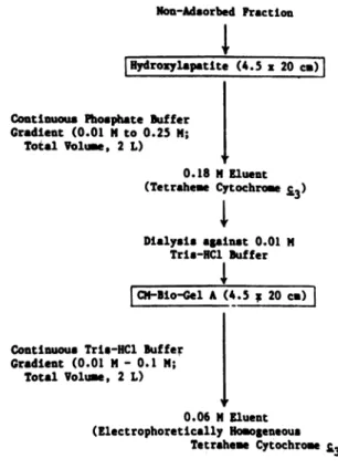
Non-Adsorbed Fraction
iHydroxylapatite
(4.5 x 20 c)Continuous Phosphate Buffer Gradient (0.01 N to 0.25 M;
Total Volume, 2 L)
0.8NEunI
0.18 tl Eluent (Tetraheme Cytochrome £3) Dialysis against 0.01 MTrio-RC1
BufferI
Continuous Trio-HC Buffer Gradient (0.01 M- 0.1 M;
Total Volume, 2 L)
I
0.06 Mgluent (Electrophoretically HomogeneousTetraheme Cytochrome
£.3)
Yields: 28.0 mg
from
300 £wet-weight
nitrate-grown
cells 29.7 mg from 300 £ wet-weight sulfate-grown cells FIG. 1. Purification scheme for tetraheme cytochromeC3. Thescheme shown is from the DEAE-Bio-Gel Asteponwards.
again at 13,200x g for 30
min.
The pellet wasdiscarded,
and thesupernatant
obtained was termed thecrude extract. The crude extract was subjected to furthercentrifugation
at 140,000 x g for 2 h at4°C.
Clear supernatantcontaining
soluble proteins was then used in the purification work described below.
As will become clear in the following
sections,
identical
cytochromes were isolated from supernatant fractionspre-pared from nitrate-grown and
sulfate-grown
cells under nearly identical purification conditions.Therefore,
acom-mon procedure will be described for bothcases. The super-natant fraction prepared was first dialyzed against 10 mM
Tris-HCI
for 24 h with three changes of the dialysisbuffer.
Dialyzed preparation was then loadedontoaDEAE-Bio-Gel
A column (6 by 40 cm) equilibrated with 10 mMTris-HCI.
The same buffer was used to remove the nonadsorbed proteins, which were found to contain the tetraheme cyto-chromeC3. A continuousTris-HCI gradient (0.01to 0.4M)
ina total volume of 4 liters wasthereafter applied to elute the
remaining proteins. Two cytochrome
bands
were resolved during the elution. The first one, eluted at0.07 MTris-HCl, was found to contain the dodecahemecytochrome
c,andthesecond one, eluted at 0.12 M Tris-HCl, wasfoundtocontain the so-called
split-Soret
cytochrome c. These twofractions,
along with the tetraheme cytochromeC3 fractiondescribed
previously, were subjected to furtherpurification
as shownin Fig. 1 through3.
RESULTS
Since the homologous
cytochromes
isolated fromnitrate-grown and sulfate-nitrate-grown cells exhibited nearly identical
J. BACTERIOL.
on September 11, 2019 by guest
http://jb.asm.org/
FractionContaining Dodcaheme Cytochrome ;
_-I._-
-1
lydrolepetiSte (3.Sx20c)
Contimoe Pbospbate Buffer Gradient (0.005 N -0.2 N; TotalVolume, 1.5 L) a A .2 0 08N fluent
(dodecaheme cytdchromer,fraction,
concentrated
to 5al by ultrafiltrati6n)l
5zfraction ContaingDoc Cytochrme c
(eluted
by 0.05NTria-KI, diluted
5 foldswithd.d. 120)D!U-io-UelA (4.5x20cm)
Contiious Trio-lCISuffer
Gradient (0.01 -0.1 N; TotalVolum, 2 L) A .2 0.06Nfluent (nectrophoreticallyDomogene o m Cytochrome )
Yields:
14.5og
from
300gwet-weight
Nitrate-Grown
Cells
19.0mg from 300 gwet-wight Sulfate-Grown Cells
FIG. 2. Purification scheme for dodecaheme cytochromec.The
schemeshown is from theDEAE-Bio-GelAstep onwards. .8
A
.21
Fraction Containing the4Split-Soret'
Cytochrome cV
tHydrozylapatite (4.5 x25 c-)I
Continuous Phoaphate Buffer
Gradient (0.01 M-0.25 M; Total Volume, 2 L)
0.18 N fluent
("split-Soret cytochrome c fraction,
concentrated to S alby ultrafiltration)
IUltrosel Uc-4 (S x90 im)l
I
Fraction Containing Split-Soret Cytochrome c (eluted by 0.05KM, diluted 5 folds with d.d. 120)
JDl-Bio-Gel(44A Sx 20 Cl)
Continuu Trio-WIC Suffer Gradlent (0.01 N - 0.15M;
Total Volume, 2 L)
0.12 tlZluent
(Electrophoreticallykmeneous
aSplit-Soret Cytochrame c)
Yields:
40.2mg from 300 gwet-wi_ght
Nitrate-Grow
Cells30.7 mg from 300 g wet-weight Sulfate-Grown Cells
FIG. 3. Purification scheme for split-Soret cytochrome c. The scheme shown is from the DEAE-Bio-Gel A steponwards.
I~~~ ~ ~~ i
.'I
: :1 ..<:t
300 400 goo600 WAVELENGTH (nm) 300 400 goo 600 WAVELENGTN (am) 700B.
700 300 40 500 600 700WAVELENGTH
(mm)FIG. 4. Absorption spectra of (A) tetrahemecytochrome C3,(B) dodecaheme cytochrome c, and(C) split-Soretcytochrome cfrom D. desulfuricans ATCC27774. Symbols: , oxidized; , dithionite reduced. The protein concentration of the three cyto-chromeswas(A)0.0145mg/ml, (B)0.015mg/ml,and(C)0.095mg'
ml. Thebuffer usedwas0.1Mpotassiumphosphate buffer,pH7.6. Forreductibn of thecytochrome,afewcrystals of sodium dithionite wereadded.
physical
and
chemical properties, no discrimination wasmade betweencytochromes isolated from thetwo kinds of cells.
Absorption spectroscopy. All three proteins
purified
dis-played typical c-type cytochrome UV-visible
absorption
spectra(Fig. 4).Intheoxidized state, the
absorption
spectra ofpurified cytochromesrevealed maximaatca.409and 530nmand a shoulder at approximately 350 nm. Ofthe three,
the spectra of the split-Soret cytochrome c revealed a
maximum at 280 nm that was absent from those of the tetraheme cytochrome C3 and the dodecaheme
cytochrome
c, possibly due to their low aromatic amino acid contents
(see Table 3). When the
cytochromes
were reduced with sodiumdithionite, newabsorption
maximaappeared
at ca. 552, 523,and420nm,which represent theoa,
P,
and Soret(-y)
bands,respectively.
For thesplit-Soret
cytochrome
c, aI
Cs
Ion September 11, 2019 by guest
http://jb.asm.org/
Downloaded from
5548 LIU ET AL.
TABLE 1. Absorptionmaxima andpurity indexes (R) of the three multiheme c-typecytochromes purifiedfrom D.desulfuricans ATCC 27774
Absorptionmaxima(nm)
Cytochrome R
Oxidizedstate Reduced state
Tetraheme cytochrome C3 532,409.2, 350, _a 551.7, 523.2, 418.5, -2.90
Dodecaheme cytochrome c 530, 409.5, 351, 551.7, 523.0,418.5, 2.20
Split-Soret cytochrome c 533,407.7, 350, 280 552.2, 523.0,424.0,415b 0.94
a-,Absenceof absorption maximum.
bShoulderattheSoretabsorption band.
shoulder at415 nm for the Soret band was also observed,
which ledustousethenamesplit-Soretfor thiscytochrome.
Each ofthese three cytochromes showed a characteristic
absorptionpeakratio(R),which is definedasthe ratio of the
absorbanceatthe a-bandposition in the reducedstate tothe A280 in theoxidized state. The Rvalues and the absorption maxima of the threecytochromes purifiedare giveninTable
1.
MW, subunitstructure,andhemecontent.The MW of the
tetraheme cytochrome C3 was determined by both
SDS-PAGE and thesedimentation equilibrium method tobe ca.
13,500. It isthereforeclear that the cytochrome exists in the monomeric form. Heme content was determined by the
pyridine hemochromogenmethodto be 4.36 hemecgroups per cytochrome molecule. Therefore, the tetraheme
cyto-chromeC3from D. desulfuricansATCC 27774appearstobe similar to the well-characterized tetraheme cytochrome C3
found in othersulfate-reducingbacteria.
A minimal MW of 40,800 was obtained for dodecaheme
cytochrome c by SDS-PAGE, with samples treated exten-sively with 5% SDS and 5%2-mercaptoethanol. Removal of thecovalently bonded heme cgroupsbymercuric chloride,
whichwaspreviously shown by otherstodecreasethe MW ofthe octaheme cytochrome C3 to halfof the original MW (9), priortothetreatmentforSDS-PAGE did nothave any
effectonthe minimal MW determined. Hemecontentof the dodecaheme cytochrome c was estimated by the pyridine
hemochromogen method to be 12.15 heme c groups per
40,800 MW. The cytochrome thereforewascalled the
dode-cahemecytochrome c(MW40,800).
The MW of the native split-Soret cytochrome c was
determined by the sedimentation equilibrium method to be 51,540; minimal MW determined by SDS-PAGEwas26,300,
and only one protein band was observed. These results
indicate that thesplit-Soret cytochrome isadimerconsisting
oftwoequal-sized (ifnotidentical) subunits. The number of hemecgroupspermoleculewascalculated from the alkaline
pyridine hemochrome data to be 3.61 heme c groups per
51,540 MW. It was therefore deduced that the split-Soret
cytochrome c is a tetraheme protein containing 2 heme c
groups persubunit. Our unpublished dataonthe determina-tionofheme redox potentials alsoindicate the presence of
twodistinctheme species.
TABLE 2. Physical propertiesof threec-typecytochromes purified fromD.desulfuricans ATCC27774
Hemecontent Subunit
Cytochrome MW (no.ofhemec tutr
groups/molecule) structure
Tetraheme cytochromeC3 13,500 4.36 Monomer Dodecaheme cytochromec 40,800 12.15 Monomer Split-Soret cytochromec 51,540 3.61 Dimera
aTwoequal-sized subunits ofMW26,360.
Data obtained from the MW determinations and heme content analyses for the three cytochromes purified are
given
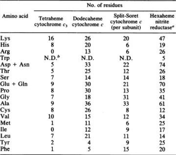
inTable 2.Amino acid composition.
Nearly
identical data were ob-tained for the homologous cytochromes isolated fromni-trate-grownandsulfate-growncells. These results are shown in Table
3,
together with data for the hexaheme nitritereductase presentin the sameorganism(21). It is clear that these four multihemec-type cytochromes haddistinct amino acidcompositions,whichsuggests thattheyareproductsof differentgenesand areprobablynotrelated in terms of their
biosynthesis. It was noted that even though the
performic
acid oxidation method forcysteine determination maylead
to underestimation, the number of cysteine residues
ob-tainedwashigh enoughtoaccountfor4, 12, and4covalently
bonded heme c groups in the case of the tetraheme
cyto-chrome C3, the dodecaheme cytochrome c, and the
split-Soret cytochrome c, respectively. Although the number of histidine residues was enough for the
histidine-heme-his-tidineligand arrangementfor the four heme cgroups in the tetraheme cytochrome C3, insufficient histidine residues were observed to account for the same type of ligand
arrangementforallhemecgroupsin either the dodecaheme cytochrome c or the split-Soret cytochrome c. The exact
ligand arrangementsfor the heme cgroups in the lattertwo
cytochromeswill be investigated further. The considerably higheraromatic amino acid content of the split-Soret
cyto-TABLE 3. Amino acidcompositionof the four multiheme c-type
cytochromesfromD.desulfuricansATCC 27774
No. of residues
Aminoacid Tetraheme Dodecaheme Split-Soret Hexaheme
cytochrome c nitrite
cytochromeC3 tochrome
cccy(perosbnt
reductea
(persubunit) reductasea
Lys 16 26 20 47 His 8 20 6 19 Arg 0 13 6 26 Trp N.D.b N.D. N.D. S Asp+ Asn 5 33 22 74 Thr 5 25 12 26 Ser 7 14 14 18 Glu+ Gln 9 30 21 70 Pro 8 30 13 35 Gly 7 18 31 41 Ala 9 36 33 61 Cys 8 26 8 12 Val 10 15 12 34 Met 1 11 6 25 Ile 0 12 9 17 Leu 7 21 11 14 Tyr 2 4 9 25 Phe 1 5 15 20
aDatataken fromLiuand Peck(21). bN.D., Not determined.
J. BACTERIOL.
on September 11, 2019 by guest
http://jb.asm.org/
MAGNETIC FIELD
(T)
FIG. 5. EPR spectra oftetraheme cytochrome C3from
sulfate-grown (a) and nitrate-sulfate-grown (b) D. desulfuricans ATCC 27774. Experimental conditions: temperature, 10 K; microwave power, 50 ,uW;field modulation, 1 mT.
chrome c than of the other two explains the presence of the
280-nm absorption band in the former but not in the latter two.
EPRand NMR spectroscopic data on tetraheme cytochrome C3. The tetraheme cytochrome C3 purified in our study revealed EPR spectroscopic features very similar to those
obtainedforthe tetraheme cytochrome C3 from other
sulfate-reducing bacteria. As shownin Fig. 5, the spectra recorded at 10 K were quite complex and showed several
superim-posed signals in the
gmax
region,with a broadcomponentat3.20 andprominent but poorlyresolved features at 2.97 and 2.89. Aderivativepeaktentativelyassigned to a
gmed
of 2.31andtwobroadcomponents at veryhigh fields
(g,in
1.56 and 1.39) were also observable. These sets of resonances were present for the spectra of tetraheme cytochromes isolatedfromboth sulfate- and nitrate-grown cells.These results, in
conjunction with EPR spectroscopic features previously reported forother tetraheme cytochromes (18), clearly indi-cate that thefour heme c groups are notquite equivalent.
Theanisotropic g values are characteristic ofrhombic
dis-tortedhemec moieties with histidinyl axial ligands.
NMR spectra of the tetraheme cytochrome C3 isolated
from the nitrate-grownand sulfate-growncellsareshown in
Fig. 6. The spectra aretypical of low-spin ferric multiheme
cytochromes which reveal several well-resolved three-proton intensity resonances at very low field. These reso-nancesbelongtothe methyl groups associated with the heme c moieties and are shifted to low field by contact and
pseudocontact interactions dueto theparamagneticlow-spin
ferric ironpresent at thehemecores. From 12 to 14of the 16
heme methyl group resonances were readily detected. The
remaining heme methyl resonances were in the crowded
region below 10ppm and were notreadily assigned. Partof
this region, which also contains the resonances due to the
aromatic amino acidside chains, is shown in Fig. 6B in an expanded scale. The NMR spectroscopic features were
nearly identicalin the aromaticregion, and the slight differ-enceswhichwereapparent in the very low fieldregionwere
probably duetodifferencesinsolution conditions in the two
samples. It is known that resonances shifted through
para-magnetic interactions are extremely sensitive to modifica-tionson the hemeelectrondensitydistributioninduced, for
A $!y \
30 25 20
PPM 15 10
14 10 S
PPM
FIG. 6. NMRspectraof tetrahemecytochrome C3 from
sulfate-grown (a) and nitrate-sulfate-grown (b) D. desulfuricans ATCC 27774. Experimentalconditions: temperature, 323 K; pH7.5.(A) Very low fieldregion. (B) Aromaticregion.
example, bya small pH change (28). A comparison ofthe NMR characteristics of this class of homologous proteins
haspreviously beenmade (23). Anobviouscommonfeature ofthe NMR spectra was thelargedown-field chemical shifts
experienced bythe heme methylgroup. However, striking differencesarefound in hememethylresonancedistribution
amongthe cytochromes C3 isolated from different
Desulfo-vibrio species. These differences result from the relative orientation or the electronic properties of the four hemes.
However, it is noteworthythat even whenX-ray structures areknown[e.g.,D.vulgaris (Miyazaki)and D.desulfuricans
(Norway 4)], althoughthe geometry and orientation ofthe hemes arehighly conserved(10, 12),the NMR data arequite different. Therefore, the NMR analysis of the two
cyto-chrome C3preparationsfromD. desulfuricans ATCC 27774
cellsgrown indifferent metabolic conditions confirmed that
this multihemeprotein was conserved.
DISCUSSION
Electrontransferproteins involved in theenergy-yielding
metabolism of theDesulfovibrio species ofsulfate-reducing
bacteria have been well characterized (18).Included in this arrayofproteinsareavarietyof monoheme and multiheme c-typecytochromes.
Monoheme cytochromes (methionine-heme-histidine). A low-MW(6,000 to9,000)cytochromec-553witha
relatively
on September 11, 2019 by guest
http://jb.asm.org/
5550 LIU ET AL.
high redox potential (ca. -0.05 V) has been isolated from different strains of Desulfovibrio vulgaris (4, 30). The axial (fifth and sixth) ligands of the heme iron in these
cyto-chromes have been shown to be histidine and methionine. With formate used as the source ofelectrons, cytochrome
c-553 wasshown to bereduced through theaction of formate dehydrogenase from the same organism (30). From D.
de-sulfuricansstrain Norway 4, acytochromec-553 (c-550)with
an MW of 9,200 has been isolated (6); its physiological function, however, is currently unknown.
Tetraheme cytochrome C3 (histidine-heme-histidine). The tetraheme cytochrome C3 has been isolated from a large number oftheDesulfovibrio species and was shown in all
cases to contain four heme c groups with an MW of ca. 13,000 (1, 16, 25). Thistype of cytochrome hasbihistidinyl coordinationtothe hemeirons. Redoxpotentials ofthefour hemes are quite low (rangingfrom -350 to -400 mV) and havebeen showntodiffer between species by40 to 80 mV (29). Tetraheme cytochrome C3 appears to be extensively involved in the hydrogen metabolism of sulfate-reducing bacteria (18). It hasbeen shown tobecapable ofmediating both the transfer of electrons derivingfrom the phosphoro-clastic reactiontohydrogenase for theproduction of
hydro-gen, and the transfer of electrons from
hydrogen/hydroge-nase to bisulfite reductase for the terminal reduction of sulfite to sulfide (18). In some strains, the cytochrome C3 appears to have anadditionalsulfur reductase activity (5,7).
Octaheme cytochrome c3 (histidine-heme-histidine). This cytochrome has been shown to have an MWofca. 26,000 and contain eight heme c groups per molecule (18). Its
distinct amino acid composition indicates that it is not
merelyadimeric form of the tetraheme cytochromeC3(6).In D. gigas, it was shown to be involved in the coupling between hydrogenase and thiosulfate reductase (11). The octaheme cytochromeC3(MW26,000)fromD.desulfuricans strain Norway 4, however, has been reported to be
com-posed oftwo
equal-sized
subunits of13,500 subunitMW(9). Theinteraction between thetwosubunitswasreportedtobestrong, persisting even in the presence of denaturing re-agentslike 8M ureaand6 Mguanidinehydrochloride. Only after treatment with mercuric chloride for the removal of hemecgroupscould thetwosubunits beseparated,andthey migrated as a single protein band of MW 13,500 during SDS-PAGE.
Hexaheme cytochrome c (high
spin/low
spin[histidine-heme-histidine] hemes). A hexaheme c-typecytochromewas
isolated from D. desulfuricans ATCC 27774 and shown to
contain six heme c groups per 66,000 MW (21). The six hemes appearedto have different redoxpotentials, as some
hemeswereascorbatereducible, while otherswerereduced only bydithionite. This hexaheme cytochrome was charac-terized as the dissimilatory nitrite reductase catalyzing the
six-electron reduction of nitrite to ammonia (21). The en-zymeexhibited both high-spin and low-spinhemesignalsin EPRstudies(19),which isdistinct fromwhat wasfoundwith
thetetraheme cytochrome C3 (18). A large cytochrome (MW
70,000) with a reduced a-band at 553 nm had been briefly
reported to be presentina strainof D. vulgaris (30). Whether
it is related to the hexaheme nitrite reductase found in D.
desulfuricans ATCC27774 remains to be clarified.
Our present work adds two new members to the c-type
cytochromefamily oftheDesulfovibrio species. The two, a
dodecaheme cytochrome c (MW 40,500) and a dimeric split-Soret cytochrome c (MW 51,540), together with the
tetraheme cytochrome C3 (MW 13,500), were isolated from D. desulfuricans ATCC 27774. Because this organism is
capable of growth with either nitrate or sulfate as the
terminal electron acceptor
during
anaerobic respiration, it was anticipated that the electron transfer components-c-type cytochromes studied in thepresent case-would bedifferentbetweencells grown onnitrate and cellsgrownon
sulfate. This rationalization was based on the fact that the redox potentials are far more positive for the reduction of
nitrate and nitrite than are those involved in
dissimilatory
sulfate reduction. Surprisingly, however, the same c-typecytochrome components were detected in both
nitrate-grown and
sulfate-grown
cells.Although slight differencesinthe final yields of the three cytochromes purified were
observed,thesedifferences may or may nothave physiolog-ical significancein viewof themethod (proteinpurification) usedinthestudy. Itseemslikely that,similartotheenzymes involved in dissimilatory sulfate reduction, these cyto-chromes are constitutively synthesized under both growth conditions. In contrast to the present finding, however, is that thelevel ofthe hexahemenitrite reductasewasfoundto
be threetofourtimeshigherininduced(nitrate-grown)than
inuninduced (sulfate-grown)D. desulfuricans ATCC 27774 cells (24).
Based on the physical properties, spectroscopic results, and aminoacid composition, the three cytochromes isolated
from nitrate-grown and sulfate-grown cells appeared to be
identical. Furthermore, in the case of the tetraheme
cyto-chrome C3,bothNMRand EPRspectroscopyfailed to reveal anysignificantdifference betweentheproteinsisolated from
cells grown under the two conditions. Magnetic resonance methods, especially proton NMR, are veryuseful for prob-ing structural differences between similar proteins. These methods are sensitive enough to detect minute differences which may notbe apparent when otherspectroscopic
meth-odsareused.As shown inFig.5 and6,nearly identical EPR and NMR spectra were obtained for the tetraheme
cyto-chrome C3 isolated from both sources. The factthat identical c-typecytochrome components are present in nitrate-grown and sulfate-grown D. desulfuricans ATCC 27774 is in con-trast to the previous findings for a facultative anaerobe, P.
perfectomarina, for which differences in cytochrome com-ponents weredetected between aerobically grown cells and
cells actively performing denitrification (20).
It is interesting that a large numberofmultiheme c-type cytochromes are present in D. desulfuricans ATCC 27774 and in sulfate-reducing bacteria in general. A similar, al-though less dramatic, situation exists in the denitrifying bacteria. These organisms have been shown to contain at
leasttwo diheme c-type cytochromes, acytochrome c
per-oxidase(4) and adiheme cytochrome c-552(22), inaddition
tocytochrome
cdl,
thedenitrifying nitrite reductase(14, 15).The evolutionary and functional significance of multi-redox
center proteins rather than multiple redox proteins is an
interesting subject for investigation.
As asulfate-reducing bacterium which constitutively
syn-thesizes many ofthe electron transfer proteins involved in
dissimilatory sulfate reduction, nitrate-grown D. desulfuri-cans ATCC 27774 offers anadvantage forthe preparationof those metal-containing redox proteins enriched in stable isotopes, because growing this organism on nitrate avoids the wastefulprecipitationof expensive isotopes such as
s7Fe
by the sulfide produced during growth on sulfate, which is required for other sulfate-reducingbacteria. This advantage has already been used in preparing57Fe-
and61Ni-enriched
hydrogenase from nitrate-grown D. desulfuricans ATCC
27774 (17).
J. BACTERIOL.
on September 11, 2019 by guest
http://jb.asm.org/
ACKNOWLEDGMENTS
Wethank Harry D. Peck, Jr., for helpful discussions, I. Pacheco for her skillful technical help, and the personnel of the University of Georgia Fermentation Plant for growing the bacteria.
This research was supported by grants from Instituto Nacional de Investigacao e Junta Nacional de Investigacao Cientifica e Tecno-logica (A.V.X., J.M., and I.M.) and by National Science Founda-tion grant DMB-860 2789 (J. LeGall).
LITERATURE CITED
1. Ambler, R. P. 1968. The aminoacid sequence of cytochromeC3 fromDesulfovibrio vulgaris (N.C.l.B. 8303). Biochem. J. 109: 47p-48p.
2. Brewer, J.M., and R. B. Ashworth. 1969. Disc electrophoresis. J.Chem. Educ. 46:41-45.
3. Bruschi,M., J.LeGall, and K. Dus. 1970. c-Type cytochromes of Desulfovibrio vulgaris. Amino acid composition and end groupsof cytochromeC553X.Biochem. Biophys. Res. Commun. 38:607-616.
4. Elifolk, N., and R. Soinien. 1970. Pseudomonas cytochrome c peroxidase. Acta Chem. Scand.24:2126-2136.
5. Fauque, G., L. L. Barton, and J. LeGall. 1980. Oxidative phosphorylation linked to the dissimilatory reduction of elemen-tal sulfurbyDesulfovibrio. CI$BAFound. Symp. 72:71-86. 6. Fauque, G., M. Bruschi, and J. LeGall. 1979. Purification and
somepropertiesof cytochromeC553(550) isolated from Desulfo-vibrio desulfuricans Norway. Biochem. Biophys. Res. Com-mun.86:1020-1029.
7. Fauque,G., D. Herve, and J. LeGall. 1979.Structure function relationships in hemoproteins: the role of cytochrome C3 in the reduction ofcolloidal sulfur bysulfate-reducingbacteria. Arch. Microbiol. 121:261-264.
8. Fuhrhop,J.-H.,and K. M. Smith.1975.Laboratorymethods, p. 757-861. InK. M. Smith (ed.), Porphyrins and metalloporphy-rins. Elsevier Press, Amsterdam.
9. Guerlesquin, F., G. Bovier-Lapierre, and M. Bruschi. 1982. Purification and characterization of cytochrome c3 (Mr 26,000) isolated fromDesulfovibrio desulfuricansNorway strain. Bio-chem. Biophys.Res. Commun. 105:530-538.
10. Haser, R., M. Pierrot, M. Frey, F. Payan, Y. P. Astier, M. Bruschi, and J. LeGall. 1979. Structure and sequence of the multihaemcytochromeC3. Nature(London) 282:806-810. 11. Hatchikian, E.C., J.LeGall, M. Bruschi, and M.Dubourdieu.
1973. Regulation of the reduction of sulfite and thiosulfate by ferredoxin, flavodoxin and cytochrome CC3 in extracts of the sulfate reducerDesulfovibrio gigas. Biochim. Biophys. Acta 258:701-708.
12. Higuchi,Y.,M.Kusunoki,N.Yasuoka, M. Naduko, and T.Yagi. 1981. Oncytochrome C3 folding. J. Biochem. (Tokyo) 90:1715-1723.
13. Hirs, C. W. H. 1967. Performic acid oxidation. Methods En-zymol. 11:197-199.
14. Horio, T. 1958. Terminal oxidation systems in bacteria. I. Purification of cytochromes from Pseudomonas aeruginosa. Biochem. J. 45:195-205.
15. Horio, T. 1958. Terminal oxidation systems in bacteria. II. Some physical and physiological properties of purified
cyto-chromes of Pseudomonas aeruginosa. Biochem. J. 45:267-279. 16. Ishimoto, M., J. Koyama, and Y. Nagai. 1954. Biochemical studieson sulfate-reducing bacteria. IV. The cytochrome sys-temofsulfate-reducing bacteria. J. Biochem.41:763-770. 17. Kruger, H.-J.,B. H.Huynh,A. V.Xavier, D. V.DerVartanian,
I.Moura, H. D. Peck,Jr.,M.Teixeira, J.J. G.Moura,andJ. LeGall. 1982.Evidence for nickel andathree-ironcenterin the hydrogenase of Desulfovibrio desulfuricans. J. Biol. Chem. 257: 14620-14623.
18. LeGall,J., J. J.G.Moura, H. D. Peck, Jr., and A. V.Xavier. 1982. Hydrogenase and other non-sulfur proteins from sulfate-reducing and methane-forming bacteria, p. 177-248. In T. G. Spiro (ed.), Iron-sulfur proteins, vol. 4. John Wiley & Sons, NewYork.
19. Liu, M.-C., D. V. DerVartanian, and H. D. Peck, Jr. 1980. On the nature of the oxidation reduction properties of nitrite reductase fromDesulfovibriodesulfuricans. Biochem. Biophys. Res. Commun. 96:278-285.
20. Liu, M.-C., W. J. Payne, H. D. Peck, Jr., and J. LeGall. 1983. Comparison ofcytochromes from anaerobically and aerobically grown cells of Pseudomonasperfectomarinus. J. Bacteriol. 154: 278-286.
21. Liu, M.-C., and H. D. Peck, Jr. 1981. The isolation of a hexahemecytochrome from Desulfovibrio desulfuricans and its characterization as a new type of nitrite reductase. J. Biol. Chem. 256:13159-13164.
22. Liu,M.-C.,H. D.Peck, Jr.,W.J. Payne, J.L.Anderson,D.V. DerVartanian, and J. LeGall. 1981. Purification and properties ofadihemecytochrome (cytochrome c-552) from Pseudomonas perfectomarinus. FEBSLett. 129:155-160.
23. Moura, I.,A. V.Xavier, G. Fauque, J. LeGall, G.R.Moore, and B. H. Huynh. 1985. Structure homology of tetraheme cyto-chrome C3. Rev. Port. Quim. 27:212-215.
24. Peck, H. D., Jr. 1984. Physiological diversity of the sulfate-reducing bacteria, p. 309-335. In W. R. Strohl and 0. H. Tuovinen (ed.), Microbial chemoautotrophy. Ohio State Uni-versity Press, Columbus.
25. Postgate, J. R. 1954. Dependence of sulfate reduction and oxygen utilization on a cytochrome in Desulphovibrio. Bio-chem. J. 58:ix.
26. Schachman, H. K. 1959. Ultracentrifugation in biochemistry.
AcademicPress, New York.
27. Weber, K.,and M.Osborn. 1969.The reliability of molecular weight determinations by dodecyl sulfate-polyacrylamide gel
electrophoresis.J. Biol. Chem. 244:44064412.
28. Wuthrich,K. 1976. NMRinbiologicalresearch: peptides and proteins.Elsevier/North-Holland, Amsterdam.
29. Xavier,A.V., J. J. G.Moura, J. LeGall,and D. V. DerVarta-nian. 1979. Oxidation-reduction potentials of the hemes in cytochrome C3 fromDesulfovibrio gigas in the presence and absence of ferredoxinbyEPRspectroscopy. Biochimie 61:689-695.
30. Yagi,T.1969.Formate:cytochrome oxidoreductase of
Desulfo-vibriovulgaris. J. Biochem.66:473-478.
31. Zamenhof, S.,and E. Chargaff. 1957.Preparation and assay of
deoxyribonucleicacid from animal tissue. MethodsEnzymol.3: 696-704.