UNIVERSIDADE DA BEIRA INTERIOR
Ciências da Saúde
Biosynthesis of small noncoding RNAs in
Rhodovulum sulfidophilum: optimization of the
fermentation process
Jane Risete Pires dos Santos Dias
Dissertação para obtenção do Grau de Mestre em
Ciências Biomédicas
(2º ciclo de estudos)
Orientador: Professor Doutor Luís Passarinha
Co-orientador: Professora Doutora Fani Sousa
Aos meus pais, pelo amor.
Acknowlegments
My academic journey in Covilhã, including this dissertation, has been of intense learning for me, and I would like to express my gratitude to the people who have supported me.
First, to my supervisors, Professor Doctor Luís Passarinha and Professor Doctor Fani Sousa, I would like to acknowledge for the trust placed on me for this work and for the dedication, scientific expertise, continuous guidance, enthusiastic encouragement, as well as the constructive criticisms and suggestions made during the guidance of this work. It was a privilege to work with you.
I would like to express my sincere gratitude to Doctor Patricia Pereira and Doctor Augusto Pedro for the engagement in this project. Thank you for the immediate availability to guide me in this work and for the assistance in keeping my progress on schedule. I really appreciate the way how you shared your knowledge with me.
To my lab colleagues, Inês Rodrigues and Bruno Batista, and to my research group, for the friendship, companionship, and help, specially to Diana Duarte for the enthusiastic way how she shared her knowledge with me. Your passion and dedication for bioreactors are captivating and made my learning process pleasant.
I would also like to extend my thanks to the CICS, to the University of Beira Interior and to Covilhã.
To the friends that CICS gave me, Marina, Dalinda, Carla, Diana, Marta, Rita and Catarina, for always be there for me. Thank you for the “coffee time”, for always being so kind, for the advices, and for all the great moments spent together. I am grateful for having had the opportunity to meet people as adorable and brilliant as you.
To my “friendfamily” in Covilhã for all the support since the first day in Covilhã. You are lit. “Nobody exists truly without friends”.
Finally, and more important to me, I would like to thank my lovely family.
First, to my greater love, my mom Ricardina, for being my daily support and inspiration, and for being always by my side even miles away.
To my little sister and best friend, Alcione, for believing more in me than anyone else and for understanding my absence during these years, and to my stepdad, Alcides, for the love and good advices.
Last but not least, to my older brothers, Dany and Kleidir, to my aunties, Ana Cristina and Filomena, and to my grandmothers for always blessing me with your love.
Resumo Alargado
Durante um longo período de tempo, a contribuição dos ácidos ribonucleicos (RNAs) nas funções celulares foram amplamente subvalorizadas, limitando-se a serem descritos como intermediários no processo celular em que uma sequência de ácido desoxirribonucleico (DNA) codifica uma proteína. A investigação em torno destas moléculas, contudo, permitiu identificar pequenas moléculas de RNA não codificantes (ncRNA) que participam em funções celulares regulatórias. Em muitos casos, a expressão diferencial destes RNAs é reconhecida como
hallmark de muitas patologias humanas incluindo vários cancros, as doenças
neurodegenerativas, as doenças cardiovasculares e a diabetes. Assim, assumem-se como potenciais biomarcadores para diagnóstico e prognóstico, mas também como novos alvos e agentes terapêuticos. O uso de RNAs para fins clínicos tem exigido à comunidade científica uma investigação centrada nestas moléculas por forma a decifrar corretamente a sua estrutura, função e interações. Assim, a necessidade de aceder a grandes quantidades de RNA aumentou e levou à procura de novos métodos para a produção de RNA. A combinação da tecnologia de DNA recombinante e de processos de fermentação de microrganismos, já amplamente utilizada para a produção recombinante de proteínas, tem-se mostrado adequada para a biossíntese de RNA em quantidades que, dificilmente, seriam obtidas por outras metodologias de forma sustentável. Um dos principais objetivos deste método é desenvolver estratégias para expressar o produto alvo de forma mais eficiente, ou seja, maior produtividade a menor custo. Para tal, a indústria biotecnológica tem apostado em fermentações de elevada densidade celular cujo sucesso assenta na otimização do bioprocesso. Inúmeros fatores devem ser considerados incluindo o organismo hospedeiro, as condições de fermentação, mas também as estratégias para maximizar seu crescimento e produção. A Rhodovulum sulfidophilum (R. sulfidophilum), bactéria marinha fototrófica facultativa, tem-se mostrado um potencial hospedeiro para a biossíntese recombinante de pequenos RNAs regulatórios, em parte, graças ao baixo nível, intracelular e extracelular, de RNAses que permitem manter a integridade dos RNAs recombinantes. Ao longo dos anos, poucos estudos têm sido levados a cabo com o propósito de maximizar o crescimento deste hospedeiro, deixando-a pouco apelativa para a indústria biotecnológica quando comparada com a Escherichia coli, cujas estratégias de crescimento já se encontram claramente definidas. Assim o objetivo da dissertação apresentada, centra-se em aumentar a densidade celular das fermentações de R. sulfidophilum e consequentemente incrementar a produtividade de ncRNAs. Para tal, foram realizadas fermentações batch em mini-bioreatores, nas quais o efeito do tamanho do inóculo, da disponibilidade de oxigénio e da temperatura foram estudadas, tendo como referência valores aplicados em fermentações em erlenmeyers. Além disso, tendo em consideração o impacto que a fonte de carbono pode ter na formação de biomassa, a glucose e o glicerol foram comparados em diferentes outputs deste bioprocesso. Relativamente ao inóculo, foram testados quatro tamanhos diferentes (13%,
18%, 24% e 30%) o que permitiu estabelecer 18% como o tamanho de inóculo mais adequado às fermentações em bioreator devido aos maiores níveis de biomassa alcançados. Foram, igualmente, testadas temperaturas diferentes para as fermentações, 25, 30 e 37 ºC, e avaliada a sua influência no perfil de crescimento. A temperatura que favoreceu a obtenção de níveis de biomassa mais elevados foi a de 30 ºC sendo, por isso, selecionada como temperatura ótima para as fermentações de R. sulfidophilum em bioreator. A glucose (10 g/L) e o glicerol (20, 10, 5 e 2,5 g/L) foram testados como fonte principal de carbono nas fermentações em bioreator e suas concentrações no meio extracelular foram quantificadas ao longo do tempo por HPLC acoplado com um detetor por índice de refração. Ambas as fontes de carbono, apresentaram um perfil de consumo semelhante, onde começaram a ser efetivamente metabolizados pela bactéria após 40 h de fermentação. Contudo a utilização de glicerol como principal fonte de carbono resultou num aumento considerável de biomassa final (5,94 g/L) quando comparado com a glucose (3,30 g/L). Em termos do produto alvo, foi estudada quantitativamente a produção de RNA total e qualitativamente a produção de ncRNAs. A produção de RNA total, em amostras normalizadas pela massa de células obtida, foi analisada ao longo do tempo, evidenciando-se, para ambas, um pico de produção às 60 horas de fermentação correspondendo a 537 ± 49 μg/mL para a glucose e 446 ± 58 μg/mL para o glicerol. Paralelamente, a análise qualitativa da expressão de ncRNAs demostrou um pico de produção às 60 h para as duas fontes de carbono, sugerindo que a produção recombinante de RNA em R. sulfidophilum acompanha o perfil de produção de RNAs homólogos. Considerando todos os resultados obtidos, é possível concluir que a substituição da glucose por glicerol para a produção recombinante de ncRNAs em R. sulfidophilum em fermentações batch à temperatura de 30 ºC e com 18 % de inóculo é promissora, uma vez que resultou numa maior produção volumétrica graças ao simples incremento da biomassa. Os resultados em batch deixam uma porta aberta para o estudo de novas estratégias de fermentações (fed-batch) e para otimização das fermentações recorrendo a design experimental no qual os ensaios são planeados estrategicamente analisando o efeito sinergético de inputs alvos, para se obter maior informação sobre o efeito de mais do que um parâmetro na produtividade do biotarget.
Abstract
Biosynthesis of noncoding RNAs (ncRNAs) in microorganisms has stood out as a cost-effective and favourable method for natural RNA production, and Rhodovulum sulfidophilum (R.
sulfidophilum), a marine phototrophic bacterium, has been studied as a potential host for this
bioprocess. Then, this work intends to optimize, in a mini bioreactor platform, the fermentation process of R. sulfidophilum strain DSM 1374 as a recombinant host to produce ncRNAs. So, the effect of the inoculum size, temperature and oxygen availability was studied, and the best outcome was achieved in fermentations at 30 ºC with 18% inoculum where fully aerobic-dark conditions are provided. Under such conditions the effects of the main carbon source were analysed in which glucose (Si = 10 g/L) was replaced by glycerol in R. sulfidophilum fermentation. Glycerol metabolization was analysed when using differents initial concentrations (Si = 20, 10, 5, and 2.5 g/L) and the consumption of both the carbon sources was assessed by HPLC-RID. Briefly, the optimized conditions in bioreactor scale yielded 1.8 times more biomass when glycerol (Si = 10 g/L) was the main carbon source, and 2.3 times more biomass compared to the optimized conditions in shake flask scale. Both carbon source lead to a maximum of total RNA production at 60 h, being of 537 ± 49 μg/mL in glucose and 446 ± 58 μg/mL in glycerol. Noteworthy, the electrophoretic qualitative analysis of small ncRNAs biosynthesis demonstrated a higher concentration also at 60 h in both glucose and glycerol fermentation. The results from the batch fermentations suggest a more efficient utilization of glycerol by R. sulfidophilum which reflects in higher biomass and reasonably higher ncRNA productivity. With a view of increasing the overall biomass and productivity, fed-batch experiments were carried out through constant glycerol feed. The results proved that a feed solution containing only glycerol does not lead to biomass increasing, as is expected, highlighting the interaction of different growth limiting factors. All these results together leave an open door for the study of glycerol as a substrate for recombinant biosynthesis of ncRNAs in
R. sulfidophilum, specifically for the development of new feed strategies adequate to the
nutritional requirements of the bacteria and that allow increasing the biomass and the productivity of ncRNAs.
Keywords
Table of Contents
CHAPTER 1 – INTRODUCTION ... 1 1.1 Noncoding RNAs ... 1 1.2 Sources of RNA... 2 1.2.1 In vitro transcription ... 3 1.2.2 Chemical synthesis ... 3 1.2.3 Recombinant biosynthesis ... 31.3 RNA biosynthesis: the hosts ... 5
1.3.1 RNA biosynthesis in Escherichia coli ... 5
1.3.2 RNA biosynthesis in Rhodovulum sulfidophilum ... 7
1.4 Rhodovulum sulfidophilum ... 9
1.4.1 Taxonomic and morphologic characterization ... 9
1.4.2 Growth and flocculation ... 11
1.4.3 Fermentation conditions and metabolism ... 12
1.4.4 RNA biosynthesis: Rhodovulum sulfidophilum versus Escherichia coli ... 14
1.5 Biosynthesis in bioreactors ... 16
1.5.1 Factors affecting biosynthesis ... 16
1.5.2 Fermentation strategies ... 18
1.5.3 Microbial growth ... 19
CHAPTER 2 - MOTIVATION AND OBJECTIVES... 21
CHAPTER 3 - MATERIALS AND METHODS... 23
3.1 Materials ... 23
3.2 Bacterial cultivation ... 23
3.2.1 Bacterial strain and culture media ... 23
3.2.2 Fermentation conditions... 24
3.3 Bioprocess analysis ... 25
3.3.1 Cell growth ... 25
3.3.2 Glucose, glycerol, and organic acids assessment ... 26
3.3.3 Nucleic acids methodologies ... 27
3.3.3.1 Intracellular RNA isolation and quantification ... 27
3.3.3.2 Genomic DNA quantification ... 28
3.3.3.3 Agarose gel electrophoresis ... 28
3.3.3.4 Urea-PAGE ... 28
CHAPTER 4 - RESULTS AND DISCUSSION ... 29
4.1 Batch fermentation of R. sulfidophilum ... 29
4.1.2 Importance of the carbon source type and concentration in the growth profile of R.
sulfidophilum ... 36
4.1.3 Biosynthesis evaluation ... 39
4.1.4 Bioreactor scale versus shake flask scale... 43
4.2 Fed-Batch fermentation of R. sulfidophilum ... 45
CHAPTER 5 – CONCLUSION AND FUTURE PRESPECTIVES ... 49
CHAPTER 6 - BIBLIOGRAPHY ... 51
List of figures
Figure 1 - RNA classification and representative members of RNA classes.
Figure 2 - RNA sources and their main characteristics. The advantages are represented in green and the disadvantages in red.
Figure 3 - Natural and synthetic RNA modifications, highlighting the advantages of natural modifications. Adapted from [21].
Figure 4- The process of recombinant ncRNA biosynthesis for analytical and clinical applications.
Figure 5- Plasmid design for recombinant RNA production in E. coli. (A) and (B): Secondary structure of recombinant RNA chimeric where sequences in black represent the scaffold and sequence in orange represent the inserted RNA. Scissors represent the excision site. (C) Permuted intron exon plasmid where black sequences are the 3´and 5´half intron and the orange sequence is the inserted RNA. Adapted from [29, 39].
Figure 6 - Plasmid design for recombinant RNA production in R. sulfidophilum. Adapted from [48].
Figure 7- Morphology of R. sulfidophilum colonies in agar plate under aerobic conditions after 96 h incubation at 30 ºC. Photo acquired during the course of this experimental work.
Figure 8 - Fermentation process of R. sulfidophilum in shake flask experiments. Adapted from [44].
Figure 9- Schematic mechanism of the aerobic process of energy generation, biomass formation and product synthesis in microbial cultures. Adapted from [79].
Figure 10 - Fermentation strategies in bioreactor. Arrows represent addictions and removals to the system.
Figure 11 - Typical bacterial growth curve: the phases and its respective specific growth rate. Adapted from [89].
Figure 12 – Cultivation of R. sulfidophilum. (A) Cultivation in solid media; (B) Inoculum in shake flask; (C) Fermentation in bench-top mini-bioreactor.
Figure 13 - Standard curve for the relationship between DCW of R. sulfidophilum DSM 1374 versus OD600.
Figure 14 - Standard curve for HPLC-RID quantification of (A) glucose, ranged from 1 to 12 g/L, (B) glycerol, ranged from 0.02 to 15 g/L, and (C) acetic acid, ranged from 0.001 and 5 g/L. Figure 15 – Effect of the inoculum size on growth of R. sulfidophilum DSM 1374 in mini-bioreactors cultivation in GLU10 medium, under aerobic conditions, in the dark at 30 ºC. Figure 16 - Influence of temperature on the growth of R. sulfidophilum DSM 1374 in mini-bioreactors cultivation in GLU10 medium under aerobic conditions in the dark. The data presents the average of 2 independent cultivations, and the bars in figure indicate standard deviation.
Figure 17 – Effect of oxygen availability on the growth of R. sulfidophilum DSM 1374 in mini-bioreactors cultivation in GLU10 medium under aerobic conditions in the dark at 30 ºC. The data presents the average of 2 independent cultivations, and the bars in figure indicate standard deviation.
Figure 18 - Monitoring panel of mini-bioreactor fermentation through Iris V5.3 software. Blue line: Oxygen percentage over the fermentation period.
Figure 19 – Typical growth profile of R. sulfidophilum DSM 1374 in mini-bioreactors cultivation in GLU10 medium with 18% inoculum under aerobic-dark conditions at 30 ºC. The data presents the average of 4 independent cultivations. Bars in figure indicate standard deviation.
Figure 20- Typical chromatogram obtained by HPLC-RID for the quantification of glucose in fermentations at GLU10 medium. Retention time of glucose: 9.6 min.
Figure 21 - Glucose consumption in mini-bioreactor fermentation of R. sulfidophilum DSM 1374 in GLU10 medium under aerobic conditions in the dark at 30 ºC. The data presents the average of 2 independent cultivations. Bars in figure indicate standard deviation.
Figure 22- Typical chromatogram obtained by HPLC-RID for the quantification of glycerol in fermentation, using GLY10 and GLY20 media. Retention time for glycerol: 13.3 min.
Figure 23 - Glycerol consumption in in mini-bioreactor fermentation of R. sulfidophilum DSM 1374 in (A) GLY10 medium and (B) GLY20 medium under aerobic conditions in the dark at 30 ºC. The data presents the average of 2 independent cultivations. Bars in figure indicate standard deviation.
Figure 24- Electrophoretic analysis of RNA biosynthesis from GLU10 fermentation. A) Agarose gel electrophoresis analysis of intracellular RNA after RNA extraction. Samples containing gDNA, rRNAs and sRNAs. B) Agarose gel electrophoresis analysis of intracellular RNA after RNA extraction and DNase treatment. Samples containing rRNAs and sRNAs.
Figure 25 - Electrophoretic analysis of RNA biosynthesis from GLY10 fermentation. (A) Agarose gel electrophoresis analysis of intracellular RNA after RNA extraction. Samples containing gDNA, rRNAs and sRNAs. (B) Agarose gel electrophoresis analysis of intracellular RNA after RNA extraction and DNase treatment. Samples containing rRNAs and sRNAs.
Figure 26 - Time-course analysis of intracellular Total RNA biosynthesis in R. sulfidophilum in GLU10 and GLY10 Total RNA production measured spectrophotometrically. Error bars indicate standard deviations calculated from 2 independent samples.
Figure 27 - Polyacrylamide gel electrophoresis of RNA samples from GLU10 and GLY10 medium. Figure 28 - Time-course analysis of gDNA contamination in RNA samples from R. sulfidophilum fermentation in GLU10 and GLY10. gDNA measured by RT-PCR. Error bars indicate standard deviations calculated from 2 independent samples.
Figure 29 – Growth profile of R. sulfidophilum DSM 1374 in mini-bioreactors cultivation in GLY10 medium with 18% inoculum in comparison with shake flask cultivation in GLU10 medium with 13% inoculum, both under aerobic-dark conditions at 30 ºC. The data presents the average of 2 independent cultivations. Bars in figure indicate standard deviation.
Figure 30 – Comparison of the time-course analysis of intracellular RNA biosynthesis in R.
sulfidophilum in Bioreactor scale (medium GLY10) and Shake flask scale. Total RNA production
measured spectrophotometrically. Error bars indicate standard deviations calculated from 2 independent samples.
Figure 31 - Batch fermentation of R. sulfidophilum DSM 1374 in mini-bioreactors under aerobic conditions in the dark at 30 ºC in (A) GLY5 and in (B) GLY2.5. The data presents the average of 2 independent cultivations. Bars in figure indicate standard deviation.
Figure 32- Constant fed-batch fermentation of R. sulfidophilum DSM 1374 in mini-bioreactors under aerobic conditions in the dark at 30 ºC having GLY5 media as batch media.
List of Tables
Table 1- Examples of Recombinant RNAs typically produced in E. coli
Table 2- Recombinant RNA produced in R. sulfidophilum through Hammerhead ribozymes plasmid design
Table 3- Summary of taxonomic and morphologic characteristis of R. sulfidophilum [54, 55] Table 4- Summary of carbon sources, electron donor and other substrates for R. sulfidophilum cultivation [54, 55].
Table 5- Semi-defined medium composition for R. sulfidophilum DSM 1374 growth in aerobic dark condition [52].
Table 6- Comparison of E. coli and R. sulfidophilum through all the stages of recombinant RNA biosynthesis. Green tag represents the host with the best rating.
Table 7 – Liquid media designations according to the carbon source type and concentration Table 8 - Summary of fermentations outcomes of R. sulfidophilum DSM 1374 from cultivation in mini-bioreactors in GLU10 medium under aerobic conditions in the dark. Condition 1 to 4: (Black tags) effect of inoculum size; Conditions 5 and 6 (blue tags): effect of temperature; Condition 7 (grey tags): effect of oxygen availability.
Table 9 - Summary of the effect of the carbon source (type and concentration) on the growth profile of R. sulfidophilum in mini-bioreactors.
Table 10 - Outcomes from R. sulfidophilum DSM 1374 fermentation from shake flask and bioreactor scale.
List of Acronyms
µ Specific growth rate
µg Microgram
µL Microliter
ATP Adenosine Triphosphate
DEPC Diethyl Pyrocarbonate
DNA Deoxyribonucleic acid
DNase Deoxyribonuclease
DSP Downstream Processes
E. coli Escherichia coli
gDNA Genomic DNA GTA Gene Transfer Agent
h Hours
HPLC High Performance Liquid Chromatography
iRNA RNA interference
k Reduction rate per carbon
L Liter
lncRNA long ncRNAs
mg milligram
min Minutes
miRNA MicroRNAs
mL milliliter
mRNA messenger RNA
NaCl Sodium chloride
ncRNAs non-coding RNAs
O2 Oxygen
OD Optical Density
PAGE Polyacrylamide Gel Electrophoresis
pO2 Oxygen pressure
R. sulfidophilum Rhodovulum sulfidophilum
RID Refractive Index Detector
RNA Ribonucleic acids
RNases Ribonucleases
rpm Revolutions per minute
rRNA ribosomal RNAs
S Substrate concentration
Sf Final substrate concentration
Si Initial substrate concentration
siRNA Small interfering RNAs
sRNA Small RNA
TES Trace elements solution
tmax Time to achieve the higher biomass
tRNA transfer RNAs
TRNA Total RNA
USP Upstream Processes
UV Ultraviolet
V Volt
X Biomass
Xmax Maximum biomass
Work presented in this thesis has resulted in:
Poster presentation at the XIII CICS-UBI Symposium, Covilhã (July 2018): Jane Dias, Diana
Duarte, Fani Sousa, Luís Passarinha: Optimization of fermentation conditions of Rhodovulum
Chapter 1 – Introduction
1.1 Noncoding RNAs
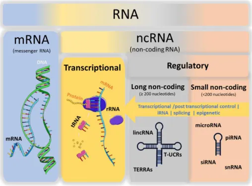
Unlike the central dogma of molecular biology, recent evidence suggests that the majority of the human genome, and even the genome of other complex organisms, is transcribed into RNAs that do not code for proteins. These RNAs are classified as non-coding RNAs (ncRNA) and include the housekeeping RNAs, also known as transcriptional ncRNAs, and the regulatory ncRNAs [1, 2].
The transcriptional ncRNAs, including transfer RNAs (tRNAs) and ribosomal RNAs (rRNAs), are constitutive RNAs that do not directly yield coding proteins but are infrastructurally involved in the processes of protein expression (Figure 1) [1, 2].
Regulatory ncRNAs, on the other hand, are mostly transcribed in a location and time dependent manner [2]. Based on the size they can be classified into small ncRNAs (shorter than 200 nucleotides) and lncRNAs (200 nucleotides or longer) (Figure 1) [2, 3]. Regulatory RNAs control crucial cell processes such as proliferation, differentiation, apoptosis, development and stress response [4]. Hence, they determine most of our complex characteristics and play a significant role in disease.
Figure 1 - RNA classification and representative members of RNA classes.
The small ncRNAs group includes well-documented ncRNAs which have triggered the RNA-centered research, such as micro RNAs (microRNA) and small interfering RNAs (siRNAs) but also
small nucleolar RNAs (snoRNAs), small nuclear RNAs (snRNAs), and PIWI-interacting RNAs (piRNAs) (Figure 1) [1, 2].
Small ncRNAs are involved, mainly, in post-transcriptional gene regulation by mean of translational repression, RNA interference (iRNA) or DNA methylation, and basically define which genes are turned on and which are turned off [1]. For instance, the subclass of microRNAs itself is estimated to regulate 30% of all human genes [5, 6]. In most of the cases, differential or abnormal expression of small ncRNAs is related to pathology scenarios, including cancer [7– 10], neurodegenerative diseases [11–14], cardiovascular diseases [15–17] and diabetes [18]. This is an indicative of their potential usage as novel diagnostic, prognostic, predictive biomarkers and, most recently, as novel therapeutic agents and targets [19–21].
All research behind both the discovery of new RNA drugs and the RNA-based therapy itself requires easy and cost-effective access to large amounts of RNA agents that meet the requirements established by regulatory authorities [21]. In one way or another, all these “requirements” are related to, or even dependent on, the RNA sources.
1.2 Sources of RNA
The rise of the RNA-centered research, mostly justified by the discoveries of novel roles of noncoding RNAs, such as microRNAs and small interfering RNAs, imposes the access to large amounts of affordable ncRNA agents.
Currently, RNAs obtained through synthetic methods, such as in vitro transcription and chemical synthesis (Figure 2), account for the major source of RNA [22]. However, in recent years large efforts have been made to develop recombinant methods to produce natural RNAs
in vivo, using prokaryotes hosts (Figure 2) [21].
Figure 2 - RNA sources and their main characteristics. The advantages are represented in green and the disadvantages in red.
1.2.1 In vitro transcription
In vitro transcription is a widely used enzymatic approach for template-directed synthesis of
RNA molecules. It is based on the engineering of a DNA template (oligonucleotides, PCR products or plasmid) that includes a bacteriophage promoter sequence (usually from the T7 bacteriophage) upstream of the sequence of interest, followed by the transcription using the corresponding RNA polymerase [23, 24]. Despite this method has RNA yields in order of milligrams and presents commercial convenience, it is a very laborious and costly approach that usually leads to RNA products with heterogeneous 3’ and 5’ ends. Such heterogeneity appears as a consequence of transcription termination can occur out of the desired site, for example, few bases before the 3’ end or bases beyond the template length, and might be a hindrance to its future applicability [25] (Figure 2).
1.2.2 Chemical synthesis
Chemical synthesis of RNA is a fast RNA production approach mainly based on phosphoramidite chemistry and commonly has a high yield of pure RNA. Yet, the main advantage of this strategy concerning the therapeutic applicability is the possibility to add chemical modifications to enhance metabolic stability and other pharmacokinetic features [14]. Unfortunately, this method has also some limitations in terms of length of the desired RNA that can be synthesized, and in terms of ensuring the synthesis of an RNA with a desired sequence [25]. Furthermore, the cost sharply increases with the adding of chemical modifications or with the increase of the RNA length [25] (Figure 2).
1.2.3 Recombinant biosynthesis
Recombinant RNA biosynthesis is a method in which natural RNAs are produced in vivo, mostly in microorganisms, and appears as a strategy to overcome some of the problems faced by the synthetic methods. This method is expected to provide large quantities of biological non-coding RNA agent with proper folding and natural modifications that are critical for RNA higher-order structure, stability, activity, and safety [22] (Figure 2).
When compared with the synthetic methods previously mentioned, we can safely say that the synthetic methods are most expensive and laborious and have their drawbacks with respect to sequence requirements, variations in yield, non-templated nucleotide additions and/or the limitation on the maximum length of the oligonucleotide. Notwithstanding, artificial RNAs have a wide applicability, ranging from structural studies to therapeutic application [23, 26]. However, through the view of therapeutic application, these methods face a problem: they produce “artificial” RNA. These artificial RNAs do not experience the same post-transcriptional modifications by which “natural” RNAs are subject [27].
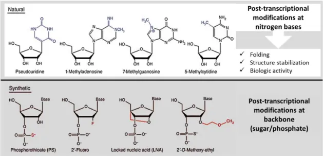
Actually, while in vivo natural RNAs go through modifications mostly on the nitrogenous bases, artificial ones undergo modifications on the backbone, either on the phosphate group or on the sugar ring (Figure 3).
The post-transcriptional modifications that occur in natural RNAs are often related to their biological function and hold biochemical and physiological functions [27]. Thus, the absence of natural modification, or the presence of artificial ones, may interfere with the natural folding and consequently with the right performance and biologic activity. Not to mention the likelihood of alteration in the safety properties offered by artificial RNAs [21]. For this reason, and for the cost-effectiveness advantages, there is a growing interest in the use of biosynthesized RNA based on recombinant methods [22].
Figure 3 - Natural and synthetic RNA modifications, highlighting the advantages of natural modifications. Adapted from [21].
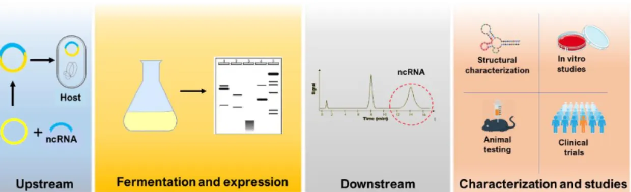
Recombinant RNA biosynthesis follows almost the same footprints of recombinant protein biosynthesis (Figure 4). Within upstream processes (USP) a vector with strong transcription signals and carrying the gene that codes the desired ncRNA is inserted into a host. The host’s cell machinery then promotes the fermentation-based expression of the gene [28, 29]. Within the downstream processes (DSP) the product, in this case, ncRNA, is separated from all types of impurities, either host impurities, process contaminants or even product-related impurities [28, 30, 31]. Together, USP and DSP aim to obtain RNA products that are suitable for analytical and clinical applications [32].
Noteworthy, the design of a biosynthesis process includes the combination of a considerably high number of options. The right combination of options, for example, in terms of the host, vector, fermentation conditions and purification methods determines the success of the
biosynthesis process [33–35]. In the recent methods for RNA biosynthesis, the options in terms of host microorganisms have been narrowed down to two: Escherichia coli (E. coli) and
Rhodovulum sulfidophilum (R. sulfidophilum). However, the choice of one rather than another
influences all the process stages from the plasmid construction to the purification strategies.
Figure 4- The process of recombinant ncRNA biosynthesis for analytical and clinical applications.
1.3 RNA biosynthesis: the hosts
There are many available production systems for recombinant biomolecules, such as microorganisms, mammalian cells, and insects. Microorganisms, like bacteria or yeast, are considered the most promising systems for this purpose and so, devoting efforts have been made in the field of fermentation-based manufacturing of biomolecules [28]. In fact, microorganisms offer many advantages in the production of recombinant biomolecules since they have well-characterized genomes, there are some versatility of the vectors that can be used, and there is availability of different host strains, and the process usually presents cost-effectiveness when compared with other cell systems [28].
As above mentioned, up until the moment E. coli and R. sulfidophilum have been the bacteria hosts used for recombinant RNA biosynthesis. Depending on the host chosen so is the plasmid design, the fermentation condition, and the DSP [36].
1.3.1 RNA biosynthesis in Escherichia coli
The historical development of microbial physiology studies, molecular genetics, and engineering genetic has always been based on the gamma-proteobacteria E. coli resulting in unique accumulation of information about this specie [37].
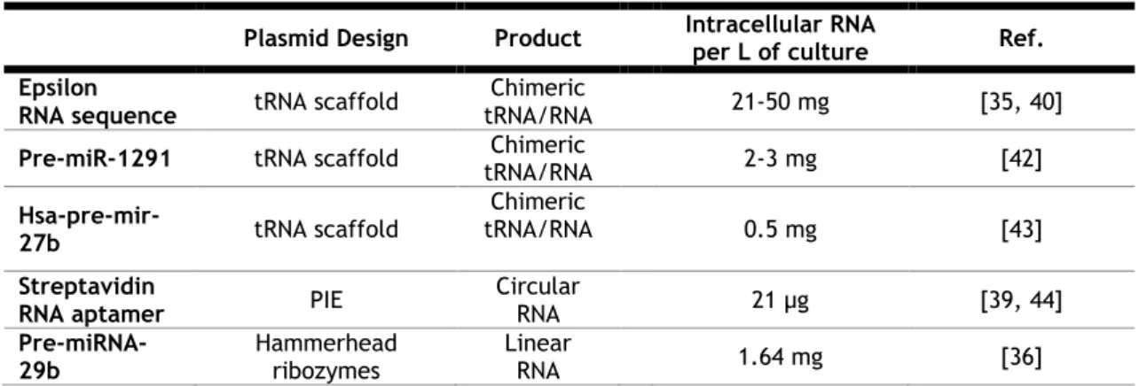
Then, it is not surprising that E. coli was the first-choice in what regards to expression systems for recombinant RNA production. In fact, there are some advantages of using E. coli as the host microorganism, since: (i) it has an unparalleled growth rate; (ii) can easily be achieved a high cell density; (iii) the growth conditions are simple and undemanding and (iv) its transformation with exogenous DNA is fast and easy [38]. Table 1 summarizes the RNAs produced in E. coli, as well as the strategy used in each case, and the respective RNA yield.
In what concerns to methods for RNA production in E. coli, Ponchon and Dardel [35], taking advantage of the natural “anti-degradation” characteristics of tRNA molecules, developed a specific technology to biosynthesize RNAs. In this innovative technology, the RNA to be produced is basically inserted into the anticodon stem of a tRNA scaffold. This biostructure plays a role of protective shielding, in which the recombinant RNA is “disguised as a natural RNA and thus hijacks the host machinery, escaping cellular RNases” [35] (Figure 5A).
Figure 5- Plasmid design for recombinant RNA production in E. coli. (A) and (B): Secondary structure of recombinant RNA chimeric where sequences in black represent the scaffold and sequence in orange represent the inserted RNA. Scissors represent the excision site. (C) Permuted intron exon plasmid where black sequences are the 3´and 5´half intron and the orange sequence is the inserted RNA. Adapted from [29, 39].
This method seems to work well since it allowed to obtain large amounts of RNA yielding up to 50 mg/L of culture of pure RNA [40] (Table 1). Despite all these advantages, it is important to depict two disadvantages that may not be attractive for the large scale biosynthesis and industrial environment: first, this method is only feasible for RNA molecules with stem-loop secondary structures and, second, it is necessary a cleavage step to release the RNA from the chimeric structure, which requires the use of enzymes [40, 41].
Table 1- Examples of Recombinant RNAs typically produced in E. coli.
Plasmid Design Product Intracellular RNA per L of culture Ref. Epsilon
RNA sequence tRNA scaffold
Chimeric
tRNA/RNA 21-50 mg [35, 40]
Pre-miR-1291 tRNA scaffold tRNA/RNA Chimeric 2-3 mg [42]
Hsa-pre-mir-27b tRNA scaffold
Chimeric
tRNA/RNA 0.5 mg [43]
Streptavidin
RNA aptamer PIE
Circular RNA 21 µg [39, 44] Pre-miRNA-29b Hammerhead ribozymes Linear RNA 1.64 mg [36]
Based on the tRNA scaffold strategy, some variants of this method were tested over the years including the co-expression of the tRNA-fused RNA with an interacting protein [45] and the use of a scaffold of tRNA/miRNA [46]. Besides the tRNA-based method, other strategies have been developed for RNA biosynthesis in E. coli, especially for ncRNA biosynthesis. Some examples are rRNA scaffolds [47] (Figure 5B) which follows the same principle of tRNA scaffolds, and Permuted intron-exon (PIE) sequence from T4 bacteriophage (Figure 5C) that produces circular target RNAs [44]. All these methods have advantages and disadvantages and despite they are all different strategies, all aim to prevent the E. coli nucleases attack to the recombinant RNA.
1.3.2 RNA biosynthesis in Rhodovulum sulfidophilum
During the last years, the peculiar characteristics of the bacterium Rhodovulum sulfidophilum have aroused the interest on its applicability as a host for recombinant RNA production. For instance, Suzuki and co-workers proposed a method for recombinant RNA biosynthesis in R.
sulfidophilum [48]. Among all the characteristics of this bacterium, two of them stood out in
what concerns to its feasibility for recombinant RNA production. The first one is that it secretes nucleic acids into the extracellular medium and, the second one, is that it does not produce RNAses to the culture medium and even the intracellular level of RNases is also low [48]. In this method, the RNA sequence is flanked on both sides by hammerhead ribozymes (Figure 6) that catalyze the cleaving of its own phosphodiester backbone after transcription without the intervention of co-factors or enzymes [49].
Figure 6 - Plasmid design for recombinant RNA production in R. sulfidophilum. Adapted from [48].
Unlike what happens when E. coli is the host, herein recombinant RNAs are produced intracellularly and extracellularly without any modification such as circularization or chimeric structures [48].
Under such transcription signal (Figure 6), the maximum level of extracellular production found for a Streptavidin RNA aptamer, was 45 ng/L of culture after 16h of anaerobic-light cultivation
(which corresponds to the last lag phase) and the total RNA production (intracellular and extracellular) was nearly 7 ug/L of culture. Despite cultivation under aerobic-dark conditions provided best results, the values still unpractical for industrial scale-up or laboratory applications [48].
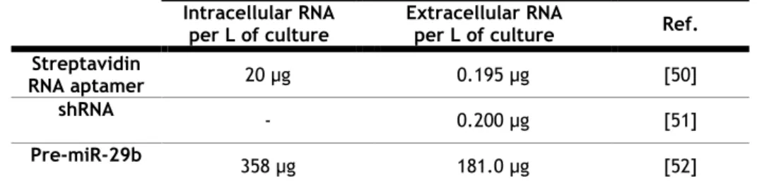
In order to improve the productivity, modifications were carried out in the promotor and allowed a 4 times increase of the extracellular Streptavidin RNA aptamer production and about a 3 times increase on the amount of total RNA production (intracellular and extracellular) when compared to the wild-type promoter [50, 51] (Table 2). With such modifications the time course of RNA production is maintained until the late stationary phase, contrarily of what happens with the wild-type promoter where the maximum extracellular RNA production occurs in the last lag phase [50].
With the purpose of generalizing this method, another type of RNA, short hairpin RNA (shRNA), with a different secondary structure, was produced under the same conditions and the result in terms of RNA productivity was quite similar [51] (Table 2).
Table 2- Recombinant RNA produced in R. sulfidophilum through Hammerhead ribozymes plasmid design
Intracellular RNA per L of culture
Extracellular RNA
per L of culture Ref. Streptavidin
RNA aptamer 20 µg 0.195 µg [50]
shRNA - 0.200 µg [51]
Pre-miR-29b 358 µg 181.0 µg [52]
More recently, Pereira and co-workers described the successful biosynthesis of hsa-pre-miR-29b in R. sulfidophilum, also based on recombinant RNA technology with the hammerhead ribozymes and under the same transcription signals [52]. The target RNA productivity was considerably higher when compared with the previously described (Table 2). Noteworthy, the growth conditions were different, as in this case aerobic conditions were used [52]. The RNA secretion, together with the high yields of the target RNA, made this method a promisor methodology for ncRNA production in R. sulfidophilum [52]. Therefore, it is important to extend and consolidate the knowledge of the characteristics and growth conditions of R.
1.4 Rhodovulum sulfidophilum
1.4.1 Taxonomic and morphologic characterization
Rhodovulum sulfidophilum, (basionym Rhodopseudomonas sulfidophila and Rhodobacter
sulfidophila) whose name means sulfide loving small red egg [53], is a purple non-sulfur gram
negative alpha-proteobacteria [54]. As all species of the Rhodovulum genus, they are morphologically characterized by being rod-shaped cells with 0.6 - 0.9 μm in width and 0.9 - 2.0 μm in length, usually movable by polar flagellum [55].
Rhodovulum species are metabolically versatile since they are facultative anaerobic
photoautotrophic. Therefore, in addition to growing anaerobically using light as an energy source, they also have the ability to use organic compounds aerobically in the dark as an energy source [54]. Depending on the growing condition, anaerobic or aerobic, they can be yellowish-brown to pinkish-red, respectively (figure 7). They are also known as “anoxygenic bacteria” since it is unable to produce O2 during photosynthesis due to their inability to use water as an
electron donor. Instead, they are capable of assimilating nitrogen, carbon dioxide and other organic compounds [56].
Figure 7- Morphology of R. sulfidophilum colonies in agar plate under aerobic conditions after 96 h incubation at 30 ºC. Photo acquired during the course of this experimental work.
Interestingly, unlike other facultative photosynthetic species, R. sulfidophilum synthesize a complete photosynthetic apparatus even under fully aerobic growth conditions, as well as under anaerobic growth conditions in the light, conflicting with the patterns of expression regulation in bacteria [56]. The photosynthetic membrane system is of the vesicular type and the pigments consist in bacteriochlorophyll α and carotenoids, most probably due to the spheroidene group [53, 54]. Table 3 summarize typical R. sulfidophilum characteristics.
Rhodovulum sulfidophilum, as the name implies, are cells that are tolerant to large
concentrations of sulfide, and unlike all other related species of purple non-sulfur bacteria, converts sulfide and thiosulfate to sulfate without the intermediate accumulation of sulfur [54]. In many aspects, R. sulfidophilum show high similarity with species of Rhodobacter genus and one of the most similar species is Rhodobacter Capsulatus which is used in many studies as a model to understand R. sulfidophilum features [53, 57]. Although R. sulfidophilum shares lots of features with Rhodobacter family, they can be undoubtedly distinguished based on their natural habitat. While Rhodobacter bacteria are found in freshwater and terrestrial environments, R. sulfidophilum is found only in marine and hypersaline environments [53].
Table 3- Summary of taxonomic and morphologic characteristis of R. sulfidophilum [54, 55]
During the last years, three distinct strains have been identified: W4, the type strain (DSM 1374, ATCC® Number 35886™, Taxonomy ID 1188256) which was first isolated from marine mud flats, W12 (DSM 2351) [53, 54, 58], PS88 which is well recognized by their high flocculation ability [58, 59] and P5 recognized as an hydrogen-producing photosynthetic strain [60].
This bacterium has been used in many fields by taking advantage of their metabolic characteristics. For example, it can be used as a cheap option of live feed at the aquaculture industry [61, 62], or as a eco-friendly means for hydrogen production [63, 64] or even as cheap tool for bioremediation including sardine wastewater treatment [65], crude oil degradation [62], among others [66]. Its peculiarity also leads to its use as a model for the study of mechanisms underlying anoxygenic photosynthesis [56, 57] and oxidative sulfur metabolism [67]. Furthermore, diversity in metabolic activities and the production of valuable products such as enzymes, proteins, and nucleic acids are great advantages of this bacterium in biotechnology [66].
Characteristic Description
Cell shape Ovoid to rod-shaped
Cell size 0.6-0.9 µm wide and 0.9-2.0 µm long
Cell Motility Motile or non-motile
Flagellum type Polar, varying from 0 to 4 in number
Gram classification Gram-negative
Intra-cytoplasmatic membrane system Vesicular type
Natural habitat Marine and hypersaline environments
Photopigments Bacteriochlorophyll α and carotenoids
Colonies color Yellowish-brown to pinkish-red
Aerobic growth in the dark Present
1.4.2 Growth and flocculation
Multiplication of Rhodovulum sulfidophilum cells occurs by binary fission and the increase in cell density is accompanied by active secretion of polymers, including sugars, proteins and nucleic acids, and the establishment of structured communities of cells called flocs [58]. Over the years, many efforts have been made to understand the mechanisms underlying nucleic acids secretion in R. sulfidophilum and although the mechanism is not yet unveiled, many cases of nucleic acid secretion in bacteria have been used as comparative models [68–71].
The first supposition to explain the presence of nucleic acids in the extracellular medium of a cell culture is autolysis. However, the presence of nucleic acids in R. sulfidophilum extracellular medium does not appear to be linked to autolysis since cell viability seems to be maintained [68, 69]. But even if the presence of nucleic acids in the extracellular medium was a consequence of autolysis, it would not be a common autolysis like the one that occurs in the stationary phase of bacterial growth but rather a programmed autolysis where partial cell death might be induced by a high cell density, as happens in the case of Streptococcus pneumoniae [68, 69]. The cell lysis of Streptococcus pneumoniae is induced by the extracellular concentration of a secreted peptide pheromone by a quorum sensing mechanism [68, 69]. Quorum sensing describes one particular form of cell-to-cell communication, in which genes are expressed in a cell-density-dependent manner after a critical concentration of signal molecules (autoinducers) has been reached [70].
Another possible explanation for the existence of nucleic acids in the culture medium could be a horizontal exchange of genetic information between prokaryotes [71]. A Gene Transfer Agent (GTA), was discovered in Rhodobacter capsulatus as a system of genetic exchange [71]. The GTA resembles bacteriophages and works in a process similar to the phage-mediated gene transfer in which the nucleic acids are packed within the GTA [71]. In many other species of bacteria, including R. sulfidophilum, clusters of genes homologous to the GTA genes of
Rhodobacter capsulatus have been found [71]. In studies aiming to unveil the mechanism of
nucleic acid secretion on R. sulfidophilum it was found that this bacterium produces a GTA-like particle that indeed packs nucleic acids but lacks the gene transfer activity [71]. Like
Streptococcus pneumoniae, the release of GTA in Rhodobacter capsulatus was reported to be
a consequence of a cell density dependent-lysis of a subpopulation of cells in a process controlled by quorum sensing [71]. It is rational to risk that this is the mechanism underlying the presence of nucleic acids in the extracellular medium of R. sulfidophilum. However, there are evidences that do not corroborate this hypothesis since the nucleic acids present in the extracellular medium of R. sulfidophilum are not packed and the existent GTA-like particles have not a transfer activity [71].
Another example of a bacterial nucleic acid secretion system is from Pseudomonas aeruginosa in which the active DNA secretion has the main purpose of forming biofilms [69]. Similarly, in
the case of R. sulfidophilum, the nucleic acids secretion plays a key role in the establishment of biofilms [69]. Some studies had shown that secreted nucleic acids are essential not only for the maintenance of flocculated cells but also to the establishment of cell to cell contact, existing in the extracellular medium as a cell-to-cell interconnecting compound [69]. Furthermore, enzymatic deflocculation experiments demonstrated that despite flocculated cell are made of a miscellany of exopolymers (proteins, nucleic acids, and others), only nucleases can truly lead to deflocculation. This finding elucidated the role of nucleic acids on the flocculation of R. sulfidophilum. Actually, RNA and DNA were determined to be the major component in the extracellular polymers mix [72, 73]. Noteworthy, the flocculation mechanism is affected by the growth conditions since in rich mediums the flocculation appears to be decreased or even absent [68, 69].
In terms of characterization of the extracellular exopolymers and focusing on nucleic acids, the RNA fraction released into the culture medium contains mainly non-aminoacylated fully mature-sized tRNAs, what suggests an intracellular processing before secretion, and fragments of 16S and 23S rRNAs. The sequence of extracellular DNA fragments showed no differences from the intracellular genomic DNA sequence [68, 69].
In terms of stability of the extracellular nucleic acids, it is easy to understand the fact that they can maintain the stability on the extracellular medium as part of the floc. However, even in the soluble form they maintain the stability which is a consequence of the very low, or even undetectable, level of nucleases on the culture medium of R. sulfidophilum [74].
1.4.3 Fermentation conditions and metabolism
Three important characteristics of this bacterium must be considered regarding R.
sulfidophilum cultivation. It is a halophilic, mesophilic and facultative aerobic
photoautotrophic system [53]. The first two characteristics are clearly a consequence of the natural habitat. As marine bacteria, the presence of sodium chloride (NaCl) and mild temperatures are imperative for optimal growth. When first isolated it was settle down that it grows in 0.5 to 7.5 % of sodium chloride with 30 to 35 ºC as the optimal temperature [53, 54]. The pH of the culture could vary from 5 to 9 [55]. However, the optimal pH must be adapted according to the electron donor and the substrates used which can be exemplified by the reported shift in the optimal pH range from 6.5 to 8.0 on sulfide and from 5.0 to 7.5 on malate [54].
As mentioned before, R. sulfidophilum can be cultivated under both anaerobic light conditions and aerobic dark conditions. In both cases, extracellular nucleic acids are produced. The growth in anaerobic light conditions can be photolithotrophic or photoorganotrophic. For these two phototrophic conditions, the method of Hiraishi and Ueda [53] is commonly applied with or without slight modifications. In this method, the bacteria grow anaerobically at 30°C in
screw-cap test tubes or bottles filled with the medium under incandescent illumination (ca. 5,000 lux) [53]. Under such conditions, the culture is yellowish-brown [53].
The growth in aerobic dark condition is chemotrophic and the aerobic condition is commonly provided by rotary shaking. Under such conditions, the culture turns into red [58]. The set of organic compounds used by R. sulfidophilum as electron donor or carbon source depends both on the growth conditions, being anaerobic or aerobic, and on the strain. Methanol, citrate, mannitol, benzoate and tartrate are examples of organic compounds not used by the bacteria [53, 54, 58]. On the other hand, glucose, fumarate, lactate, pyruvate and glycerol are examples of organic compounds used by the bacteria in both aerobic and anaerobic conditions and by all the strains [53, 54, 58]. In terms of nitrogen sources, the ammonium salts are documented as the best source [54]. Growth factors requirements include B-complex vitamins such as biotin, niacin, and thiamin, however, the need for these growth factors can be well satisfied with yeast extract. Besides this, casamino acids and peptone are recognized as essential to its growth [53, 54, 58]. Table 4 summarizes some of the growth requirements.
Table 4- Summary of carbon sources, electron donor and other substrates for R. sulfidophilum cultivation [54, 55].
Category
Sulfur sources Sulphate, Thiosulphate, Cysteine
Vitamins requirement Biotin, Niacin and Thiamin / Yeast extract
Nitrogen and amino acids source Ammonium salts, peptone, casamino acids,
Carbon source (some examples) Glucose, Fumarate, Lactate, Pyruvate, Glycerol
Pereira and coworkers [52] studied the best conditions for R. sulfidophilum growth and overexpression of hsa-pre-miR-29b. A semi-defined medium was tested (Table 5) with different sodium chloride concentrations (170, 342, 513, and 850 mM) and the ideal temperature for growth (25, 28, 30, 32, and 35 °C) was also tested. The study demonstrated that 513 mM is the ideal sodium chloride concentration and that under such salinity the ideal temperature is 30 ºC. The growth conditions established by Pereira and coworkers (2016) for the growth of R.
sulfidophilum in shake flask experiments are illustrated in Figure 8.
In recombinant biosynthesis the growth environment in terms of growth medium composition and physical parameters (pH, temperature, and agitation) is decisive not only to achieve high cell densities but also to guarantee high productivity of the recombinant product [75]. As example, some nutrients may enhance the productivity by acting as precursors for a product or even by preventing the degradation of the product [75, 76]. Along this same line of thinking,
the growth environment must thus be developed concomitantly with the production outcomes since they will affect each other.
Table 5- Semi-defined medium composition for R. sulfidophilum DSM 1374 growth in aerobic dark condition [52].
Medium Component
Concentration TES composition Concentration
Triptone 10.0 g/L FeSO4·7H2O 20.0 mM
Polypeptone 5.0 g/L MnCl2·4H2O 20.0 mM
Yeast extract 0.5 g/L CoSO4· 7H2O 20.0 mM
NaCl 513.0 mM CuCl2·2H2O 2.0 mM K2HPO4 23.0 mM ZnSO4·7H2O 2.0 mM KH2PO4 7.0 mM H3BO3 9.7 mM Glucose 278.0 mM NiCl2·6H2O 0.2mM CaCl2 0.3 mM Na2MoO4·2H2O 0.2 mM MgSO4 0.8 mM (NH4)2SO4 7.6 mM Trace elements solution 1 mL
Figure 8 - Fermentation process of R. sulfidophilum in shake flask experiments. Adapted from [44].
1.4.4 RNA biosynthesis: Rhodovulum sulfidophilum versus Escherichia coli
Based on the data from Tables 2 and 3 it is safe to say that, in terms of recombinant RNA productivity, higher values are achieved when E. coli is the host. However, in a manufacturing bioprocess, productivity is not the only parameter to be evaluated. In fact, the host choice affects not only the productivity but all the other stages of the production process, including the USP and DSP. Therefore, the host choice may also depend on plasmid design, fermentation
conditions, and purification methodologies, which can be more adequate to the target recovery.
In terms of plasmid design, R. sulfidophilum offers more advantages since E. coli requires a plasmid design capable to avoid RNases degradation. In addition, the strategies for RNA protection from RNases either require a post-production procedure, as enzymatic cleavage or biosynthesize the RNA product in a nonlinear form. On the other hand, considering the fermentation time, medium and conditions, E. coli offer advantages since it grows faster and the growth medium and conditions are undemanding when compared with R. sulfidophilum [48, 50–52].
Considering the isolation and recovery of the target product, R. sulfidophilum offer much more advantages. Beside the isolation of intracellular RNAs, R. sulfidophilum offers the possibility to recover the secreted RNAs without the need of lysis procedures that usually involve hazardous chemicals and affect the RNA integrity and stability [36, 52].
Table 6 specifies the manufacturing process and the host that offer more advantages in each stage. The two stages in which R. sulfidophilum loses out to E. coli are closely related to the growth characteristics of this bacteria that, when compared with E. coli, is slow and fastidious. This slow growth implies that fermentations reaches low cell density or take longer to reach considerable cell density which in turn affects the RNA productivity. Hence, an optimization in the fermentation process of R. sulfidophilum could overcome this drawback and level up the biosynthesis of ncRNA.
Table 6- Comparison of E. coli and R. sulfidophilum through all the stages of recombinant RNA biosynthesis. Green tag represents the host with the best rating.
Manufacturing parameters E. coli R. sulfidophilum
Plasmid design
Fermentation requirements RNA Isolation and recovery Contaminants
RNA Productivity
RNA integrity and stability
TOTAL score 2 4
Based on this comparison, so far E. coli should be the choice when the aim is to obtain large amounts of RNA but R. sulfidophilum should be the choice when the RNA stability and integrity are a major concern [36].
1.5 Biosynthesis in bioreactors
Many high-value products, including therapeutic agents, are microbial fermentation by-products. Thus, microbial fermentation is an important practice for biotechnology and for the development of bioprocesses [28].
In a laboratory environment, shake flasks fermentations are the most common system for bacterial cultivation. However, it is limited in that it lacks the control of many parameters that play a key role in microbial growth and production. On the other hand, bioreactors fermentation technology is a more complex system that aims to control growth environments. Bioreactors control many parameters that affect the fermentation process like temperature, pH, dissolved oxygen, stirring among others [77].
1.5.1 Factors affecting biosynthesis
Nutrients, temperature, and pH are factors of extreme importance to the cell growth and to the biosynthesis and must be a concern when running an optimization of the fermentation process of a microorganism.
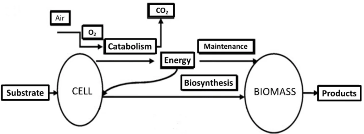
Nutrients must satisfy the elemental requirements in terms of energy supply for cell maintenance and biosynthesis. In an aerobic fermentation, this process is represented by Figure 9 [78].
Figure 9- Schematic mechanism of the aerobic process of energy generation, biomass formation and product synthesis in microbial cultures. Adapted from [79].
The conversion of a carbon source into biomass and energy consists of its transport to the cell and subsequent degradation in simpler molecules by central metabolic pathways [80, 81]. Carbohydrates, such as glucose, can be catabolized by different pathways (such as Glycolytic, Entner-Doudorof, and Pentose-Phosphate). These pathways lead not only to energy production in the form of Adenosine Triphosphate (ATP) and reduced coenzymes but also to the biosynthesis of monomers of different macromolecules constituting the cells (proteins, nucleic acids, lipids and polysaccharides) [80, 81]. On the other hand, lipids, proteins, and other
carbohydrates can be converted to several intermediates of carbohydrates catabolic pathways and slip into the catabolic pathway through a multitude of side doors [82].
A good example is glycerol (1,2,3-propanetriol, glycerin), a by-product of transesterification of vegetable oils and animal fats, which share with glucose the same metabolic pathway from glyceraldehyde-3-phophate to pyruvate and the respective derived products. Yet, likewise with glucose, other pathways can be used by bacteria to its metabolization. Its degree of reduction per carbon, k, is significantly higher (C3H8O3: k = 4.67) than for sugars such as glucose (C6H12O6: k = 4) or xylose (C5H10O5: k = 4) [76, 83, 84]. The high k provides a distinct advantage over more oxidized carbohydrate-based carbon sources in the production of reduced chemicals. Actually, by the view of industrial microbiology, glycerol has been considered a promising carbon source. Besides being cheap, many microorganisms can naturally utilize glycerol thanks to its abundant occurrence in nature. One of the promising applications for the use of glycerol is its bioconversion to high value compounds through microbial fermentation assuming as a promising carbon source to the industrial microbiology [76, 83, 84].
Oxygen is a crucial substrate of aerobic fermentations which is needed for growth, maintenance and metabolic pathways, including product synthesis. Therefore, oxygen must be continuously provided by a gas phase to assure the correct oxygen supply to the cells. The concentration of dissolved oxygen in the culture medium depends on the oxygen transfer rate from the gas to the liquid phase, and on the oxygen uptake rate which represents oxygen consumption by the microorganism [79]. Agitation is required to ensure homogeneous distribution of the nutrients. In shake flasks scale, oxygen transport by aeration and agitation are accomplished by the action of the shaker apparatus. In bioreactors, oxygen is commonly supplied as compressed air and distributed by a gas distributor, and mechanical devices are used to improve mixing of the culture medium [79]. The oxygen is transferred from a suspended gas bubble into a liquid phase, where it is taken up by the microorganism and finally transported to the site of reaction inside the cell where it is used to energy generation, biomass production and biosynthesis (Figure 9). During the fermentation process, the oxygen requirements depend on many factors such as the metabolic activity, the changes in the medium viscosity and with the foam formation [78]. For example, oxygen uptake rate commonly presents an increase during the exponential stage and a decrease during the stationary stage as a function of the metabolic activity [79].
Temperature, like essential chemical elements and organic substrates, play a key role in the bacterial growth and thus in the productivity in terms of natural and recombinant by-products. Since it affects all chemical and biochemical process it is a potential limiting factor for bacterial growth and, at some point, govern substrates consuming kinetic. However, while the temperature is always a factor in microbial growth, respiratory rate, and organic carbon assimilation, it is not always the only factor or even a dominant one [85].
Likewise, each microorganism has an optimal pH in which they experience ideal growth. However, pH optimization in recombinant product-formation should be a compromise between the microorganism optimal pH and the pH that assure the stability of the target recombinant products [76].
1.5.2 Fermentation strategies
Basically, fermentation can follow three different strategies: Batch, fed-batch and continuous fermentation. Batch fermentation represents a simple and robust strategy characterized by no addition or removal of nutrients (Figure 10). The initial medium composition is not altered during fermentation resulting in the interruption of growth, product formation, and substrate utilization after a certain period. After the initial nutrients are consumed, the culture stops growing or, in some cases, microorganism metabolism may shift and begin to consume other metabolites in the culture broth, what in turn may alter the patterns of biomass formation and productivity [76, 86].
Otherwise, continuous fermentation guarantee that growth and product formation last more, since continuous nutrient medium is added to the fermentation while cells and products are also withdrawn (Figure 10) until a moment that the system reaches a steady state where substrates, products, and cells concentration are constant. The idea of continuous fermentation seems promisor for bioprocess intending to obtain a large quantity of a fermentation by-product. However, besides the higher probability of mutation and contamination, in cases of expression systems based on plasmids more instability is reported [76, 86].
The fed-batch strategy appears as an intermediate between batch and continuous fermentation. Fed-batch starts as a batch approach and then the metabolism is controlled by a feed (Figure 10) that can follow different strategies. The feed can be either the initial medium or a concentrated solution of the growth limiting substrate(s) and can also follow different feed profiles (constant, exponential and stepwise). Besides the increase in the fermentation duration, the overall reactor productivity is increased [76, 86].
The decision towards a fermentation strategy must take in account many factors such as economic balances, type of product, desired productivity, type of microorganism and its vulnerability to mutation, stress exposure and metabolic shifts [87].
1.5.3 Microbial growth
The typical growth of a bacterial culture usually follows a pattern like the growth curve shown in Figure 11. The curve can be divided into six well defined phases [88, 89]: The lag phase indicates the time that the bacteria require to adapt to the new environment. It is characterized by long generation time, zero growth rate and maximum rate of metabolic activity. The acceleration phase indicates the end of the adaptation period and the beginning of cell generation. It is characterized by decreasing generation time and increasing growth rate. The exponential phase, also known as log phase, is represented by minimal but constant generation time and maximum rate of substrate utilization. Declining phase represents a slowing in the growth because of gradual decrease in substrate concentration as well as increased accumulation of toxic metabolites. This phase is characterized by increased generation time and decreased growth rate. The stationary phase indicates stagnation of the microbial population generally because of depletion of the substrate, maximum physical crowding, a higher concentration of toxic metabolites and/or balance between growth and death rate of biological cells. The endogenous decay is the phase in which death rate exceeds growth rate. The phase is characterized by endogenous metabolism and cell lysis and is usually the inverse of the exponential growth phase.
Figure 11 - Typical bacterial growth curve: the phases and its respective specific growth rate. Adapted from [89].
The growth rate of a batch culture under the exponential phase is generally believed to follow the first order kinetic model in which the growth rate is proportional to the microbial mass in the system. Mathematically expressed by Equation 1 where µ represents the specific growth rate (h-1), t is the time (h), and X is the biomass concentration (g/L) [88–90].
𝑑𝑋
𝑑𝑡 = 𝜇𝑋 (1)
Summing up, the main goal of the biosynthesis processes in microorganism is the transformation of substrates into the desired metabolic products. This requires well-controlled conditions, which can be supplied by bioreactors platforms, where several factors should be considered such as nutritional control, the type of process (batch, fed-batch, continuous), temperature, pH, and oxygen supply control. The process outcomes, in terms of growth and productivity should suggest a direction for the process optimization [28, 76].

![Figure 6 - Plasmid design for recombinant RNA production in R. sulfidophilum. Adapted from [48]](https://thumb-eu.123doks.com/thumbv2/123dok_br/18068137.864256/29.892.178.747.677.924/figure-plasmid-design-recombinant-rna-production-sulfidophilum-adapted.webp)
![Table 4- Summary of carbon sources, electron donor and other substrates for R. sulfidophilum cultivation [54, 55]](https://thumb-eu.123doks.com/thumbv2/123dok_br/18068137.864256/35.892.172.755.561.690/table-summary-carbon-sources-electron-substrates-sulfidophilum-cultivation.webp)
![Table 5- Semi-defined medium composition for R. sulfidophilum DSM 1374 growth in aerobic dark condition [52]](https://thumb-eu.123doks.com/thumbv2/123dok_br/18068137.864256/36.892.136.712.229.913/table-defined-medium-composition-sulfidophilum-growth-aerobic-condition.webp)