Cátia Sofia Pereira Frias
Licenciatura em Biologia
Dissecting neuronal development
deficits by inflammation: from
morphology to cytoskeleton dynamics
Dissertação para obtenção do Grau de Mestre em
Genética Molecular e Biomedicina
Orientador: Dora Maria Tuna de Oliveira Brites,
Investigadora Coordenadora e
Professora Catedrática Convidada,
Faculdade de Farmácia, Universidade de Lisboa
Co-orientador: Adelaide Maria Afonso Fernandes Borralho,
Professora Auxiliar,
Faculdade de Farmácia, Universidade de Lisboa
Júri:
Presidente: Prof. Doutor José Paulo Nunes de Sousa Sampaio
Arguente: Doutora Cláudia Alexandra dos Santos Valente de Castro
Vogal: Prof. Doutora Adelaide Maria Afonso Fernandes Borralho
i
Cátia Sofia Pereira Frias
Licenciatura em Biologia
Dissecting neuronal development
deficits by inflammation: from
morphology to cytoskeleton dynamics
Dissertação para obtenção do Grau de Mestre em
Genética Molecular e Biomedicina
Orientador: Dora Maria Tuna de Oliveira Brites,
Investigadora Coordenadora e
Professora Catedrática Convidada,
Faculdade de Farmácia, Universidade de Lisboa
Co-orientador: Adelaide Maria Afonso Fernandes Borralho,
Professora Auxiliar,
Faculdade de Farmácia, Universidade de Lisboa
Júri:
Presidente: Prof. Doutor José Paulo Nunes de Sousa Sampaio
Arguente: Doutora Cláudia Alexandra dos Santos Valente de Castro
Vogal: Prof. Doutora Adelaide Maria Afonso Fernandes Borralho
iii Dissecting neuronal development deficits by inflammation: from morphology to cytoskeleton dynamics
Copyright Cátia Sofia Pereira Frias, FCT/UNL, UNL
v Part of the results discussed in this thesis was presented in the following meetings:
Cátia Frias, Adelaide Fernandes, Lorene Lanier, Dora Brites. Changes in Neuronal Development by Inflammation: from Neuritic Outgrowth to Synaptogenesis. 12th Meeting of the
Portuguese Neuroscience Society. Lisboa, Portugal, May 26th-28th 2011 [Poster];
Adelaide Fernandes, Cátia Frias, Lorene Lanier, Dora Brites. Acute-phase cytokines IL-1beta and TNF-alpha alters neuronal neuritic outgrowth and synaptogenesis. 10th European Meeting
vii
AGRADECIMENTOS
Quero agradecer, primeiramente, à Professora Catedrática Dora Brites, orientadora desta dissertação. Agradeço a oportunidade de realizar o meu projecto de mestrado no grupo
Neuron Glia Biology in Health and Disease, por me ter recebido de forma calorosa, abrindo-me
as portas ao mundo científico, e por se ter interessado sempre por este trabalho. O seu rigor científico e espírito crítico, aliados ao seu elevado grau de exigência, aos seus vastos conhecimentos e à sua inteligência, são deveras inspiradores, tendo sido fulcrais para o desenvolvimento desta dissertação e para o meu desenvolvimento enquanto investigadora. Trabalhar consigo foi um privilégio e, mais uma vez, obrigada.
À Professora Doutora Adelaide Fernandes, co-orientadora deste trabalho, agradeço todo o apoio, toda a disponibilidade, encorajamento e dedicação demonstrados ao longo deste ano. És um exemplo, pelas tuas capacidades científicas e pela tua inteligência. Agradeço a tua paciência, as tuas sugestões, o teu acompanhamento e a tua compreensão, que foram essenciais para que este trabalho fosse finalizado. Obrigada por me teres recebido de forma tão calorosa e simpática, mas mais ainda por me teres ensinado tanta coisa.
À Professora Doutora Margarida Castro Caldas, orientadora interna desta dissertação, e ao Professor Doutor José Paulo Sampaio, coordenador do Mestrado em Genética Molecular e Biomedicina, agradeço a prontidão e a eficiência com que sempre esclareceram as minhas dúvidas.
À Professora Doutora Maria Alexandra Brito e ao Professor Doutor Rui Silva, agradeço a forma como me receberam no grupo. Para além da inteligência e conhecimentos científicos, saliento o espírito crítico e o rigor tão patentes em todas as discussões.
Às Doutoras Sofia Falcão e Ana Rita Vaz agradeço a simpatia com que me receberam. Agradeço, também, as sugestões feitas, as mensagens de ânimo e os conhecimentos partilhados ao longo deste ano.
Às meninas doutorandas, Andreia, Inês, Filipa e Ema, agradeço a vossa alegria, o vosso apoio, os vossos conhecimentos e a vossa maturidade. Vocês são todas um exemplo a seguir, pela vossa inteligência e garra! Espero poder ver-vos a conquistar este ―monstrinho‖ do vosso
viii
parecidas! Ema, agradeço-te toda a ajuda que me deste ao longo deste ano, todas as tuas sugestões e a tua enorme simpatia. Agora, toca a aproveitar o sol carioca, cara!
Às minhas companheiras de viagem, Ana Filipa e Inês, agradeço o vosso apoio, amizade e a disponibilidade que sempre tiveram para os meus desabafos. Foi um longo caminho, com alguns buracos pelo meio, mas que conseguimos transpor de forma meritória. Espero que o vosso futuro esteja pintado com muito sucesso, muita alegria e muitas concretizações, tanto profissionais como pessoais. Finalmente, conquistámos esta batalha, meninas!
Às novas meninas de mestrado, dou-vos as boas vindas e espero que esta jornada que agora se inicia seja timbrada pelo crescimento pessoal e científico, terminando num grande sucesso.
A todas as pessoas com quem partilhei a Cave escura, muito obrigada pelo vosso companheirismo (mesmo quando pedia para desligar o ar condicionado) e por me terem recebido tão bem. Aproveito para desejar a maior das sortes ao André e ao Miguel para a fase seguinte: o doutoramento.
Ao meu grupinho de mestrado, Ana Filipa, Pedro, Francisco e Marcos, agradeço a paciência que tiveram ao longo daquelas viagens intermináveis para a FCT, no meio de senhores a cantar Queen ou de muitos ―Ayo, I’m tired of using technology‖ e ―Fireflies‖. Vocês
fizeram-me rir durante meses a fio, o que ajudou a enfrentar os dias longos na sala 101. Desejo-vos a maior das sortes nesta fase que termina e que o vosso futuro seja brilhante.
Aos meus grandes amigos, André, Inês, João, João, José e Andreia, agradeço a vossa constante preocupação e o vosso ombro amigo. Foram fundamentais para o meu bem-estar, para o meu conforto. Momentos com vocês são da mais pura alegria. Vocês fazem todos parte da pessoa que sou hoje. André, meu padrinho, uma palavra especial para ti por seres quem és e por sentires orgulho naquilo em que me tornei. Espero retribuir a todos o carinho, o companheirismo, a alegria, a amizade.
À minha enorme família, agradeço o apoio e toda a preocupação. Sinto um enorme orgulho em ter-vos a todos na minha vida. Novecentos quilómetros não são suficientes para apagar este carinho especial que nutro por todos vocês! Somos quem somos por causa da nossa família e vocês ajudaram-me a crescer sempre. Uma especial palavra à minha prima
ix fez-me seguir este caminho das Neurociências e a avó sempre me fez acreditar que devemos seguir os nossos sonhos, tendo-me mostrado que a determinação e a força de vontade levam-nos onde ambicionamos ir.
Aos meus pais, os meus fantásticos pais, João e Maria, agradeço por me terem tornado na pessoa que sou hoje. Sou o que sou graças a vocês. Mantivemo-nos sempre unidos, independentemente de todos os obstáculos que apareceram no nosso caminho. Tenho imenso orgulho em ser vossa filha. Pai, somos muito parecidos e revejo-me em muita coisa do que o pai representa para mim, justiça, persistência, lealdade, força de vontade. Mãe, muito da minha personalidade vem de si, a sensibilidade, a amabilidade, a alegria, a gentileza. Obrigada por terem sempre acreditado em mim e pelo apoio incondicional.
Ao Miguel, o meu Miguel, o homem que me dá sempre força, ânimo, alento. Obrigada pelo teu apoio incondicional, pela tua amizade, pela tua compreensão, pelo teu carinho, pela tua paciência, pelo teu amor. És o companheiro que muitos gostavam de encontrar para partilhar esta viagem. Miguel, ouvir-te dizer que tens orgulho em mim torna a minha vida mais brilhante. Agradeço-te por nunca me teres deixado ir abaixo nos piores momentos, por me fazeres sentir uma pessoa especial, por todo o amor que me dás e continuas a dar ao fim de quase quatro anos. O amor está em cada sorriso, em cada palavra, em cada adversidade. Agora é a minha vez de te apoiar nesta fase que se inicia. Sabes que tentarei retribuir da melhor forma humanamente possível tudo o que sempre fizeste por mim.
xi
ABSTRACT
Neuroinflammation, a response of the nervous system to injury, results in the release of pro-inflammatory mediators, as interleukin-1β (IL-1β) and tumor necrosis factor-alpha (TNF-α).
Exposure of nerve cells to a neuroinflammatory environment was shown to change the normal neurodevelopment, which can be linked to the appearance of neurological disabilities. In this work, we aimed to assess the effects of moderate levels of IL-1β and TNF-α in the
establishment of neuronal arborization, growth cone morphology and synaptogenesis.
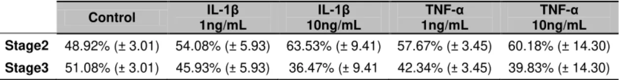
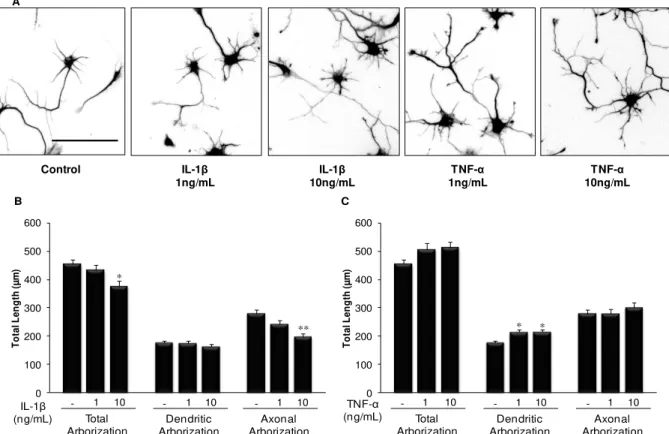
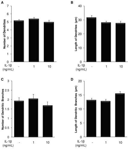
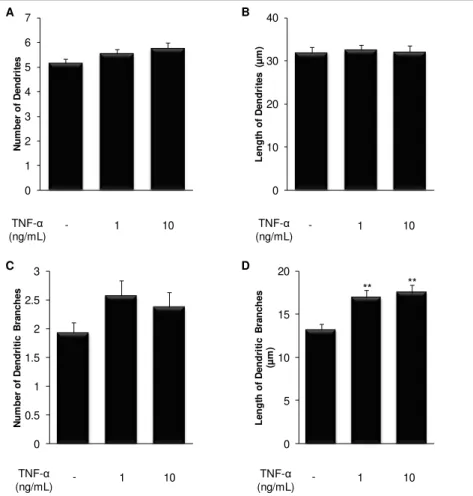
An early exposure of embryonic hippocampal neurons to cytokines delay neuronal development, with an increase in the number of non-polarized cells, stage 2 of development. When analyzing stage 3 neurons, IL-1β showed to decrease total arborization, in particular at the axonal level, while TNF-α increased dendritic arborization. In fact, IL-1β reduces dendritic
and axonal length and the number of axonal branches, whereas it increases the extent of dendritic and axonal branches, probably to compensate the other effects. In contrast, TNF-α
increases the number of primary dendrites and dendritic branches, as well as their length. By next analyzing microtubule dynamics as the ratio of acetylated- (old) vs. tyrosinated-tubulin
(newly-formed), we found that IL-1β and TNF-α induce microtubule stabilization, which may be
related to a deficient axonal outgrowth. In addition, both cytokines reduced the area of growth cones, with an increase in the immunofluorescence of F-actin, indicating alterations at the cytoskeleton which may compromise axonal elongation and branching. Regarding neuronal connectivity, we demonstrated that both cytokines not only reduced the density of dendritic spines and synapses, but also the maturity of dendritic spines, suggesting a reduction in the synaptic strength.
These findings establish a relation between neuroinflammation in fetal life and the emergence of neuronal damage, similar to those observed in neurodevelopmental disorders, as schizophrenia.
xiii
RESUMO
A neuroinflamação, resposta do sistema nervoso a danos, resulta na libertação de mediadores pró-inflamatórios, como a interleucina (IL)-1β e o factor de necrose tumoral
(TNF)-α. A exposição de células nervosas a ambientes neuroinflamatórios induz mudanças no normal
neuro-desenvolvimento, podendo levar ao aparecimento de incapacidades neurológicas. Assim, neste trabalho pretendemos elucidar os efeitos de níveis moderados de IL-1β e TNF-α
na formação da arborização neuronal, morfologia do cone de crescimento e sinaptogénese. Uma exposição inicial dos neurónios de hipocampo às citocinas atrasa o seu desenvolvimento, aumentando a percentagem de neurónios não polarizados, estadio 2 de desenvolvimento. A análise de neurónios já polarizados, estadio 3, demonstrou que a IL-1β
diminui a arborização total, em particular no axónio, enquanto que o TNF-α aumenta a arborização dendrítica. De facto, a IL-1β reduz o comprimento das dendrites e do axónio, bem como o número de ramificações axonais, embora aumente a extensão dos ramos axonais e dendríticos, provavelmente para compensar os outros efeitos. Relativamente ao TNF-α, este
aumenta o número de dendrites primárias, suas ramificações e extensão. Analisando a nível axonal a dinâmica dos microtúbulos (rácio tubulina-acetilada, antiga vs. tubulina-tirosinada,
recém-formada), observámos que ambas as citocinas induzem uma estabilização selectiva dos microtúbulos, o que pode originar deficiências no crescimento axonal. A IL-1β e o TNF-α
induzem ainda uma redução da área do cone de crescimento, estrutura que direcciona o movimento axonal, em paralelo com um aumento da F-actina, indicando alterações do citoesqueleto e possível comprometimento da arborização axonal. Relativamente à conectividade neuronal, as citocinas reduzem o número de espículas dendríticas e de sinapses, atrasando a maturação das espículas, o que sugere uma redução na força sináptica. Assim, os nossos dados apontam para uma relação entre a neuroinflamação no período embrionário e o estabelecimento de danos neuronais semelhantes aos observados em doenças do neuro-desenvolvimento, como a esquizofrenia.
xv
TABLE OF CONTENTS
ABBREVIATIONS ... xxi
I. INTRODUCTION ... 1
1. Development of Hippocampal Neurons... 1
1.1. Neuronal Growth Cones ... 3
1.2. Neuritic Arborization ... 5
1.3. Synapse ... 6
2. Cytoskeleton Dynamics in Neuronal Development ... 9
2.1. Microtubules ... 10
2.2. Microtubule-associated proteins ... 11
2.3. Microfilaments ... 12
3. Neuroinflammation: from a beneficial to a detrimental effect ... 14
3.1. Cytokines as neuroinflammation mediators ... 16
3.2. Neuroinflammation during development ... 17
4. Aims ... 19
II. MATERIALS AND METHODS ... 23
1. Materials ... 23
2. Animals ... 23
3. Hippocampal neuronal cell culture and treatment ... 23
4. Immunocytochemistry... 24
5. Analysis of neurites ... 25
6. Analysis of axonal microtubules ... 25
7. Analysis of growth cones ... 25
8. Analysis of dendritic spines and synapses ... 25
9. Statistical analysis ... 26
III. RESULTS ... 29
1. Treatment of Immature Hippocampal Neurons With Pro-Inflammatory Cytokines Alters Neuritic Output... 29
xvi
3. IL-1β Has a Higher Impact at the Axonal Level of Immature Hippocampal Neurons, while
TNF-α Only Increases Axonal Branches Length ... 32
4. Cytokines Affect the Distribution of Microtubules Along the Axon of Immature Hippocampal Neurons ... 34
5. Exposure of Immature Hippocampal Neurons to Cytokines Decreases Axonal Growth Cone Area and Alters the Composition of Growth Cone Cytoskeleton... 36
6. Cytokines Not Only Reduce Spinogenesis and Synaptogenesis, But Also Dendritic Spine Maturation ... 39
IV. DISCUSSION ... 45
Future Perspectives ... 50
xvii
INDEX OF FIGURES
Fig. I.1. Developmental stages of mouse hippocampal cultured neurons. ... 2
Fig. I.2. The growth cone cytoskeleton in primary embryonic hippocampal neurons. ... 4
Fig. I.3. Synapses allow the directional connection of neurons. ... 6
Fig. I.4. Schematic representation of different dendritic spine morphologies. ... 8
Fig. I.5. Schematic representation of dendritic and axonal cytoskeletons. ... 13
Fig. I.6. Neuroinflammation is a complex response of neural cells to a detrimental stimulus... 15
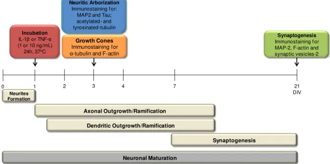
Fig. II.1. Schematic representation of the experimental model. ... 24
Fig. III.1. Treatment of immature hippocampal neurons with pro-inflammatory cytokines alters the neuritic output. ... 30
Fig. III.2. Treatment of immature hippocampal neurons with IL-1β reduces the length of primary dendrites, but increases the extent of their branches.. ... 31
Fig. III.3. Treatment of immature hippocampal neurons with TNF-α has a marked effect at the dendritic level... 32
Fig. III.4. Treatment of immature hippocampal neurons with IL-1β has a marked impact at the axonal level. ... 33
Fig. III.5. Treatment of immature hippocampal neurons with TNF-α only affects the length of axonal branches. ... 34
Fig. III.6. Treatment of immature hippocampal neurons with IL-1β affects the distribution of microtubules along the axon. ... 35
Fig. III.7. Treatment of immature hippocampal neurons with TNF-α affects the distribution of microtubules along the axon. ... 36
Fig. III.8. Treatment of immature hippocampal neurons with pro-inflammatory cytokines alters the growth cone area and cytoskeleton. ... 38
Fig. III.9. Treatment of immature hippocampal neurons with IL-1β decreases the density of dendritic spines and synapses, as well the rate of dendritic spine maturation. ... 40
xix
INDEX OF TABLES
xxi
ABBREVIATIONS
ADP adenosine diphosphate
AMCA amino-methyl-coumarin-acetate
AMPA α-amino-3-hydroxy-5-methyl-4-isoxazolepropionic acid ATP adenosine triphosphate
BDNF brain-derived neurotrophic factor BSA bovine serum albumin
Cdc42 cell division cycle 42 CNS central nervous system CO2 carbon dioxide
C-domain central domain DIV days in vitro ECM extracellular matrix
EGTA ethylene glycol tetraacetic acid E16 embryonic day 16
FBS fetal bovine serum Fig. figure
F-actin filamentous actin
FITC fluorescein isothiocyanate G-actin globular actin
GTP guanosine triphosphate HBSS Hanks' balanced salt solution
HEPES 4-(2-Hydroxyethyl) piperazine-1-ethanesulfonic acid HSA human serum albumin
IL-1 interleukin-1
IL-1ra interleukin-1 receptor antagonist IL-1α interleukin-1alpha
IL-1β interleukin-1beta LPS lipopolysaccharide LTP long-term potentiation
MAPs microtubule-associated proteins MAP2 microtubule-associated protein 2 MEM minimum essential medium miRNAs microRNAs
MgCl2 magnesium chloride PBS phosphate buffer saline
pH negative logarithm for hydrogen ion PHEM PIPES, HEPES, EGTA, MgCl2 buffer
xxii
PPS paraformaldehyde in PHEM buffer with sucrose; P-domain peripheral domain
PSD postsynaptic density
Rac1 Ras-related C3 botulinum toxin substrate 1 RhoA Ras homolog gene family member A SEM standard error of the mean
SV2 synaptic vesicles 2
TNFR tumor necrosis factor receptor TNF-α tumor necrosis factor-alpha
I. INTRODUCTION
1
I.
INTRODUCTION
The formation of a functional nervous system, in which neurons are accurately connected to each other, is one of the major steps of embryogenesis. This defined process involves a sophisticated neuronal polarization and the appropriate extension of axons and dendrites directed by external guidance cues and intracellular signalling pathways, with the ultimate goal of synapse formation. Hence, understanding how the neuronal development can be negatively affected by perinatal risk factors has a captivating importance, since changes in this early neurodevelopment have been linked with several detrimental outcomes, as mental disorders and cognitive deficits.
1. Development of Hippocampal Neurons
A mature neuron is a highly polarized and specialized cell characterized by elongated protrusions named dendrites, which can appear in a variable number, and a single axon. Two neurons and their interconnecting dendritic-synaptic-dendritic field are considered to be the basic functional unit in the brain involved in information processing (Baslow, 2011). Dendrites receive electrochemical signals at postsynaptic sites. Then, the electrochemical signals are transmitted to the axon, through the cell body or soma that contains the nucleus and the majority of organelles. The axon is responsible for the transport of the electrical signal from the cell body to the presynaptic terminal, which then will transmit the signal to the postsynaptic partner at the synapse. The process that leads to a fully developed and polarized neuron follows an intrinsic program of various steps with great morphological changes critical for neuronal network assembly and signal propagation (Brandt, 1998; Tahirovic and Bradke, 2009). Furthermore, in order to form the correct connection between neurons, axons and dendrites grow as a response to molecular signals, encompassing one of the major steps of embryogenesis, besides the maintenance of neuronal polarization (Geraldo and Gordon-Weeks, 2009).
One of the earliest and best studied in vitro systems to evaluate the development and
maturation of nerve cells uses the hippocampal neuronal culture from rodent embryonic brain. This system has shown that neurons develop their characteristic morphology through a stereotypic sequence of events with distinct intermediate steps (Fig. I.1.). In fact, hippocampal neuronal cultures allow the study of neuronal development and synaptogenesis, enabling the comprehension of the events that lead to the differentiation of both pre- and postsynaptic compartments (Verderio et al.,
1999). Hippocampus has a central role on learning and memory processes, and several cognitive impairments have been linked to lesions in this cerebral region (Chauvière et al., 2009; Finke et al.,
2011). Indeed, oxidative damage in rat hippocampus after injection of corticosterone induced marked deficits in memory processes (Sato et al., 2010). Furthermore, neuronal network dysfunction has been
I. INTRODUCTION
2
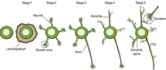
Fig. I.1. Developmental stages of mouse hippocampal cultured neurons. Initially, neurons are round cells that extend lamellipodia (stage 1) and develop minor processes called neurites (stage 2). One of these neurites starts to grow more rapidly and becomes the axon (stage 3), while the remaining neurites will develop into dendrites (stage 4). Finally, dendritic spines develop and mature at dendritic shafts leading to the establishment of synapses with the axonal presynaptic partner (stage 5), originating a mature neuron.
Upon seeding, hippocampal neurons start as simple and symmetric spheres that spread lamellipodia all around the cell body. The lamellipodia stably attach to the substrate and this is considered the stage 1 of neuronal development (Tahirovic and Bradke, 2009). Next, these round cells start to acquire several minor processes with similar lengths, named neurites, which are formed by an active outgrowth from the cell body (Verderio et al., 1999; Tahirovic and Bradke, 2009). Neurites
exhibit a dynamic behavior and frequently elongate in a saltatory manner, with periods of sudden advances and retractions or pausing, also displaying turning and branching (Dehmelt and Halpain, 2004a; Tahirovic and Bradke, 2009). These neurites are decorated with dynamic growth cones at their tips (stage 2), which are bulbous and highly motile structures responsible for sensing the surrounding environment. At this early developmental state, all neurites are assumed to be similar and the regulation of polarity in vivo is induced by extracellular signals (e.g. nerve growth factor), which trigger
intracellular signaling events (Tahirovic and Bradke, 2009). In the absence of external cues, as in the culture condition, all neurites compete to become the axon (Craig and Banker, 1994). As the polarization of a neuron is deeply associated with intense cytoskeleton rearrangements, alterations in its dynamics occur prior to morphological changes and polarization, which become retained in the future axon. There is also a cytoplasmic flow of cargos containing limiting factors for axonal growth (as cytoskeleton regulators) into the future axon preceding the beginning of neuronal polarization (Witte and Bradke, 2008). These events are required for the formation of the axon, the initial step in the establishment of neuronal polarization and, subsequently, in the loss of the cellular symmetry. The symmetry breakage has to be precisely regulated, since only one of the neurites is selected to become the axon, while the remaining processes develop into dendrites. Once the selected neurite starts to grow more rapidly and to develop into an axon, its growth is reinforced and internal inhibitory cues prevent the growth of the other neurites, being the stage 3 of development achieved (Andersen and Bi, 2000; Tahirovic and Bradke, 2009). Indeed, several intra- and extracellular signals have been
Stage 1 Stage 2 Stage 3 Stage 4 Stage 5
Axon Growth cone
Neurite Dendrite
Dendritic spine Lamellipodium
I. INTRODUCTION
3 shown to be involved in the selection of the future axon, as small GTPases, and netrins and brain-derived neurotrophic factor (BDNF), respectively (Polleux and Snider, 2010). After the establishment of neuronal polarity, the axon can extend considerably and start branching. Next, the remaining shorter neurites start to grow and acquire the morphology of typical dendrites, characteristic of the stage 4 of neuronal development (Witte and Bradke, 2008). As neuronal development proceeds, neurons become connected due to synapses formation (stage 5) with the purpose of establishing a proper neuronal circuitry. In this final stage, presynaptic axonal tips connect with postsynaptic partners at the dendritic trees in order to transmit the electrochemical signal from one neuron to the other. After neuronal polarization and maturation, axonal and somatodendritic compartments display distinct patterns of protein segregation, due to the proteins sorting into different vesicles at the trans-Golgi (Song et al., 2009). Nevertheless, a physical barrier exists at the initial segment of the axon and it is
important to maintain the differential molecular segregation of both membrane and cytoplasmic proteins (Song et al., 2009).
Thereby, during neuronal development, several signals and pathways converge aiming the formation of a functional nervous system.
1.1. Neuronal Growth Cones
I. INTRODUCTION
4
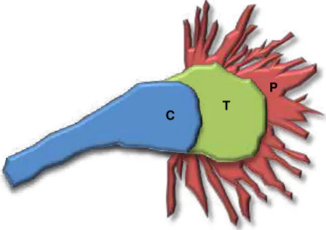
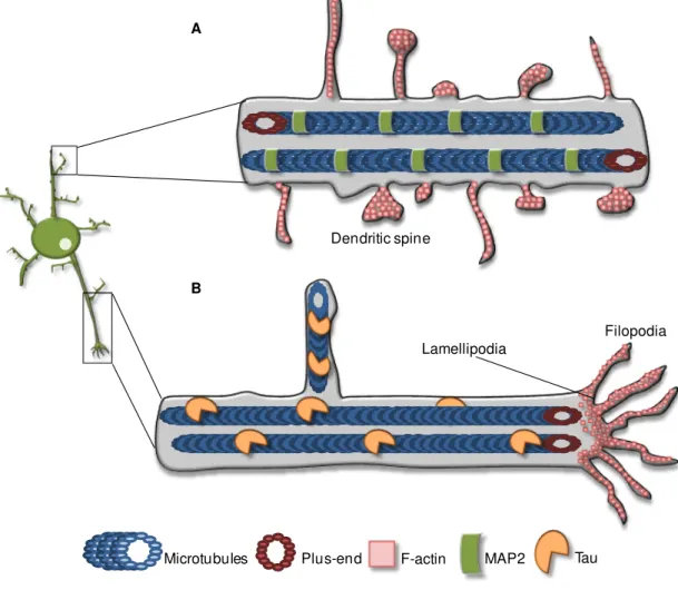
Fig. I.2. The growth cone cytoskeleton in primary embryonic hippocampal neurons.
The cytoskeletal filaments are organized into three different regions. The microtubules emerge from the axon shaft and splay out into the central domain (C; blue). Here, there is a relatively sparse network of actin filaments of unknown organization. The transition region (T; green) contains a dense meshwork of cross linked actin filaments, while the peripheral domain (P; red) is dominated by filopodia with bundle actin filaments oriented with their polymerizing ends at the distal tips.
Filopodia are probably the first to detect the guidance cues, since their dynamic expansion and retraction places them in the front line to sense the surrounding environment (Gallo and Letourneau, 2004; Gordon-Weeks, 2004). Moreover, filopodia, as well as the surface of the growth cone, express several receptors that bind to extracellular molecular signals, ultimately eliciting the guided axonal outgrowth (Myers et al., 2011). Therefore, filopodial length is important in determining the
environmental area that a growth cone can directly probe. As a transient structure, the growth cone is only present during the time elapsed between the beginning of neuronal polarization and synaptogenesis, as well as during axon sprouting and regeneration following axonal injury (Avwenagha et al., 2003). Interestingly, during neuronal polarization cytoskeleton dynamics at the
growth cone plays an important role. Indeed,the actin cytoskeleton becomes more dynamic and less stable in the growth cone of one neurite and the microtubules at the neuritic shaft become stabilized and aligned to form a bundle, initiating the future axon (Dehmelt and Halpain, 2004a; Witte and Bradke, 2008). By contrast, the growth cones of the remaining neurites are static and exhibit a rigid actin cytoskeleton, which blocks the microtubules protrusion (Tahirovic and Bradke, 2009).
In order to achieve a directional growth, extracellular molecules, as the ones present in the extracellular matrix (ECM), are thought to induce a selective stabilization of actin cytoskeleton in both lamellipodia and filopodia, since actin destabilization alters growth cone turning (Challacombe et al.,
1996). In fact, ECM proteins can provide a cellular substratum to the axon outgrowth and they can also bind to soluble molecules at the growth cones, influencing intracellular signaling pathways and, consequently, the axonal outgrowth (Myers et al., 2011). Also, in the developing axon, specific micro
RNAs (miRNAs), non-coding oligoribonucleotides that regulate the gene expression at the post-transcriptional level, are translated in response to extracellular guidance cues at growth cones, influencing the axonal pathfinding (Hengst and Jaffrey, 2007). Furthermore, microtubule dynamic instability is also required for growth cone turning in response to guidance cues. Indeed, attenuation of microtubule dynamics blocks in vitro growth cone turning due to microtubule restriction to C-domain,
which decreases or abolishes the interaction with actin network (Williamson et al., 1996; Challacombe et al., 1997). Additionally, the release of the microtubule-stabilizing drug taxol in one side of the growth
cone induces attraction and turning toward the site of the cue, while microtubule-destabilizing drug nocodazole induces repulsion, steering the growth cone away from that side (Buck and Zheng, 2002).
C T
I. INTRODUCTION
5 Thus, there is dynamic microtubule reorganization in the growth cone in the direction of a guidance cue and microtubule stabilization in the direction of the turn (Gordon-Weeks, 2004).
1.2.
Neuritic Arborization
Both axonal and dendritic development and stabilization is due to several complex processes extremely organized at the molecular level. Indeed, there is an intricate network of molecules involved in various processes as signal transduction, synthesis of macromolecules, cytoskeleton rearrangements and protein intracellular trafficking, all of them regulated by both intrinsic programs and extracellular cues (Urbanska et al., 2008). Furthermore, the neuronal development and maturation
are also controlled by miRNAs, since they are able to regulate the expression of proteins involved in the extension of neuritic processes and dendritogenesis (Loohuis et al., 2011). The axonal
arborization provides neurons with the ability to make synaptic contacts with a multitude of targets, being a decisive factor for the interconnection, a characteristic of the nervous system (Hall and Lalli, 2010). For an interstitial axonal branch to emerge, the primary axonal growth cone should pause, becoming larger. After the pausing period, a new primary growth cone then emerges to direct the growing axon, whereas the pausing one remains behind (Szebenyi et al., 1998; Kalil et al., 2000). The
formation of axonal branches is characterized by the local splaying of the bundled microtubules and the breakdown of longer microtubules (Kalil et al., 2000). These shorter microtubules start to explore
new directions and to invade the branches formed from the paused growth cone, allowing the ramification development from the axonal shaft (Kalil et al., 2000). Thus, cytoskeleton, specially the
microtubule compartment, assumes an important role in the development of axonal complexity.
On the other hand, dendrites receive electrochemical signals transmitted by axons from other neurons, being the preferential localization of postsynaptical sites. Hence, dendrites and their branching complexity are intimately related with synaptic integration (Häusser et al., 2000). The
development of the dendritic tree is associated with high rates of branch addition and retraction, but the mature dendritic arborization is less plastic and has lower rates of branching (Wu et al., 1999).
Nevertheless, the dendritic trees preserve some degree of plasticity in the mature nervous system (Urbanska et al., 2008). In this context, the contribution of extracellular signals, as BDNF and
semaphorins, may be critical to direct dendritic arborization development, stability and plasticity (Urbanska et al., 2008). Moreover, neuronal activity may count to the growth of dendritic trees. Indeed,
modification of the α-amino-3-hydroxy-5-methyl-4-isoxazolepropionic acid (AMPA) receptor activity decreases the complexity of the dendritic arborization and the lifetime of the dendritic branches (Haas
et al., 2006). Curiously, anomalies in dendritic arborization structure are related with several mental
retardation syndromes and even neurodegenerative conditions such as Alzheimer’s disease
I. INTRODUCTION
6
1.3. Synapse
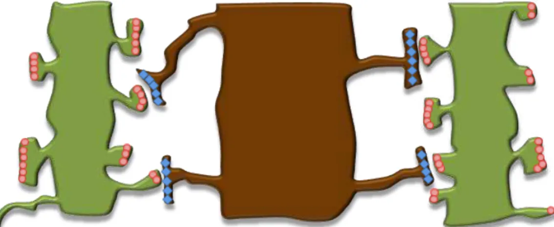
Synapses are highly specialized and asymmetrical junctions responsible for the directional transfer of electrochemical signals from a presynaptic neuron to a postsynaptic cell. In vertebrates, synapse formation, or synaptogenesis, essentially occurs from embryonic development to early postnatal life, characterized by its complex nature, involving a myriad of hierarchical signals (Waites et al., 2005). However, the formation of new synapses also occurs throughout adulthood and is thought
to contribute to learning and memory, despite the progressive decline of synapses with age (Waites et al., 2005; Harms and Dunaevsky, 2007).
Synapse formation engages coordinated changes due to ligand-receptor interactions, intracellular signaling cascades and subsequent cytoskeletal rearrangements, and intrinsic processes that contribute to the pre-establishment of synaptic machinery prior to synaptic contacts (Shen and Cowan, 2010). A multitude of signaling molecules, including secreted factors and cell-adhesion molecules, are involved in synaptogenesis specificity. Prior to synapse formation, secreted molecules involved in growth cone guidance act diffusely from local sources and guide axons to their targets, which are normally dynamic dendritic filopodia that extend from the dendritic shaft (Shen and Cowan, 2010). Upon approach to the target site, a presynaptic growth cone slows its advance, makes a physical contact and transforms itself into a rudimentary synaptic ending (Munno and Syed, 2003). Then, priming factors derived from surrounding glia and neurons promote the maturation of both target neurons and innervating axons, as well as the competence of dendrites and axons to undergo synaptogenesis (Waites et al., 2005). The premature contacts between axons and dendrites are
allowed by adhesive factors, being then stabilized by the cooperative action of adhesive and inductive factors, which are involved in the specialization of both pre- and postsynaptical compartments (Waites
et al., 2005). So, several mechanisms permit the establishment of proper connections, upon growth
cone guidance, allowing axons and dendrites to find their appropriate synaptic targets (Fig. I.3.).
I. INTRODUCTION
7 Synaptic development is characterized by a prolonged maturation phase with the purpose of properly integrate individual neurons into a functional network. Perhaps the most dramatic change in the postsynaptic side is the appearance and maturation of dendritic spines. Dendritic spines are small protrusions emerging from dendrites and the postsynaptic regions of most excitatory neuronal synapses, exerting an important role in several brain functions, such as memory and learning at the hippocampus (Mattila and Lappalainen, 2008). The development of these spines usually involves filopodia-like precursors that dynamically grow, with the intention of reaching the presynaptic partner, being very abundant during early development, but decreasing as neurons maturate (Fiala et al.,
1998). The filopodial precursor stabilizes and matures into a dendritic spine if the signal is proper, whereas it shrinks back to the dendritic backbone in the absence of an appropriate signal (Sekino et al., 2007). Generally, dendritic spines consist of a head linked to the dendrite by a stalk or neck and
I. INTRODUCTION
8
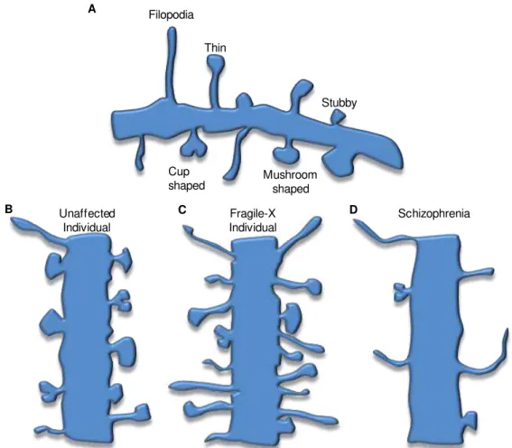
Fig. I.4. Schematic representation of different dendritic spine morphologies. The immature morphologies are characterized by longer necks and smaller heads (e.g. filopodia and thin shapes), whereas the mature forms have lower height and larger heads (e.g. cup and mushroom shapes) (A). Dendritic shafts usually present several types of dendritic spines which may suffer morphological changes during plasticity (B). Spine density and morphology is altered in cases of mental retardation, as fragile-X syndrome (C). In such case, increased immature spines and high density appear along the dendritic shaft of neurons from both temporal and visual cortices. On the other hand, schizophrenic patients exhibit lower spine density along the dendritic shaft, but the dendritic spines are also more immature (D). The alterations observed in both conditions may trigger the cognitive deficits verified in patients. (Based in Irwin et al., 2001; von Bohlen und Halbach, 2010 and Faludi and Mirnics, 2011).
Interestingly, a high degree of neuronal plasticity does not end during the early neuronal development and persist even after neuronal polarization and synaptogenesis. As Gomis-Rüth and colleagues (2008) have demonstrated, neurons fully integrated in a neuronal network can convert a mature dendrite to a functional axon upon a proximal axon cut, whereas a more distal injury leads to the re-growth of the axon (Gomis-Rüth et al., 2008). Additionally, synaptic plasticity, characterized by
reconfigurations in the structure and functionality of synapses, is important in normal brain function and induces changes in synapse number, shape and strength and, ultimately, in neuronal connectivity. In fact, the inability to undergo these plastic changes can be a cause for several neurodegenerative and psychological disorders (Munno and Syed, 2003; von Bohlen und Halbach, 2010; Faludi and Mirnics, 2011). However, neuronal plasticity may not only depend on neurons. Indeed, glial cells have been indicated as active controllers of dendritic outgrowth and dendritic spine morphologies, therefore contributing for the non-stationary state of the central nervous system (CNS) (Procko and Shaham,
Filopodia
Cup
shaped Mushroomshaped Stubby Thin
Unaffected Individual
Fragile-X Individual
A
I. INTRODUCTION
9 2010). Moreover, the development of synapses is facilitated by the presence of glial cells, as they help the guidance and the growth of both axons and dendrites in the CNS (Pfrieger, 2009).
2. Cytoskeleton Dynamics in Neuronal Development
Cytoskeleton has a vital role in the normal cell function and takes part in several activities from cell shape and locomotion to intracellular organelle transport. Within eukaryotic cells, the cytoskeleton is composed by three organized types of polymeric protein filaments: microtubules, intermediate filaments and microfilaments. These long protein polymers, that exhibit distinct properties, interact with each other and are dynamic structures. The regulation of cytoskeleton filaments is achieved by several proteins, such as microtubule-associated proteins (MAPs), actin-binding proteins and motor proteins. In neurons, all cytoskeletal components are crucial to provide structural organization, to establish and maintain neuronal polarity, to serve as tracks for intracellular transport and to allow different cellular morphologies (Tahirovic and Bradke, 2009; Polleux and Snider, 2010; Dent et al.,
2011a). In fact, the formation of a polarized neuron is essential for the integration and proper propagation of synaptic information in the adult CNS, which is dependent on cytoskeleton rearrangements (Witte and Bradke, 2008; Barnes et al., 2008; Tahirovic and Bradke, 2009). Moreover,
cytoskeleton has also a substantial role in neurite elongation, growth cone turning, advance and branching, and all of these processes are involved in neuronal development (Dehmelt and Halpain, 2004a; Gallo and Letourneau, 2004). So, an intricate interplay between the different compartments of cytoskeleton and their respective proteins exists during neuronal development.
From a classic point of view, the microtubule cytoskeleton has been thought to be important for cell division and organelle trafficking, while the dynamic actin cytoskeleton was known to generate force during cell contraction and dispersion. In more recent times, this classic vision has been contested and a cooperative relation between these two cytoskeleton complexes has emerged, as actin filaments have been implied in both cell division and trafficking, as well as microtubules have been addressed with roles in cellular morphology generation and plasticity (Dehmelt and Halpain, 2004a; Dent et al., 2011b). Furthermore, the precise control of neuronal morphogenetic program
I. INTRODUCTION
10
2.1.
Microtubules
Microtubules are formed by polymers of α- and β-tubulin heterodimers associated head-to-tail to form a hollow and elongated cylinder normally composed by 13 protofilaments (Lodish et al., 2004).
The microtubules are polar structures given the intrinsic polarity of the tubulin heterodimers and their linearly disposition. These polymers undergo post-translational modifications, as tyrosination, present in recently synthesized and dynamic microtubules, and acetylation, which occurs in more elderly and stable microtubules (Fukushima et al., 2009). These structures have the ability to grow and shrink
through end-polymerization of heterodimer subunits using the energy derived from the hydrolysis of
GTP bound to β-tubulin (Gordon-Weeks, 2004). Therefore, the rate of growth or shrinkage depends on the kinetics of subunit addition or subtraction, which is different in the two ends. The microtubule’s
fast-growing end, with the β-subunit exposed, is named the ―plus end‖ and it is the preferential local to tubulin assembly, while the slow-growing end, with the α-tubulin uncovered, is denominated the
―minus end‖ and is usually attached to a microtubule organizing center or capped (Gordon-Weeks, 2004). Microtubule cytoskeleton in vitro cultured cells suffers a dynamic instability characterized by
cycles of relative slow and continuous growth interrupted by catastrophes, phases of rapid shortening due to heterodimer dissociation probably caused by the loss of the GTP-β-tubulin cap at the end of the microtubule (Kirschner and Mitchinson, 1986; Cassimeris et al., 1988). However, this rapid length loss
does not lead to microtubule complete depolymerization because it occurs in parallel to a phase of sustained growth (Gordon-Weeks, 2004, Geraldo and Gordon-Weeks, 2009).
Microtubules exhibit a typical compartment-specific distribution in the axons of developing neurons with their plus-ends oriented toward the distal end (Fig. I.5.). In contrast, dendrites display a mixed microtubules orientation, i.e. some have their plus-ends toward the cell soma, while others have
their plus-ends pointing toward the peripheral zone. The uniform axonal distribution is thought to be the default state in neurites, as it exists when minor neurites start to emerge, where they are usually found in bundles (Brandt, 1998; Gordon-Weeks, 2004). As one of the principal cytoskeletal components, microtubules are involved in the maintenance of neuronal morphology and in the establishment of both dendritic and axonal arborizations. In neurites, a gradient of microtubule instability is observed, with distal microtubules undergoing a higher turnover than the microtubules in the middle of the neurite or near the cell body (Bamburg et al., 1986). However, studies with taxol and
nocodazole have shown that the spontaneous neurite initiation depends on both the presence and dynamic properties of microtubules (Letourneau and Ressler, 1984; Witte et al., 2008). Therefore,
changes in microtubule dynamics are sufficient to induce modifications in normal neuronal development. Indeed, stabilization of microtubules is required to the formation of the axon and, consequently, to neuronal polarization rising. Witte and colleagues (2008) have shown that low doses of taxol lead to the formation of multiple axons, while local microtubules stabilization, by UV-mediated photoactivation of caged taxol at the tip of one random minor neurite, promotes axon formation (Witte
et al., 2008). This stabilization favors microtubules to distally advance with their dynamic plus-ends,
I. INTRODUCTION
11 At both dendritic and axonal growth cones, microtubules are disposed in a similar way with their plus ends oriented distally towards the P-domain (Heidemann et al., 1981; Baas et al., 1988). Here, as
microtubules enter the C-domain, they defasciculate and some can even cross this domain as single microtubules (Gordon-Weeks, 2004). Furthermore, dynamically unstable microtubules invade the P-domain and explore the actin network, where they become stabilized, leading to axon specification (Geraldo and Gordon-Weeks, 2009; Dent et al., 2011a). In response to guidance cues, microtubule
dynamic instability is required for growth cone turning and consequently to the directional axonal outgrowth (Tanaka et al., 1995; Challacombe et al., 1997) Moreover, in the absence of dynamic
microtubules, the remaining microtubules do not suffer the typical cycle of bundling and splaying, what is deeply associated with the forward movement of the growth cone (Tanaka et al., 1995). In addition,
the attenuation of microtubule dynamics in growth cones has also shown to impair lamellipodial protrusion (Gallo, 1998).
Microtubules are also important to dendritic spine morphology and function, even though the central role of the actin filaments in these excitatory postsynaptical sites (Dent et al., 2011b). Thus,
modifications in microtubule dynamics are related to changes in the normal dendritic spine formation and further maturation (Gu et al., 2008; Jaworski et al., 2009). Moreover, the polymerization and
depolymerization of microtubules is coincident with the extension and retraction of transient spine heads protrusions, respectively (Hu et al., 2008). This study has also shown that an increase of
neuronal activity is related to a higher number of spines with a longer-invasion by microtubules (Hu et al., 2008). Additionally to structural support, microtubules are important to cellular trafficking, because
they provide tracks for motor proteins, as kinesin and dynein, allowing the cargo transportation to specific cellular parts, as dendritic spines and synaptic terminals. This transportation to and from pre- and post-synaptical sites is critical for synaptic function, as synapses are highly vulnerable to transport impairments (Gendron and Petrucelli, 2009).
2.2. Microtubule-associated proteins
Microtubule associated proteins (MAPs) are a family of proteins known by their role in regulation of microtubules polymerization, stability and organization (Gendron and Petrucelli, 2009). However, besides the regulation of microtubule networks in the axons and dendrites, there are evidences for a larger range of functions for MAPs, including binding to F-actin, recruitment of signaling proteins and regulation of microtubule-mediated transport (Dehmelt and Halpain, 2004b). Microtubule associated protein 2 (MAP2) and Tau are structural MAPs, i.e. have the ability to alter microtubule structures,
acting to reduce catastrophe periods and to increase rescue phases upon binding along the outer ridges of the protofilaments (Al-Bassam et al., 2002; Dehmelt and Halpain, 2004a). Hence, they do not
prevent microtubule dynamic instability. Instead, they simply alter the dynamic behavior of this cytoskeleton compartment, creating a partial stable but dynamic state important for cell growth and transport.
I. INTRODUCTION
12
disposed in a proximo-distal gradient in the process that will develop into the axon, with the higher concentration at the transition from the axonal shaft to the growth cone (Kempf et al., 1996). Thus,
Tau is gradually segregated into the future axon and is able to adjust microtubules assembly and stabilization in this process (Fig. I.5.) (Dehmelt and Halpain, 2004a). The distinctive distribution of Tau in neurons is regulated by its degree of phosphorylation, as this post-translational modification of certain residues detaches Tau from microtubules, decreasing its affinity for them. Therefore, phosphatases (e.g. tyrosine phosphatases) are implicated in the complex regulation of the intracellular function and localization of this MAP (Mandell and Baker, 1996). In contrast, MAP2 is essential for initiation of neurites and is segregated into the emerging dendrites, exhibiting a somatodendritic distribution. In fact, the suppression of MAP2 synthesis inhibits the initial formation of neurites in cultured neurons (Cáceres et al., 1992). The differential distribution of these two MAPs is maintained
in the mature neuron, i.e. MAP2 is exclusively present in the somatodendritic region, whereas Tau
only exists inthe axon (Dehmelt and Halpain, 2004a).
MAPs were also found to interact with both actin filaments and microtubules. While actin filaments and tubulin polymers do not interact without MAPs, their presence is sufficient to induce interactions between the two cytoskeletal compartments (Griffith and Pollard, 1978). Thus, MAPs might mediate the crosstalk between microtubules and actin filaments. However, it is suggested that both microtubules and microfilaments bind to the same domain in MAP2 and Tau proteins, because microtubules exclude the binding of microfilaments and vice-versa (Correas et al., 1990).
2.3. Microfilaments
Microfilaments consist in two helical and separated strands of filamentous actin (F-actin), each one a polymer of globular-actin (G-actin). As microtubules, microfilaments are intrinsically polar because all actin subunits are directed toward the same end of the filament. Conventionally, the terminal actin subunit exposing adenosine diphosphate (ADP) -G-actin is designated the minus-end or the pointed-end, while the opposite end, the plus-end or the barbed-end, is the fast-growing end of the filament and the local for the binding of adenosine triphosphate (ATP)-G-actin (Lodish et al., 2004).
Hence, the barbed-end is the preferential local for actin subunits addition, whereas the pointed-end is the local for actin subunit dissociation. Microfilaments are highly dynamic structures that can rapidly suffer cycles of assembly and disassembly and the actin polymerization is favored by stimulation of ADP/ATP exchange of G-actin (Witte and Bradke, 2008). Actually, actin filaments undergo a process called treadmiling, by which they are able to exert net movement, consisting in the assembly of filaments at their fast-growing ends and disassembly at their slow-growing ends (Brandt, 1998).
Actin cytoskeleton is deeply involved in neurite development, breakage of the initial symmetry in neurons, i.e. neuronal polarization, and alterations in actin dynamics are also associated with changes
in neuronal morphology. Initiation of neurites is induced by a reduction in the tensile forces mediated by actin cytoskeleton (Georges et al., 2008). Indeed, the use of cytochalasin B, which reduces tensile
forces by depolymerization of actin filaments, increases the length of both axons and dendrites (Lafont
I. INTRODUCTION
13 causes axonal retraction, showing that the turnover of microfilaments is required for axonal extension (Gallo et al., 2002). One feature of the future axonal growth cone in immature neurons is a lower actin
cytoskeleton stability, that can be the cause of microtubule protrusion and, consequently, of neurite outgrowth (Bradke and Dotti, 1999). Furthermore, the disruption of the actin network of one individual growth cone is sufficient to induce its neurite to develop as an axon, and the application of actin depolymerizing drugs is enough to produce neurons with multiple axons (Bradke and Dotti, 1999).
Fig. I.5. Schematic representation of dendritic and axonal cytoskeletons. (A) Dendrites display mixed microtubules orientations, i.e. some microtubules have their plus-end directed to the cell soma, whereas others exhibit their plus-end toward the distal end of the dendrite. The dendritic microtubules are stabilized by MAP2. The dendritic spines are, essentially, composed by actin filaments, which contribute to their dynamics. (B) In the axon, microtubules are stabilized by Tau and disposed in a regular and uniform way, with their plus-ends pointing to the distal end. Usually, microtubules are retained in the C-domain of the growth cone and actin filaments occupy the P-domain, with both filopodia and lamellipodia.
In order to achieve a directional growth, extracellular cues are thought to induce a selectively stabilization of actin cytoskeleton in both lamellipodia and filopodia, the preferential locals for actin polymerization (Fig. I.5.) (Gallo and Letourneau, 2004). Actually, the extension of filopodial tips is determined by the rate of F-actin polymerization and the retrograde transport of the polymerized filaments on the way to the bottom of the filopodium (Mallavarapu and Mitchinson, 1999). During axon guidance, actin polymerization is favored in areas contacting with positive or attractive guidance cues,
Dendritic spine
Tau MAP2
F-actin
Microtubules Plus-end
Lamellipodia Filopodia
A
I. INTRODUCTION
14
inducing growth cone turning (Shen and Cowan, 2010). Moreover, actin structures redirect and stabilize microtubules in the direction of turning, since actin destabilization by low concentrations of cytochalasin B alters growth cone turning and microtubules fail to retract from the side in contact with an inhibitory signal (Challacombe et al., 1997). Furthermore, the loss of actin filaments at the leading
edge of the growth cones is related to growth cone collapse (Fan et al., 1993; Fournier et al., 2000;
Avwenagha et al., 2003). Hence, F-actin can be a major intracellular target of extracellular guidance
cues that modulate growth cone behavior.
The heads of dendritic spines, mainly the postsynaptic density zone, are enriched in actin cytoskeleton, which is important for both formation and motility of dendritic spines (Fig. I.5.) (Dunaevsky et al., 1999; Capani et al., 2001). Moreover, the high level of plasticity observed in spines
may be due to the rapid turnover rates of actin cytoskeleton. Concerning synaptogenesis, the use of the actin-depolymerizing agent latrunculin A leads to an almost complete loss of synapses in the first week of hippocampal neuronal cultures, as well as to a decrease in the synaptophysin clusters number and size, revealing an important role of F-actin in the development and maintenance of young synapses (Zhang and Benson, 2001).
Despite the morphological and plastic roles of actin cytoskeleton, these filaments are also involved in the transportation of certain cargos, because myosins, a family of motor proteins, directly bind to actin filaments and transport a cargo in their tails (Lodish et al., 2004). Actin cytoskeleton can
even serve as a substrate for the microtubule anterograde movement (from the cell soma to the presynaptic terminal), when there are no longer microtubules available to play this role (Hasaka et al.,
2004). Indeed, the transport of microtubules at the growth cone may be dependent on actin based-mechanisms (Myers et al., 2006).
3. Neuroinflammation: from a beneficial to a detrimental effect
For a long time, the CNS was considered an immune privileged site, by the absence of a typical and classical immune response (Di Filippo et al., 2008). Likewise, it was also thought that the brain
was not largely affected by systemic or immune inflammatory reactions (Lucas et al., 2006).
Nowadays, it is widely accepted that an interaction between immune and nervous systems exists, involving a bidirectional cross talk. Indeed, the CNS is provided with an active immune surveillance and it can be the host of inflammatory responses to different types of injuries (Di Filippo et al., 2008).
Neuroinflammation, the inflammation of the CNS, consists in a set of events, including the activation of the resident neural cells, as microglia and astrocytes, which together with neurons release a panoply of inflammatory molecules (Fig. I.6.) (Chavarria and Alcocer-Varela, 2004; Infante-Duarte et al., 2008;
I. INTRODUCTION
15 Fig. I.6. Neuroinflammation is a complex response of neural cells to a detrimental stimulus.
Upon stress, injury or sepsis, the resident neural cells, as microglia (blue), astrocytes (orange) and neurons (green) produce inflammatory mediators (purple). These molecules influence the normal state of the CNS and are capable of recruiting circulating immune cells, leading to neuroinflammation.
The segregated inflammatory mediators are responsible for the expression of chemokines and adhesion molecules, the activation of endogenous glial cells and the stimulation of astrogliosis, in addition to the recruitment of immune cells to the site of inflammation (Rothwell and Luheshi, 2000; Whitney et al., 2009). Indeed, the CNS can be invaded by circulating immune cells, which contribute to
the brain inflammatory state. Hence, neuroinflammation is seen as a complex cellular and molecular response of neural cells to a detrimental stimulus, such as injury, stress or sepsis (Semmler et al.,
2008; Whitney et al., 2009). The main goals of neuroinflammation are the defense against these
insults, the clearance of dead and damaged neurons, as well as the return of the CNS to homeostasis (Whitney et al., 2009). However, when neuroinflammation is not controlled, its beneficial role is
overcome and may then turn into damage. In that case, the recruited cells become activated and release even more inflammatory mediators, establishing a positive feedback and leading to neuronal injury with changes in neurogenesis (Das and Basu, 2008; Whitney et al., 2009). Therefore,
neuroinflammation, if in excess, may contribute to neurodevelopmental impairments. In addition, oligodendrocytes are extremely vulnerable to inflammatory molecules which may result in white matter damage and the emergence of long-term neuromotor, cognitive, and behavioral limitations, or to degenerative disorders, such as Alzheimer’s and Parkinson’s diseases (Lucas et al., 2006; Aktas et al., 2007).
Most attractive is the recent hypothesis that an early infection could produce a latent or hidden change in the immune system that could be unmasked by a second immune challenge. This ―two-hit hypothesis‖ was first proposed for schizophrenia etiology, suggesting that the first hit, which may
occur during the embryonic life, ―primes‖ the nervous system for the second one, accelerating the
disease symptoms (Maynard et al., 2001). Therefore, the concept of ―glial priming‖ has been proposed
to account for the ―two-hit hypothesis‖ (Bilbo and Schwarz, 2009). The authors describe that, after a
first insult in the early-life, a subset of glial cells become primed and display an activated morphology.
Sepsis Injury Stress
I. INTRODUCTION
16
Hence, when facing a second immune challenge in later-life, the primed glia, which do not chronically produce inflammatory mediators, over-release cytokines within the brain exaggerating the immune response and producing damaging consequences (Bilbo and Schwarz, 2009).
3.1. Cytokines as neuroinflammation mediators
Cytokines are secreted polypeptides with several addressed roles in immune responses. Previously, cytokines were thought to only act in the peripheral system, but nowadays a crescent number of data have found that these molecules can exert several actions in the brain, including in the development of neurons. Pro-inflammatory cytokines, as tumor necrosis factor-alpha (TNF-α) and
interleukin-1beta (IL-1β), are synthesized by neural cells and take part in the normal intercellular
communication, assuming an important role for the maintenance of homeostasis. The pro-inflammatory cytokines can act as neurotrophic factors during the normal development of the brain,
e.g. IL-1 is regarded as an important growth factor in brain formation (Giulian et al., 1988; Zhao and
Schwartz, 1998). Additionally, these cytokines can induce the production of anti-inflammatory mediators, therefore creating a feedback loop, in order to control the immunologic state (Morganti-Kossman et al., 2002). Normally, the levels of such cytokines are low, but their expression is rapid and
radically increased in situations of neuroinflammation. When this elevation of expression is sustained, pro-inflammatory cytokines can become pathological, deregulating cytokine release, and can even cause neuron and oligodendrocyte death (Donnelly and Popovich, 2008). In reality, when neuroinflammation happens in the perinatal period during the development of the nervous system, the intrinsic developmental program of the brain can be altered by the exposure of the fetus to inflammatory mediators, what may eventually result in lasting neuronal disorders (Degos et al., 2010).
Yet, the detrimental effect of cytokines may rely on the type and level of cytokines produced. In other words, the low physiological rates of cytokine expression may be important for the cross-talk between the neural cells during development, but the overexpression observed during the neuroinflammatory stage may compromise neuronal survival and plasticity.
IL-1 is a potent pro-inflammatory cytokine with the capability to orchestrate inflammatory responses, since it can induce the expression of several inflammatory molecules that intensify the immune response. Moreover, IL-1 has the ability to enhance responses in cells with low levels of its own receptor, for the reason that it activates a complex signaling cascade that amplifies the initial signal (Rothwell and Luheshi, 2000). This cytokine exists in two partially homologous isoforms, IL-1α
and IL-1β, both translated as a proform that it is further cleaved by cell-surface-bound calpain and caspase-1 or IL-1β converting enzyme, respectively, in order to produce the mature forms (Mrak and Griffin, 2001; Basu et al., 2004). The two isoforms bind with high affinity to a specific membrane
receptor, the IL-1 receptor type I, which contains a cytoplasmic domain engaging intracellular signaling pathways (Di Filippo et al., 2008; Spulber and Schultzberg, 2010). It is described that the binding of
IL-1β to its receptor may induce modifications at the cytoskeleton level, through the activation of p38 mitogen-activated protein kinases (Temporin et al., 2008). However, an endogenous receptor
I. INTRODUCTION
17 actions of the cytokine (Lucas et al., 2006). All these components are present within the brain, albeit at
low rates in the normal and healthy CNS (Basu et al., 2004). Following CNS damage, IL-1 is regarded
as an important initiator of the immune response and, consequently, is rapidly produced and released specially by activated microglia (Rothwell and Luheshi, 2000). There is accumulating evidence suggesting a detrimental role of high levels of IL-1β in neuronal function following an in vivo insult.
Indeed, it was reported that IL-1β may even exacerbate the initial damaging stimulus of conditions
such as ischemia and/or excitotoxicity (Loddick and Rothwell, 1996; Allan et al., 2000; Boutin et al.,
2001).
Other important pro-inflammatory cytokine, TNF-α, is predominantly produced by glial cells and
in a lower rate by neuronal cells, also having an important role in the initiation and regulation of the inflammatory process (Whitney et al., 2009; Ziebell and Morganti-Kossmann, 2010). This cytokine is
synthesized as a membrane-bound precursor that is subsequently cleaved by the TNF converting enzyme, in order to produce the active cytokine, which levels are quickly increased in response to CNS damage (Perry et al., 2001). TNF-α can bind to two different cell-surface receptors, p55 (tumor
necrosis factor receptor-1; TNFR1) and p75 (TNFR2) (Lucas et al., 2006; Whitney et al., 2009). The
cytotoxic actions leading to neuronal apoptosis exerted by TNF-α are mediated through p55, while the actions upon binding to p75 are generally proliferative and may involve anti-apoptotic signals (Muñoz-Fernández et al., 1998). Therefore, opposing and different effects of TNF-α may be due to the distinct
signaling pathways triggered by the different receptors. Nevertheless, as IL-1β, TNF-α was shown to
mediate neuronal damage after an insult, and exert a variety of effects, from neuronal loss to neurotoxicity and brain edema (Shohami et al., 1997; Ådén et al., 2010; Kendall et al., 2011). Besides,
TNF-α was also demonstrated to act synergistically with IL-1β, enhancing neuronal injury and
neurotoxicity (Chao et al., 1995).
3.2. Neuroinflammation during development
During pregnancy, the inflammatory state of the fetus may be influenced by the substances derived from the uterine environment that can cross the placenta. Indeed, the injection of lipopolysaccharide (LPS) to pregnant rats at day 18 of gestation, mimicking a pre-natal infection condition, triggers the production of cytokines in the fetal brain (Cai et al., 2000). These fetuses
present elevated mRNA levels for pro-inflammatory cytokines such as TNF-α and IL-1β in a dose-dependent manner (Cai et al., 2000). Consequently, intrauterine infection can have a significant
impact on the proper development of the brain. Indeed, several works have confirmed that maternal infection may influence the neurodevelopment of the fetus due to alterations in pro-inflammatory cytokine levels (Ling et al., 2002; Bell et al., 2004; Burd et al., 2010). Furthermore, neuroinflammation
in the fetal brain has been associated with neurological sequelae. High levels of pro-inflammatory
cytokines in the amniotic fluid or umbilical blood increase the risk for white matter damage and/or cerebral palsy (Yoon et al., 1996; Yoon et al., 2000). Moreover, antenatal inflammatory events have