Quim. Nova, Vol. 32, No. 9, 2347-2350, 2009
Artigo
*e-mail: [email protected]
DEVELOPMENT AND VALIDATION OF A GC-FID ASSAY FOR DETERMINATION OF FLUVASTATIN IN PHARMACEUTICAL PREPARATIONS
Serap Saglik Aslan*, Olcay Sagirli and Lale Ersoy
Department of Analytical Chemistry, Faculty of Pharmacy, Istanbul University, 34116 Beyazit-Istanbul, Turkey
Recebido em 1/12/08; aceito em 24/5/09; publicado na web em 30/10/09
A gas chromatographic method has been developed for the assay of fluvastatin sodium (FLU). FLU was silylated with N,O-bis(trimethylsilyl)triluoroacetamide-1% trimethylchlorosilane at 90 ºC for 30 min and analysed in a DB-1 column by capillary gas chromatograph with a lame ionization detector. The method was validated. The assay was linear over the concentration range at 10.0 to 50.0 mg mL-1. The limit of detection and the limit of quantitation were 1.0 and 3.0 mg mL-1, respectively. The recoveries of FLU derivatives were in the range of 99.25-99.80%. In inter-day and intra-day analysis, the values of relative standard deviation (%) and the relative mean error (%) were found between 0.20-0.80% and -0.20-0.75%, respectively. The developed method was succesfully applied to analyze the FLU content in tablet formulation. The results were statistically compared with those obtained by the oficial method, and no signiicant difference was found between the two methods. Therefore, it can be recommended for the quality control assay of FLU in pharmaceutical industry. Keywords: luvastatin; validation; pharmaceutical preparations.
INTRODUCTION
Fluvastatin (FLU), (3R,5S,6E)-rel-7-[3-(4-fluorophenyl)-1-(1-methylethyl)-1H-indol-2-yl]-3,5-dihydroxy-6-heptenoic acid (Figure 1), is an inhibitor of 3-hydroxy-3-methylgluataryl-coenzyme A (HMG-CoA) reductase and blocks the production of cholesterol in the body.1-4 FLU is used in the treatment of hypercholestrolemia,5,6 and treatment with statin drugs decreases the risk of cardiovascular diseases, in addition to mortality.7
FLU has been determined by high performance liquid chromato-graphic methods with UV detection,8 luorometric detection9-12 and mass spectrometric detection13,14 in human plasma. A few methods such as spectrophotometric,15 electrometric16,17 and electrophore-tic18,19 have been reported for the assay of FLU in pharmaceutical preparations. The antioxidant effects of FLU have also been studied in humans20-22 and animals.23,24
Gas chromatography/negative ion chemical ionization mass spectrometric method with pentaluorobenzylbromide has been used for determination of FLU in the human plasma.25 However, there is no gas chromatographic method with lame ionization detection (GC-FID) for determination of FLU in pharmaceutical preparations. Therefore, we have developed and validated a
ra-pid, simple, accurate, precise and sensitive GC-FID method with N,O-bis-(trimethylsilyl) triluoroacetamide (BSTFA)-1% trime-thylchlorosilane (TMCS). This is the irst time in the literature that this procedure was carried out. The present method provides certain advantages such as non-extraction, non-interference by excipients, being worked on easily in laboratories, as well as the economic advantage. Although GC instrument is not as common as HPLC in the pharmaceutical industry, the proposed method can be suggested as an alternative one for FLU determination in pharmaceutical preparations.
EXPERIMENTAL
Chemicals and reagents
FLU sodium and its tablets (Lescol XL80 mg) were kindly supplied from Novartis (Istanbul, Turkey). N,O-bis-(trimethylsilyl) triluoroacetamide (BSTFA) with 1% trimethylchlorosilane (TMCS) was used as silylating reagent and purchased from Sigma (Canada, USA). Solvents and other chemicals were of analytical grade (Merck, Darmstadt, Germany).
Standard solutions
Stock solution of FLU was prepared at 0.1 mg mL-1 concentra-tion in methanol and stored at 4 oC. Appropriate aliquots from stock solution were decanted to air-tight stopped glass vials and evaporated under nitrogen to obtain the residues for providing the calibration standards at 10.0, 20.0, 30.0, 40.0 and 50.0 mg mL-1 concentrations after derivatization.
Derivatization
The trimethyl silyl derivatives of standards and samples were prepared from residues obtained above, part by reacting with 100 mL of BSTFA-1% TMCS solution at 90 °C for 30 min in a block heater. The resulting solutions were cooled and injected into the GC without removing the excess of derivativing agent, and they were kept in the air-tight glass vials below -20 oC.26
Aslan et al.
2348 Quim. Nova
Stability
Long- and short term stability of trimethyl silyl derivative of FLUwere studied over a period of 24 h at room temperature and one week at 4 oC.
Instrument and chromatographic conditions
GC analysis were carried out using a capillary gas chromatograph (Shimadzu GC-14A, Shimadzu, Tokyo, Japan) coupled with a lame ionization detector on an DB-1 column (15 m x 0.25 mm I.D., 0.25 mm ilm thickness, Shimadzu, Kyoto, Japan). The injection and detector temperatures were set at 300 °C. The oven temperature was kept at 280 °C. The GC-FID injected volume was 2 mL and the solutions were injected in 1/20 split ratio. The carrier gas was nitrogen with a pressure of 1kg/cm2. Retention times and peak areas were calculated by Class CR10 programme.
Method validation
The calibration curves were constructed with six concentrations, ranging from 10.0 to 50.0 mg mL-1. The limit of detection (LOD) and limit of quantitation (LOQ) values were calculated at a signal-to-noise ratio (S/N) of 3 and 10, respectively.27 A placebo solution was prepared and injected for determining of the method speciicity. To determine the precision of the method, intra-day and inter-day analyses were carried out by analysing FLU samples at ive concentrations; 10.0, 20.0, 30.0, 40.0 and 50.0 mg mL-1. The values for the relative standard deviation (RSD) and the relative mean error (RME) were calculated for FLU solutions. Accuracy was assessed using 6 determinations of each 5 concentration levels (10.0, 20.0, 30.0, 40.0 and 50.0 mg mL-1). Accuracy was reported as percent recovery by the assay of known added of amount of analyte in the sample.27
The robustness of the method was investigated by small changes in these parameters; temperature of the injector, temperature of the detector, oven temperature and injected volume at 30.0 mg mL-1 concentration of FLU.
Assay procedure for tablets
Ten tablets were weighed and powdered. An accurately wei-ghed portion of the powdered tablets, equivalent to about 10 mg of FLU sodium was shaken mechanically with 50 mL methanol for 30 min and diluted to 100 mL with the same solvent, mixed and iltered. Appropriate aliquots from the iltrate were decanted to air-tight stopped glass vials and evaporated under nitrogen to obtain the residues for providing at 30.0 mg mL-1 concentration after derivatization. For the selectivity of assay, titanium dioxi-de, yellow iron oxide and red iron oxide were analysed as tablet excipients including no any organic one.
Reference method
Liquid chromatographic analyses were performed on a ther-mo separation products liquid chromatograph (TX, USA) which consisted of a P4000 solvent delivery system equipped with a rheodyne injection valve with a 20 μL loop, a UV3000 detector and an SN4000 automation software system. The analyses were carried out using a C18 column with a low rate of 2.0 mL min-1. 54% solution A [pH 7.2 buffer solution:methanol (3)-acetonitrile (2); 87.5 : 12.5] and 46% solution B [methanol (3)-acetonitrile (2):pH 7.2 buffer solution; 87.5 : 12.5] were used as mobile phase. UV-detection was at 305 nm.28
RESULTS AND DISCUSSION
Derivatization procedure
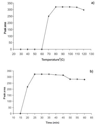
The optimum derivatization conditions were set up to obtain tri-methyl silyl derivative of FLU with BSTFA-1% TMCS solution for quantitative analyses. Different times and temperatures of heating were veriied, ranging from 15.0 to 60.0 min, and 30 to 120 oC, respectively. It was veriied based on the experimental results presented in Figure 2 that the best condition for derivatization step was heating at 90 °C for 30 min. After the experienced of adding the range of 50 to 150
mL of reagent solution, a better yield of trimethyl silyl derivative was obtained with 100 μL of that solution. The derivatization reaction did not take long, and no extraction with a toxic solvent was necessary in the procedure. The derivatives were stable for one week at 4 °C. Their stability was then investigated for 24 h at room temperature. There was no signiicant difference in the peak areas of FLU (Table 1).
Chromatographic conditions
In the literature, [18O
2]-FLU has been prepared from unlabelled FLU and used as an internal standard for gas chromatography/mass spectro-metry under negative ion chemical ionization conditions for determina-tion of FLU in human plasma.25 In that procedure, sample preparation took long time, whereas internal standard addition was not necessary in our study and sample preparation was simply, fast, and with good in
Figure 2. a) Effect of derivatization temperature on peak area of FLU (30
mg mL-1); b) effect of derivatization time on peak area of FLU (30 mg mL-1)
Table 1. Stability data for FLU derivatives at 30 mg mL-1 concentra-tion (n=6 per test)
Test Recovery (%) RSD* (%)
24 h, room temperature 99.73 0.60
One week, -4 oC 99.33 0.44
Development and validation of a GC-FID assay for determination of luvastatin 2349 Vol. 32, No. 9
reproducibility. The FLU typical chromatogram (standard solution, 30
mg mL-1) is shown in Figure 3 and its retention time was 6.0 min with good peak shape. The FLU amount was calculated using the peak areas.
Method validation
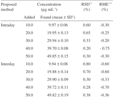
Calibration graphs were constructed by plotting the values of the peak area against concentrations. A linear correlation was obtained over the range of 10.0-50.0 μg mL-1 concentrations. The regression equation was found to be A=7.040C+106.8; r = 0.9996 (A: area, C: concentration; mg mL-1). Standard deviation on slope (Sb) was 0.087 and on intercept (Sa) was 2.691. LOD was 1.0 μg mL-1 at a signal-to-noise ratio (S/N) of 3, and LOQ was found to be 3.0 μg mL-1 at signal-to-noise ratio (S/N) of 10. The sensitivity of this study was similar when compared with those of in literature indings for the assay of FLU in pharmaceutical preparations.15-19 No peak was found at the retention time of FLUin placebo solution. The good recoveries of FLU were in the range of 99.25-99.80% in determining of the accuracy of the method (Table 2). In intra-day and inter-day analy-ses, the values of RSD (%) and RME (%) were found to be between 0.20-0.80% and -0.20-0.75% for FLU solutions, respectively (Table 3). These data indicate that the assay method is precise within the same day and on different days. The robustness of the method was investigated in view of temperatures of injection, detector and oven, in addition to the injected volume. There was no signiicant difference in the results obtained.
Assay procedure for tablets
The developed method was applied to the determination of FLU in tablets. The drug content in tablet was calculated from the regression equation of the calibration curve. The statistical values were com-pared with those obtained by the reference method,28 and there was no signiicant difference between them (Table 4). The selectivity of assay was determined by analysing of interferences by the excipients; titanium dioxide, yellow iron oxide and red iron oxide; no peaks were present in the chromatogram at the retention time of FLU.
CONCLUSION
A GC-FID method for BSTFA-1% TMCS derivatized FLU determination in pharmaceutical preparations was developed and validated. This method is precise, linear and sensitive. Preparation of the sample preparation is simple, does not take long, and is not solvent-consuming. The retention time of FLU is 6.0 min and there is no interference by excipients. The GC-FID instrument is readily available in every laboratories. Therefore, the proposed method is suitable for rutine control of the uniformity of the FLU content in pharmaceutical industry.
Figure 3. Gas chromatogram of FLU standard solution (30 mg mL-1)
Table 2. Results from recovery studies of FLU (n=6) Concentration (mg mL-1)
Recovery (%) RSD** (%) Added Found (mean ± SD*)
10.0 9.96 ± 0.06 99.60 0.60
20.0 19.85 ± 0.14 99.25 0.71
30.0 29.94 ± 0.09 99.80 0.30
40.0 39.74 ± 0.15 99.40 0.38
50.0 49.87 ± 0.17 99.74 0.34
*SD = standard deviation. **RSD = relative standard deviation
Table 3. Intra-day and inter-day variabilities of FLU (n=6) Proposed
method
Concentration (mg mL-1)
RSD** (%)
RME*** (%) Added Found (mean ± SD*)
Intraday 10.0 9.97 ± 0.06 0.60 -0.30
20.0 19.95 ± 0.13 0.65 -0.25
30.0 29.94 ± 0.10 0.33 -0.20
40.0 39.70 ± 0.08 0.20 - 0.75
50.0 49.85 ± 0.15 0.30 -0.30
Interday 10.0 9.94 ± 0.08 0.80 -0.60
20.0 19.88 ± 0.14 0.70 -0.60
30.0 29.90 ± 0.09 0.30 -0.33
40.0 39.72 ± 0.11 0.28 -0.70
50.0 49.82 ± 0.19 0.38 -0.36
* SD = standard deviation. ** RSD = relative standard deviation. ***RME = relative mean error
Table 4. Determination of FLU in tablets labelled containing 80 mg of FLU per tablet (n=6)
Statistical value Proposed method Reference method27 Means amount of drug found
(mg per tablet) 79.09 79.72
SD* 0.38 0.68
RSD**(%) 0.48 0.85
% Recovery 98.86 99.65
Aslan et al.
2350 Quim. Nova
REFERENCES
1. Kathawala, F. G.; Med. Res. Rev. 1991, 11, 121.
2. Mitani, H.; Kimura, M.; Cardiovasc. Drug Rev. 2000, 18, 284. 3. Farmer, J. A.; Torre-Amione, G.; Drug Saf. 2000, 23, 197. 4. Hamelin, B. A.; Turgeon, J.; Trends Pharmacol. Sci. 1998, 19, 26. 5. Broyles, F. E.; Walden, C. E.; Hunnighake, D. B.; Hill-Williams, D.;
Knopp, R. H.; Am.J. Cardiol. 1995, 76, 129A.
6. Tazuma, S.; Ohya, T.; Mizuna, T.; Takizawa, I.; Kunita, T.; Takata, K.; Hayashi, K.; Hino, F.; Am. J. Cardiol. 1995, 76, A110.
7. LaRosa, J. C.; He, J.; Vupputuri, S.; J. Am. Med. Assoc. 1999, 282, 2340. 8. Nakashima, A.; Saxer, C.; Niina, M.; Masuda, N.; Iwasaki, K.;
Furukawa, K.; J. Chromatogr., B 2001, 760, 17.
9. Kalafsky, G.; Smith, H. T.; Choc, M. G.; J. Chromatogr. Biomed. Appl. 1993, 614, 307.
10. Toreson, H.; Eriksson, B. M.; J. Chromatogr., A 1996, 729, 13. 11. Lanchote, V. L.; Rocha, A.; de Albuquerque, F. U. V.; Coelho, E. B.;
Bonato, P. S.; J. Chromatogr., B 2001, 765, 81.
12. Al-Rawithi, S.; Hussein, R. F.; Alzahrani, A.; Ther. Drug Monit. 2003, 25, 88.
13. Nirogi, R. V. S.; Kandikere, V. N.; Shrivaslava, W.; Mudigonda, K.; Datla, P. V.; Rapid Commun. Mass Spectrom. 2006, 20, 1225. 14. Di Pietro, G.; Coelho, E. B.; Geleilete, T. M.; Marques, M. P.; Lanchote,
V. L.; J. Chromatogr., B 2006, 832, 256. 15. Erk, N.; Pharmazie 2002, 57, 817.
16. Ozkan, S. A.; Uslu, B.; Anal. Bioanal. Chem. 2002, 372, 582. 17. Neves, M. M. P. S.; Nouws, H. P. A.; Delerue-Matos, C.; Anal. Lett.
2008, 41, 2794.
18. Dogrukol-Ak, D.; Kircali, K.; Tuncel, M.; Aboul-Enein, H. Y.; Biomed. Chromatogr 2001, 15, 389.
19. Turung, T. Q.; Dung, P. T.; Hoan, N. N.; Kim, D. J.; Lee, J. H.; Kim, K. H.; Arch. Pharm. Res. 2008, 31, 1066.
20. Hussein, O.; Schlezinger, S.; Rosenblat, M.; Keidar, S.; Aviram, M.; Atherosclerosis 1997, 128, 11.
21. Leonhardt, W.; Kurktschiev, T.; Meissner, D.; Lattke, P.; Abletshauser, C.; Weidinger, G.; Jaross, W.; Hanefeld, M.; Eur. J. Clin. Pharmacol. 1997, 53, 65.
22. Bellosta, S.; Bernini, F.; Ferri, N.; Quarato, P.; Canavesi, M.; Arnaboldi, L.; Fumagalli, R.; Paoletti, R.; Corsini, A.; Atherosclerosis 1998, 137, S101.
23. Mitani, H.; Bandoh, T.; Ishikawa, J.; Kimura, M.; Totsuka, T.; Hayashi, S.; Br. J. Pharmacol. 1996, 119, 1269.
24. Bandoh, T.; Mitani, H.; Niihashi, M.; Kusumi, Y.; Ishikawa, J.; Kimura, M.; Totsuka, T.; Isakurai, I.; Hayashi, S.; Eur. J. Clin. Pharmacol. 1996, 315, 37.
25. Leis, H. J.; Windischhofer, W.; Rapid Commun. Mass Spectrom. 2005, 19, 128.
26. Blau K.; Halket J.; Handbook of Derivatives for Chromatography, 2nd ed., John Wiley & Sons: New York, 1993.
27. ICH, Q2B. Harmonised tripartite guideline, validation of analytical procedure: methodology, IFPMA. In International conference on harmonization, Geneva, 1996.