Volume 30, número 1, 2005
www.scielo.br/eq
Spectrophotometric determination of diclofenac in
pharmaceutical preparations assisted by microwave oven
E. G. Ciapina, A. O. Santini, P. L. Weinert, M. A. Gotardo, H. R. Pezza, L. Pezza*
Instituto de Química - UNESP - P.O.Box 355, CEP 14801-970, Araraquara, SP, Brazil. * Corresponding author
E-mail address: [email protected] (L. Pezza)
Abstract: In this work, an effective and low-cost method for the determination of sodium or potassium diclofenac is proposed in its pure form and in their pharmaceutical preparations. The method is based on the reaction between diclofenac and tetrachloro-p-benzoquinone (p-chloranil), in methanol medium. This reaction was accelerated by irradiating of reactional mixture with microwave energy (1100 W) during 27 seconds, producing a charge transfer complex with a maximum absorption at 535 nm. The optimal reaction conditions values such as reagent concentration, heating time and stability of the reaction product were determined. Beer’s law is obeyed in a concentration range from of 1.25x10⁻⁴ to 2.00x10⁻³ mol l-1 with a correlation
coefficient of 0.9993 and molar absorptivity of 0.49 x10³ l mol-1 cm-1. The limit of detection (LOD) was
1.35x10⁻⁵ mol l-1 and the limit of quantification (LOQ) was 4.49x10⁻⁵ mol l-1. In the presence of the common
excipients, such as glucose, lactose, talc, starch, magnesium stearate, sodium sulphite, titanium dioxide, polyethyleneglycol, polyvinylpirrolidone, mannitol and benzilic alcohol no interferences were observed. The analytical results obtained by applying the proposed method compare very favorably with those given by the United States Pharmacopeia standard procedure. Recoveries of diclofenac from various pharmaceutical preparations were within 95.9% to 103.3%, with standard deviations ranging from 0.2% to 1.8%.
Keywords: Diclofenac; p-chloranil; spectrophotometry; microwave oven; pharmaceutical preparations.
Introduction
Diclofenac, as the sodium or potassium salt, is a benzeneacetic acid derivative, designated chemically as 2-[(2,6-dichlorophenyl)amino] benzeneacetic acid monosodium or monopotassium salt. It is a potent non-steroidal anti-inflammatory agent, extensively used for the treatment of active rheumatoid arthritis and osteoarthrosis.
Owing to the importance of diclofenac salts in pharmaceuticals and its widespread use, efforts have been made towards the development of simple and reliable analytical methods. Several methods have been reported in the literature to determine diclofenac in pharmaceutical preparations including
HPLC [1,2], liquid chromatography [3], capillary electrophoresis [4,5], LC-APCI-MS [6], differencial scanning calorimetry [7] and nuclear magnetic resonance [8]. However, these techniques are time-consuming or require expensive and sophisticated instruments and for this reason they are not suitable for routine analysis.
Spectrophotometric methods have also been described for the determination of diclofenac in pharmaceuticals [9-18], showing reasonable sensitivity with significant economical advantages over other methods. However, the most of these methods involve complicated procedures, which require several manipulation steps [9-15, 18].
electron acceptors can interact in solution to form intensively colored charge-transfer complexes. These complexes are usually characterized by absorption bands, not present in either reagents, assigned to an intermolecular charge-transfer transition [19-21]. Therefore, charge-transfer complexation reactions have been extensively applied to the spectrophotometric determination of several important drugs such as methyldopa [22], fluoxetine [23], sertraline [23], famotidine [24], perindopril [25] and some â- adrenergic blocking agents [26].
Amines are excellent electron donors and
can strongly interact with electron acceptors. Studies showed that tetrachloro-p-benzoquinone (p-chloranil), acting as an electron ð-acceptor, could be used for determination of aliphatic and aromatic amines as well as some amino acids [27-30].
Taking these considerations into account, diclofenac molecule, which has a secondary amino group, can act as an electron donor and react with p-chloranil. The proposed mechanism for the reaction between diclofenac and p-chloranil to produce the charge transfer complex is showed in Figure 1.
Figure 1. Proposed mechanism for the reaction between diclofenac and p-chloranil to produce the charge transfer complex .
Preliminary studies carried out in our laboratory showed that diclofenac reacts with p-chloranil (a ð-acceptor) at 25ºC, in a methanol medium, producing a violet complex. The formation of the violet complex was complete within 15 minutes. In order to facilitate the formation of the charge transfer complex promoting a faster color development, microwave irradiation was used.
The use of microwave oven for analytical procedures was first demonstrated three decades ago [31]. Since that time several papers have described the applications of microwave ovens for sample dissolution [32-35] and more recently for organic synthesis [36].
correlation coefficient higher than 0.9993. The proposed method was successfully applied to the determination of diclofenac in bulk pharmaceutical, tablets and injections. The results obtained by the proposed method were in excellent agreement with those given by the official method [37], proving that the method is a reliable alternative for the analysis of diclofenac in pure form and in pharmaceutical preparations.
Experimental
Instruments
A Varian - Cary 1E - UV-Visible spectrophotometer with 1.00 cm glass cells was used. All absorbance measurements were carried out at 25 ± 1ºC.
The standard procedure of the United States Pharmacopeia (USP) for the assay of diclofenac injection is based in a HPLC method [37]. Chromatographic analysis were carried out on a Shimadzu model SPD-10A liquid chromatograph equipped with a variable UV-Vis absorbance detector (Shimadzu, model SR-10A) set at 254 nm, Shimadzu LC-10 AS pumps, gradient control (Waters, model 680) and a Rheodyne 20 mL injector. A Microsorb MV column (5 mm particle size; 250 x 4.6 mm i.d.) was used. Before injection, the samples were filtered through a Milex unit (0.45 mm, Millipore). Chromatograms were recorded and the areas were measured using a Waters integrator (model 746 recording integrator).
For diclofenac tablets, the standard procedure of USP [37] is based on a potentiometric titration in glacial acetic acid media. All potentiometric measurements were carried out using a pH/ion meter (Metrohm, 692 model) and a combined glass electrode Metrohm (n. 6.0234.100). A domestic microwave oven, Panasonic 1100 Watts, Model Junior Plus Intelligent Chaos, was used for heating. The distribution of radiation in the oven cavity was performed similarly to a literature procedure [38,39].
Materials, reagents and solutions
All reagents used were of analytical reagent grade. For the preparation of the solutions and samples, deionised water and grade “A” glassware
were used throughout. The solvents used were dioxane (p.a. grade) and methanol (HPLC grade) from Mallinckrodt, Xalostoc, Mexico. p-Chloranil ( Sigma, St.Louis, USA) was used to prepare a 2.0x10 -2 mol l-1 solution in dioxane. Sodium diclofenac was
purchased from Sigma, St. Louis, USA. A 5.0x10⁻³
mol l-1 stock solution of sodium diclofenac was
prepared in methanol.
Samples
The pharmaceutical preparations were purchased locally or directly from the manufacturers and all were tested prior to the listed expiration date. Five commercial brands of injection solutions and three commercial brands of tablets containing diclofenac sodium or potassium were analyzed. The injection samples were package labeled to contain 25 mg of diclofenac per milliliter of solution and the tablets samples were 50 mg diclofenac per tablet.
Procedures
General Procedure and Analytical Curve
Aliquots (0.5 to 4.0 ml) of a stock standard solution of sodium diclofenac (5.00´10-3 mol l-1) were
transferred into a 50 ml beakers in order to obtain the analytical curve from 1.25´10-4 mol l-1 to
2.00´10-3 mol l-1. Then, 2.00ml of 2.00´10-2 mol l-1
Assay of diclofenac sodium in pure form
From of the stock standard solution of diclofenac sodium (5.0x10⁻³ mol l-1) suitable aliquot
for obtain a final diclofenac concentration of 1.0x10⁻³ mol l-1 was transferred to a 50 ml beaker.
Then 2 ml of 2.0x10⁻³ mol l-1 p-chloranil solution
was added. Finally, the solution was analyzed according to the procedure mentioned under the section 2.4.1.
Assay of pharmaceutical preparations
Tablets: Ten tablets were placed in a mortar and ground to the fine powder. An amount of the powder equivalent to 50 mg of pure diclofenac was accurately weighed and dissolved in a minimum volume of methanol. The solution was stirred for 15 minutes and then made up 50 ml with methanol. The solution was filtered in an appropriate apparatus [39] and an aliquot from this filtered solution was analyzed using the procedure previously mentioned under the section 2.4.1.
Ampoules: An accurate volume of the solution contained in the ampoules, nominally equivalent to 25 mg of pure diclofenac, was transferred to 25 ml volumetric flask with methanol. Then 4.0 ml of this solution was placed to a 10 ml volumetric flask and analyzed according to the procedure previously mentioned under section 2.4.1.
Results and Discussion
Effect of Solvent
The polarity of the solvent used in the reaction between p-acceptors with n-donors can influence on the formation of charge transfer complexes [23]. Therefore, investigations were carried out to establish the most favorable solvent for the formation of the colored product (lmax = 535 nm). The solvents studied were: ethanol, methanol, and isopropanol. Diclofenac was found to yield a violet product with p-chloranil when all these solvents were used. However, methanol was the choice solvent since that it gave maximum intensity and stability of color faster than the others.
Reagent Concentration
The influence of the concentration of the reagent on the reaction was studied in order to achieve maximum absorbance measurement value at 535 nm. Various concentrations of the reagent solution were added to fixed concentration (2.0x10 -3 mol l-1) of diclofenac. The solutions of p-chloranil
were evaluated in the following concentrations: 5.0x10⁻⁴, 1.0x10⁻³, 1.5x10⁻³, 2.0x10⁻³, 3.0x10⁻³ and
4.0x10⁻³ mol l-1. The 2.0x10⁻³ mol l-1 p-chloranil
solution was found to be sufficient for the production of maximum color intensity with reproducible results. Higher concentrations of p-chloranil did not affect the color intensity.
Microwave oven Settings and Effect of Heating time
Radiation absorbed by samples in the oven depends on their position in its cavity. The distribution of microwave radiation was determined as described in the literature [38, 39] and the samples were strategically positioned on the center of the oven cavity, point where the microwave energy is more intense.
Different heating times and power settings on the microwave oven were investigated to determine the optimal reactions conditions. In this study were used “low”, “medium” and “high” power and the best results were obtained using a “low” power output setting on the microwave because when higher powers were used occurred boiling and loss of the solution. Since set up the “low” power different heating times were studied (from 5 to 30 seconds) and 27 seconds of heating provided a temperature around 40ºC, which was sufficient for the complete color development without boiling start.
Optical Stability of Reaction Product
The stability of the colored product in methanol:dioxane (4:1) reaction medium was verified by the absorbance measurements (535 nm) to each 5 min and it was observed that the colored product obtained was stable for 60 min.
Analytical Evaluation
diclofenac. Under the proposed experimental conditions a linear response between absorbance and diclofenac concentration was verified. Beer’s law was obeyed in a concentration range from 1.25x10⁻⁴ to
2.0x10⁻³ mol l-1 (equivalent to 39.7 - 636 mg ml-1) with
correlation coefficient 0.9993. The spectrophotometric method showed a molar absorptivity of 0.49x10³ l mol-1 cm-1, indicating a good
sensitivity for the samples analyzed. The analytical curve for diclofenac-chloranil charge-transfer complex used for sample treatment showed fit linear results:
A535nm = -0.0319 + 485.4[diclofenac].
The regression equations for the described procedures were derived using the least-square method.
The limit of detection (3.SDblank/slope of
analytical curve) and limit of quantification (10.SDblank/slope of analytical curve) were 1.35 x
10-5 mol l-1 and 4.49 x 10⁻⁵ mol l-1 respectively [40].
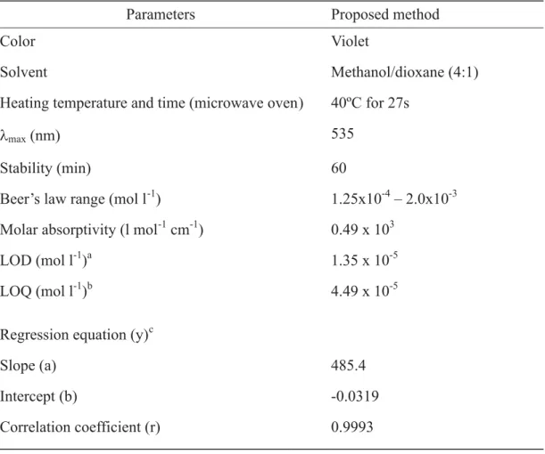
The analytical parameters and the optical characteristics for the spectrophotometric determinations of diclofenac by the proposed method are given in Table1.
Table 1. Analytical parameters for the spectrophotometric determination of diclofenac.
a limit of detection b limit of quantification
Effect of Interferences
In order to evaluate the selectivity of the developed method for the analysis of pharmaceutical preparations containing diclofenac, the effect of presence of several substances that can occur in the real samples was investigated. The excipients studied were: glucose, lactose, talc, starch, magnesium stearate, sodium sulphite, titanium dioxide, polyethyleneglycol, polyvinylpirrolidone, mannitol and benzilic alcohol. For this study, solutions containing diclofenac and each one of the excipients taken separately in concentrations equal or then-times greater than that of diclofenac were analyzsed under the same conditions described in section 2.4.3. A level of interference was considered to be acceptable if the error was not higher than + 3% relative to the expected diclofenac value.No interferences were observed in the determination of diclofenac in the presence of the excipients studied.
Application of the proposed method
In order to confirm the feasibility of the proposed method, diclofenac was determined in tablets and in injection solutions.
The results of the comparison of the proposed method with the pharmacopoeia method [37] (potentiometric and chromatographic methods for the analyses of tablets and injection solutions of diclofenac, respectively) are showed in Table 2. For all samples assayed, the results obtained by official and proposed methods were compared by applying the F-test and t-test at 95% confidence level. In all cases, the calculated F and t values did not exceed the theoretical values, indicating that there is no significant difference between either methods in concerning precision and accuracy in the determination of diclofenac in pharmaceuticals. The average recoveries obtained by the proposed method ranged from 95.9 to 103.3% for all the assayed samples.
Table 2. Diclofenac determination in commercial pharmaceuticals.
a Package labeled to contain 25 mg diclofenac/ml. b Package labeled to contain 50 mg diclofenac/tablet.
Conclusion
The spectrophotometric method proposed is simple, sensitive, rapid, low-cost, does not involve any treatment or extraction steps and gives pre-cise and accurate results. Significant improvements in the time of analysis could be attained using the microwave energy (1100 W) for 27 seconds. The proposed method was successfully applied to analysis of diclofenac in tablets and injection solutions, suggesting its use as a reliable and
advantageous alternative to other previously reported methods for routine analysis of diclofenac in these samples.
Acknowledgements
The authors would like to thank CNPq and FAPESP foundation (Brazil) for financial support.
Recebido em: 28/08/2004 Aceito em: 11/11/04
E. G. Ciapina, A. O. Santini, P. L. Weinert, M. A. Gotardo, H. R. Pezza, L. Pezza. Determinação espectrofotométrica de diclofenaco em preparações farmacêuticas assistida por forno de microondas.
Resumo: Neste trabalho é proposto um método eficaz e de baixo custo para a determinação de diclofenaco de sódio (ou potássio) na forma pura e em preparações farmacêuticas. O método é baseado na reação de diclofenaco com tetracloro-p-benzoquinona (p-cloranil) em meio de metanol. Esta reação foi acelerada pela irradiação da mistura reagente com energia microondas (1100W) por 27 segundos, produzindo um complexo de transferência de carga com um máximo de absorção em 535 nm. As condições ótimas da reação, tais como: concentração de reagentes, tempo de aquecimento e a estabilidade do produto da reação foram determinadas. A lei de Beer é obedecida num intervalo de concentração de 1,25x10⁻⁴ a
2,00x10⁻³ mol l-1 com um coeficiente de correlação de 0,9993 e absortividade molar de 0,49x10³ l mol-1 cm -1. O limite de detecção (LOD) do método foi de 1,35x10⁻⁵ mol l-1 e o limite de quantificação (LOQ) foi
4,49x10⁻⁵ mol l-1. Não foram observadas interferências de substâncias comumente encontradas junto com
diclofenaco em preparações farmacêuticas, tais como: glicose, lactose, talco, amido, estearato de magnésio, sulfito de sódio, dióxido de titânio, polietilenoglicol, polivinilpirrolidona, manitol e álcool benzílico. Os resultados analíticos obtidos a partir da aplicação do método proposto estão em muito boa concordância com aqueles obtidos pelo método padrão da Farmacopéia Americana. As recuperações obtidas para o diclofenaco a partir de um estudo com várias preparações farmacêuticas estiveram dentro de 95,9% a 103,3%, com desvio padrão variando de 0,2% a 1,8%.
Palavras-chave:diclofenaco, p-cloranil, espectrofotometria, forno de microondas, preparações farmacêuticas.
References
[1] R.T. Sane, R.S. Samant, V.G. Nayak, Drug Dev. Ind. Pharm. 13 (1987) 1307.
[2] J. Klimes, J. Sochor, P. Dolezal, J. Körner, Int. J. Pharm. 217 (2001) 153.
[3] N. Beaulieu, E.G. Loveringe, J. Lefrancois, H. Ong, J. Assoc. Off. Anal. Chem. 73 (1990) 698.
[4] M.S. Aurora-Prado, M. Steppe, M.F.M. Tavares, E.R.M. Kedor-Hackmann, M.I.R.M. Santoro, J. Assoc. Off. Anal. Chem. 85 (2002) 333.
[5] W.R. Jin, J. Zhang, , J. Chromatogr. A. 868 (2000) 101. [6] M.E. Abdel-Hamid, L. Novotony, H. Hamza, J. Pharm. Biomed. Anal. 24 (2001) 587.
[7] R. Bucci, A.D. Magri, A.L. Magri, J. Therm. Anal. Calorim. 61 (2000) 369.
[8] S.A. Abdel Fattah, S.Z. El-Khateeb, S.A. Abdel Razeg, M.S. Tawakkol, Spectrosc. Lett. 21 (1988) 533.
[9] J.C. Botello, G.P. Caballero, Talanta 42 (1995) 105. [10] M.S. Bhatia, S.R. Dhaneshwar, The Eastern Pharmacist 38 (1995) 133.
[11] Y.K. Agrawal, K. Shivramchandra, J. Pharm. Biomed. Anal. 9 (1991) 97.
[12] S. AgatonovicKustrin, L. Zivanovic, D. Radulovic, M. Vasiljevic, Analyst 116 (1991) 753.
[13] C.S.P. Sastry, A.S.R.P. Tipirneni, M.V. Suryanaryana, Analyst 114 (1989) 513.
[14] B.V. Kamath, K. Shivram, Anal. Lett. 26 (1993) 903. [15] S. AgatonovicKustrin, L. Zivanovic, M.Zecevic, D. Radulovic, J. Pharm. Biomed. Anal. 16 (1997) 146. [16] R. Bucci, A.D. Magri, A.L. Magri, Fres. J. Anal. Chem. 362 (1998) 577.
[18] B.V. Kamath, K. Shivram, G.P. Oza, S. Vangani. Anal. Lett. 26 (4) (1993) 665.
[19] R.S. Mulliken, J. Am. Chem. Soc. 72 (1950) 600. [20] R.S. Mulliken, J. Am. Chem. Soc. 74 (1952) 811. [21] R.S. Mulliken, J. Phys. Chem. 56 (1952) 801. [22] M.A. Korany, M.A. Wahby, Analyst 104 (1979) 146. [23] L.I. Bebawy, N. El-Kousy, J.K. Suddik, M. Shokry, J. Pharm. Biomed. Anal. 21 (1999) 133.
[24] B.V. Kamath, K. Shivram, S. Vangani, Anal. Lett. 25 (1992) 2239.
[25] H.E. Abdellatef, J. Pharm. Biomed. Anal. 17 (1998) 1267. [26] H. Salem, J. Pharm. Biomed. Anal. 29 (2002) 527. [27] R.E. Smith, W.R. Davis, Anal. Chem. 56 (1984) 2345. [28] F. Feigl, V. Gentil, C. Stark-Mayer, Mikrochim. Acta 51 (1957) 341.
[29] F. Feigl, Spot Tests in Organic Analysis, 7th ed., Elsevier, Amsterdam, 1966, pp. 249 and 407.
[30] E.A. Ibrahim, A.S. Issa, M.A. Abdel-Salam, M.S. Mahrous, Talanta 30 (1983) 531.
[31] A. Abu-Samra, J.S. Morris, S.R. Koirtyohann, Anal. Chem.
47 (1975) 1475.
[32] L.A. Fernando, W.D. Heavner, C.C. Gabrielli, Anal. Chem. 58 (1986) 511.
[33] L.B. Fischer, Anal. Chem. 58 (1986) 261.
[34] F. Smith, B. Cousins, J. Bozic, W. Flora, Anal. Chim. Acta 177 (1985) 243.
[35] H.M. Kingston, L.B. Jassie, Anal. Chem. 58 (1986) 2534. [36] T. Hirose, B.G. Hopek, Z. Wang, R. Yusa, B.W. Baldwin, Tetrahedron Lett. 44 (2003) 1831.
[37] United States Pharmacopeia National Formulary, USP 25, NF 20, Rockville, 2002, p. 554.
[38] H.M. Kingston, P.J. Walter, S. Chalk, E. Lorentzen, D. Link, in: H.M. Kingston, S.J. Haswell (Eds.), Microwave -Enhanced Chemistry - Fundamentals, Sample Preparation and Applications, ACS, Washington, 1997, p. 772.
[39] A. Morales-Rubio, J. Cerezo, A. Salvador, M. De la Guardia, Microchem. J. 47 (1993) 270.
[40] L. Pezza, H.R. Pezza, C.B. Melios, A. Leandro, A.G. Tininis, Anal. Lett. 33 (2000) 2901.