Cop
yright
© ABE&M t
odos os dir
eit
os r
eser
vados
.
Correspondence to:
Pedro Marques
Endocrinology Department, Instituto Português de Oncologia de Lisboa Rua Professor Lima Basto, 1099-023 – Lisboa, Portugal [email protected] Received on Nov/11/2013
Accepted on Apr/13/2014
DOI: 10.1590/0004-2730000003116
Aggressive pituitary lesion with
a remarkably high Ki-67
Lesão pituitária agressiva com Ki-67 notavelmente elevado
Pedro Marques1, Manuela Mafra2, Carlos Calado3, Anabela Martins4, Joaquim Monteiro3,Valeriano Leite1
SUMMARY
The uncommon aggressive pituitary tumors are named carcinomas when metastases are de-tected, either in the central nervous system and/or systemically. Some cases are associated with hormonal overproduction, but most are diagnosed because of local symptoms. These ne-oplasias are generally refractory to current treatments. A 51 year-old woman presented sudden onset of headache, left arm paresis and left facial hypoesthesia. Computed tomography scan and magnetic resonance imaging revealed a pituitary tumor invading the left sphenoidal and cavernous sinuses. Laboratory data excluded hormonal hypersecretion. The patient underwent transsphenoidal surgery and histological indings showed a neoplasia with Ki-67 estimated at 75%. Medical imaging excluded both a primary occult tumor and central nervous system or systemic dissemination. Three weeks postoperatively, neurological condition worsened, with new onset of ataxia, bilateral ptosis, ophthalmoplegia and an increase in the size of the lesion, leading to surgical intervention by craniotomy, followed by only a few sessions of radiotherapy, because of severe disease progression. Patient died nearly 2 months after the initial manifesta-tions. This case illustrates the aggressiveness of some pituitary lesions, the limited eficacy of current treatment modalities such as surgery or radiotherapy and the pitfalls of the current pitui-tary tumors classiication. To our knowledge, this case corresponds to one of the most aggres-sive pituitary neoplasms reported so far, with a very high Ki-67 index (75%) and short survival (2 months). Ki-67 index could be of prognostic value in pituitary tumors. Pituitary tumors World Health Organization (WHO) classiication could be revisited. Arq Bras Endocrinol Metab. 2014;58(6):656-60
SUMÁRIO
Os raros tumores pituitários agressivos são chamados carcinomas quando são detectadas me-tástases, sejam sistêmicas e/ou em sistema nervoso central. Alguns casos estão associados com superprodução de hormônio, mas a maioria é diagnosticada em função dos sintomas locais. Essas neoplasias são geralmente refratárias aos tratamentos atuais. Uma mulher com 51 anos de idade apresentou dor de cabeça de início súbito, paralisia de braço esquerdo e hipoes tesia facial esquerda. A tomograia e a ressonância magnética revelaram um tumor pi-tuitário invadindo os seios esfenoidal e cavernoso esquerdos. Os dados laboratoriais excluíram hipersecreção hormonal. A paciente foi submetida à cirurgia transesfenoidal, e os achados his-tológicos mostraram uma neoplasia com Ki-67 estimado em 75%. As imagens excluíram tanto um tumor oculto primário quanto disseminação sistêmica ou do sistema nervoso central. Três semanas após a cirurgia, a condição neurológica apresentou piora com início de ataxia, ptose bilateral, oftalmoplegia e aumento do tamanho da lesão, levando à intervenção cirúrgica por craniotomia, seguida por apenas algumas sessões de radioterapia devido à progressão grave da doença. A paciente veio a óbito depois de quase dois meses das manifestações iniciais. O caso ilustra a agressividade de algumas lesões pituitárias, a eicácia limitada das modalidades atuais de tratamento, como a cirurgia ou a radioterapia, e as limitações da classiicação atual de tumores pituitários. Até onde sabemos, esse caso corresponde a uma das neoplasias pituitárias mais agressivas descritas até hoje, com um nível muito alto de Ki-67 (75%) e sobrevida curta (2 meses). O nível de Ki-67 pode ser de valor prognóstico em tumores pituitários. A classiicação da Organização Mundial da Saúde (OMS) para tumores pituitários deveria ser revisitada. Arq Bras Endocrinol Metab. 2014;58(6):656-60
1 Endocrinology Department,
Instituto Português de Oncologia de Lisboa, Francisco Gentil, Lisboa, Portugal
2 Pathology Department, Centro
Hospitalar de Lisboa Zona Central, Lisboa, Portugal
3 Neurosurgery Department,
Centro Hospitalar de Lisboa Zona Central, Lisboa, Portugal
4 Endocrinology Department,
Cop
yright
© ABE&M t
odos os dir
eit
os r
eser
vados
.
INTRODUCTION
P
ituitary tumors account for approximately 15% ofall intracranial neoplasms (1,2). The majority is benign, non-invasive and asymptomatic. Some are de-tected incidentally by imaging exams; others are func-tioning-tumors generating hormonal syndromes (de-creasing ordered: prolactin; ACTH; GH; TSH; LH), whereas others are suspected due to local mass symp-toms. Hypopituitarism can also occur in some cases, particularly in larger tumors (3).
Pituitary carcinomas are rare conditions, accounting only 0.1 to 0.2% of all pituitary tumors (4,5). Pituitary carcinoma is deined by the presence of craniospinal and/or systemic metastases (6,7). There is no gender preference and mean of age at diagnosis is 44 years
(4).Most pituitary carcinomas develop from invasive
relapsing or previously operated or irradiated invasive adenomas (8,9). Type and grade of invasiveness do not represent a criteria for malignancy, although when prominent increase its probability (4,10). For some, proliferation indexes particularly Ki-67, have important prognostic value and should be considered as diagnos-tic criteria (10-12). Pituitary metastadiagnos-tic disease, typi-cally from breast or lung cancer, accounts for 1 to 2% of sellar masses and its differential diagnosis with pituitary tumors is challenging as they often mimic them clini-cally, imagiologically and histologically (13,14).
We present a rare case of pituitary neoplasm with extremely aggressive behavior, illustrating the pitfalls and dificulties in the classiication and management of these entities.
CASE REPORT
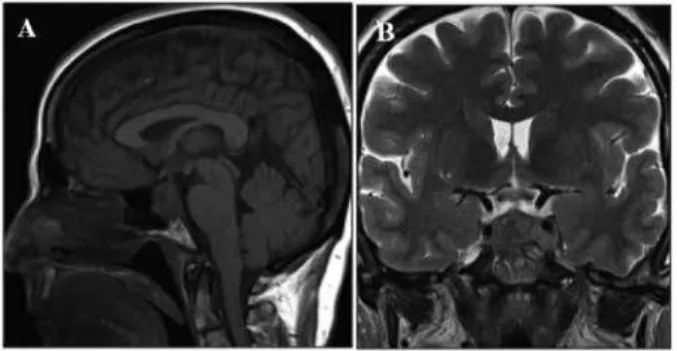
A Caucasian 51 year-old woman, non-smoker, with ir-relevant past medical history, not taking any medica-tion, was asymptomatic until February 2012, when she reported sudden onset of intense headache associated with left arm paresis and left facial hypoesthesia. Neu-rological examination identiied a minor left arm pare-sis, left facial hypoesthesia and slight left upper-lid pto-sis; no visual impairment or other neurological defects were observed. Computed tomography (CT) scan and magnetic resonance imaging (MRI) documented a pi-tuitary mass invading the sphenoidal and left cavernous sinuses, and the pituitary stalk was centered (Figure 1). Laboratory data was signiicant for mild hyponatre-mia (130 mmol/L), high creatine kinase (1,188 u/L),
central hypothyroidism (TSH = 0.05 uUI/mL [0.34-5.60]; free-T4 = 0.51 ng/dL [0.61-1.12]) and adrenal insuficiency (ACTH not measured but serum cortisol = 1.5 ug/dL [6.7 - 22.6]). Serum prolactin was 27.9 ng/mL (2.74-19.64); GH was 0.447 ng/mL (0.01-3.607) and beta-hCG < 0.5 mUI/mL (< 0.5-2.9). LH and FSH were not measured.
The patient was submitted to transsphenoidal sur-gery. A tumor with a soft consistence and necrotic com-ponent of the pituitary apoplexy type was found and partially resected. Clinical improvement was observed after surgery, particularly on the intensity of the hea-daches, but the third left cranial nerve palsy persisted. Patient was discharged under replacement therapy with levothyroxine and hydrocortisone.
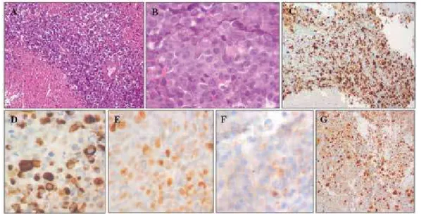
Histological indings revealed a monotonous hyper-cellular population of cohesive small round cells, with large nuclei and prominent nucleoli. Numerous mitotic and apoptotic igures were seen, as well as diffuse necro-tic areas. Ki-67 index was estimated at 75%. Immuno-histochemistry was positive for AE1/AE3 cytokeratines, nuclear p53 protein, cyclin D1 and focally positive for synaptophysin, chromogranin, neuron-speciic enolase, CD 56 and epithelial membrane antigen. There was no immunoreactivity for pituitary hormones, melanoma markers (S-100, HMB45, MelanA), cytokeratines 7, 8, 18, 19, 20, thyroid transcription factor-1, CD38, CD45, CD30, CD117, vimentin and desmin (Figure 2).
Fluo-rescence In Situ Hybridization(FISH) molecular
analy-sis was performed for c-MYC and EWS genes showing normal c-MYC and EWS gene copies in 98% and 91% of the nuclei, respectively, with no rearrangements.
Pituitary metastization from an occult tumor or sys-temic secondary lesions from pituitary tumor were ru-led out by several complementary exams which included mammography; breast, pelvic and thyroid ultrasound;
whole-body CT scan and 18F-Fluorodeoxyglucose
Po-Figure 1. Magnetic resonance imaging at presentation: sagittal (A) and
Cop
yright
© ABE&M t
odos os dir
eit
os r
eser
vados
.
Figure 2. Pituitary carcinoma photomicrographs: A: hypercellular neoplasia with extensive necrotic areas (hematoxylin-eosin stain, 20x). B: small round
cells with visible high mitotic activity (hematoxylin-eosin stain, 40x). C: immunohistochemistry for Ki67 protein using antibody MIB-1 quantiication (20x).
D: immunohistochemistry for AE1/AE3 cytokeratines (40x). E: Immunohistochemistry for p53 protein (40x). F: immunohistochemistry for Synaptophysin
(40x). G: immunohistochemistry for Cyclin D1 (20x).
Figure 3. Follow-up magnetic resonance imaging, approximately one month after the transsphenoidal surgery (March 2012): sagittal (A), coronal (B)
views and magnetic resonance angiography (C).
sitron Emission Tomography (18F-FDG-PET). Serum
tumoral markers were within normal values (Carcino-embryonic Antigen; Carcinoma Antigens 15.3/19.9; CYFRA 21.1; chromogranin A, calcitonin). An Ear--Nose-Throat specialist biopsied a non-suspicious pro-minence in the posterior wall of cavum, which was ne-gative for neoplasia.
Three weeks postoperatively headaches recurred associated with vomiting and neurological complaints (ataxic gait, ophthalmoplegia and bilateral ptosis); no visual ield defects were present and fundoscopy was normal. Postoperatively, hypopituitarism persisted with undetectable ACTH, TSH, LH, GH and low IGF-1. CT scan at that time, showed a pituitary mass with supra-sellar and lateral extension, touching the optic chiasm and involving the left cavernous sinus and tem-poral lobe; no compressive effect in the brain stem or
anomalies in the cerebral parenchyma were detected. Angio-MRI demonstrated a reduction in internal caro-tids caliber but maintained patency (Figure 3).
Cop
yright
© ABE&M t
odos os dir
eit
os r
eser
vados
.
or other secondary lesions, we highlight the 18
F-FDG--PET scan, which was negative for hypermetabolic sys-temic lesions eventually related with the pituitary mass of the patient. Moreover, as many authors describe, it is possible that metastatic invasive pituitary macroadeno-mas have silent and non-evident metastases, misleading the carcinoma diagnosis (4). In fact, autopsy studies of pituitary tumors patients revealed unknown metastases in some (4,10). On the other hand, the fatal course of these neoplasms do not allow the development of metastases, which some describe to occur in months or even years later (4,5,9). The most frequent metastatic locations includes brain (35%) and spinal cord (17%), followed by bone (14%), lymph nodes (12%), liver (10%) and lungs (6%) (9).
Pathology has a central role to the diagnosis of pi-tuitary lesions. At present, there are no histological, im-munochemical or ultrastructural markers that separate conclusively pituitary carcinoma from adenoma (15). In this case, many immunohistochemistry tests were used to exclude entities like lymphoproliferative disor-der, melanoma, sarcoma, nasopharyngeal carcinoma, primitive neuroectodermal tumors, Ewing sarcoma; genetic analysis for c-MYC and EWS also contribute to rule out more convincingly lymphomas, solid tumors (like breast cancer) or central/peripheric primitive neu-roectodermal tumors, respectively (16). So, at last, this case would it pathologically the diagnosis of carcinoma of a small cell subtype from pituitary origin. ___
The protein Ki-67 is a cell proliferation marker (17) detected by the monoclonal antibody MIB-1 and is expressed as a percentage of immunopositive nuclei in the form of a Ki-67 proliferation index (18). Literature describes a mean Ki-67 for pituitary carcinomas around 12% (± 14) (4,5,9). Thapar and cols. reported a Ki-67 index of 1.37%, 4.66% and 11.91% in noninvasive adenomas, invasive adenomas and carcinomas, respec-tively (11). Ki-67 over 10% is already criteria to admit pituitary carcinoma for some authors (4,10,11). In our patient Ki-67 was estimated at 75%, one of the highest so far described, which predicted an unfavorable course and high probability of unsuccessful treatment, as seen.
About 90% of pituitary adenomas are operated by transsphenoidal route and a. transcranial approach is usually reserved for cases with signiicant suprasellar, parasellar, retrosellar and/or subfrontal tumoral exten-sion (19). In this case, surgery as well as radiotherapy failed to control disease progression. Chemotherapy, such as temozolomide (20), was not attempted in the Figure 4. Follow-up magnetic resonance imaging after the second
surgery and before radiotherapy, approximately 2 months after
presentation (April 2012): coronal (A,C), axial (B) and sagittal (D) views.
DISCUSSION
This case report illustrates the diagnostic, therapeutic complexity and pitfalls that may characterize some pi-tuitary lesions. In spite of the fact that a fulminant cli-nical course and histological indings strongly suggest a malignant diagnosis, the case did not fulill the diag-nostic criteria of pituitary carcinoma according with
WHO classiication (6),becausethere was no evidence
of systemic or craniospinal metastization. However, the course of the disease was highly aggressive and rapi-dly progressive, despite two surgeries and radiotherapy, leading to death in approximately 2 months after the initial clinical manifestations. In addition, several his-tological indings (high Ki-67 proliferative index and p53 protein immunostaining) suggested a carcinoma and extensive tests excluded other differential diagnosis of primary or secondary pathology of sellar region (12-14). Nonetheless, a metastatic lesion from a primary occult tumor cannot be deinitively excluded, although
the negativity of 18F-FDG-PET scan and the absence
of other metastatic lesions makes such a diagnosis very unlikely.
Cop
yright
© ABE&M t
odos os dir
eit
os r
eser
vados
.
patient because of low performance status and severe disease progression.
We present an exceptionally rare case of pituitary le-sion that clearly illustrates the paradox and pitfalls betwe-en histological classiications and clinical behavior. This case report is a paradigm of the aggressiveness of some pituitary lesions. In our opinion, the WHO pituitary tu-mors classiication (6) should be revisited and the criteria for pituitary carcinoma diagnosis could be revisited.
Acknowledgments: this work was partially funded by Associação de Endocrinologia Oncológica. The authors declare that there is no conlict of interest.
Disclosure: no potential conlict of interest relevant to this article was reported.
REFERENCES
1. Raverot G, Sturm N, de Fraipont F, Muller M, Salenave S, Caron P, et al. Temozolomide treatment in aggressive pituitary tumors and pituitary carcinomas: a French multicenter experience. J Clin Endocrinol Metab. 2010;95(10):4592-9.
2. Asa SL, Ezzat S. The pathogenesis of pituitary tumors. Annu Rev Pathol. 2009;4:97-126.
3. Hall WA, Luciano MG, Doppman JL, Patronas NJ, Oldield EH. Pituitary magnetic resonance imaging in normal human volun-teers; occult adenomas in general population. Ann Intern Med. 1994;120(10):817-20.
4. Kaltsas GA, Nomikos P, Kontogeorgos G, Buchfelder M, Gross-man AB. Clinical review: diagnosis and Gross-management of pituitary carcinomas. J Clin Endocrinol Metab. 2005;90(5):3089-99. 5. Heaney AP. Clinical review: pituitary carcinoma: dificult
diagno-sis and treatment. J Clin Endocrinol Metab. 2011;96(12):3649-60. 6. Al-Shraim M, Asa SL. The 2004 World Health Organization
clas-siication of pituitary tumors: what is new? Acta Neuropathol. 2006;111(1):1-7.
7. Kaltsas GA, Mukherjee JJ, Plowman PN, Monson JP, Grossman AB, Besser GM. The role of cytotoxic chemotherapy in the
ma-nagement of aggressive and malignant pituitary tumors. J Clin Endocrinol Metab. 1998;83(12):4233-8.
8. Garrão AF, Sobrinho LG, Pedro-Oliveira, Bugalho MJ, Boavida JM, Raposo JF, et al. ACTH-producing carcinoma of the pituitary with haematogenic metastases. Eur J Endocrinol. 1997;137(2):176-80. 9. Dudziak K, Honegger J, Bornemann A, Horger M, Müssig K.
Pi-tuitary carcinoma with malignant growth from irst presentation and fulminant clinical course – Case report and review of literatu-re. J Clin Endocrinol Metab. 2011;96(9):2665-9.
10. Ragel B, Couldwell W. Pituitary carcinoma: a review of literature. Neurosurg Focus. 2004;16(4):E7.
11. Thapar K, Kovacs K, Scheithauer BW, Stefaneanu L, Horvath E, Pernicone PJ, et al. Proliferative activity and invasiveness among pituitary adenomas and carcinomas: an analysis using the MIB-1 antibody. Neurosurgery. 1996;38(1):99-106.
12. Dekkers OM, Pereira AM, Romijn JA. Treatment and follow-up of clinically nonfunctioning pituitary macroadenomas. J Clin Endo-crinol Metab. 2008;93(10):3717-26.
13. Freda PU, Wardlaw SL, Post KD. Unusual causes of sellar/para-sellar masses in a large transsphenoidal surgical series. J Clin Endocrinol Metab. 1996;81(10):3455-9.
14. Komninos J, Vlassopoulou V, Protopapa D, Korias S, Konto-georgos G, Sakas DE, et al. Tumors metastatic to the pituitary gland: case report and literature review. J Clin Endocrinol Metab. 2004;89(2):574-80.
15. Iezzoni JC, Mills SE. “Undifferentiated” small round cell tumors of the sinonasal tract: differential diagnosis update. Am J Clin Pa-thol. 2005;124 Suppl:S110-21.
16. Dedeurwaerdere F, Giannini C, Sciot R, Rubin BP, Perilongo G, Borghi L, et al. Primary peripheral PNET/Ewing’s sarcoma of the dura: a clinicopathologic entity distinct from central PNET. Mod Pathol. 2002;15:673-8.
17. Sholzen T, Gerdes J. The Ki-67 protein: from the known and the unknown. J Cell Physiol. 2000;182(3):311-22.
18. Salehi F, Agur A, Scheithauer BW, Kovacs K, Lloyd RV, Cusimano M. Ki-67 in pituitary neoplasms: a review--part I. Neurosurgery. 2009;65(3):429-37.
19. Sav A, Rotondo F, Syro LV, Scheithauer BW, Kovacs K. Biomarkers of pituitary neoplasms. Anticancer Res. 2012;32(11):4639-54. 20. Raverot G, Castinetti F, Jouanneau E, Morange I,