Cop
yright
© ABE&M t
odos os dir
eit
os r
eser
vados
.
603 Arq Bras Endocrinol Metab. 2010;54/7
original article
TSH reference values for adult
Brazilian population
Valores de referência do TSH para a população brasileira adulta
Pedro Weslley Rosario1,2, Arthur Cezar Malard Xavier1, Maria Regina Calsolari2
ABSTRACT
Objective: To establish limits of normal serum TSH for the adult (18 to 60 years) Brazilian popula-tion according to recommendapopula-tions of the Napopula-tional Academy of Clinical Biochemistry. Subjects and methods: Healthy volunteers were evaluated and those fulilling the following clinical crite-ria were selected: absence of known thyroid disease; no use of any interfering medications; no history of head and neck external radiotherapy, type 1 diabetes or autoimmune disease; no family history of thyroid disease, and absence of goiter or palpable nodules. Subjects with anti-thyrope-roxidase antibodies and/or altered free T4 were excluded. The sample consisted of 960 subjects (480 males and 480 females). Results: TSH values corresponding to the 2.5th and 97.5th percenti-les of the sample were 0.43 and 3.24 mIU/L, respectively. TSH values > 2.5 mIU/L were observed in 9.15% of the volunteers and levels > 3 mIU/L in 3.11%. Conclusion: The present study suggests an upper limit of normal TSH of approximately 3.5 mIU/L. Arq Bras Endocrinol Metab. 2010;54(7):603-6
Keywords
TSH; reference values; adult population
RESUMO
Objetivo: Estabelecer os limites de normalidade do TSH sérico em uma população adulta (18 a 60 anos) brasileira, conforme as recomendações da National Academy of Clinical Biochemistry. Sujeitos e métodos: Inicialmente foram avaliados voluntários saudáveis e selecionados aque-les sem doença tireoidiana conhecida, uso de medicamentos interferentes, passado de radio-terapia externa de cabeça e pescoço, diabetes tipo 1 ou doença autoimune, história familiar de doença tireoidiana, bócio ou nódulos palpáveis. Indivíduos com anticorpos antitireoperoxidase (TPOAb) e/ou T4 livre alterado foram excluídos. A amostra foi composta de 960 participantes (480 de cada sexo). Resultados: Os valores correspondentes aos percentis 2,5 e 97,5 da amostra foram 0,43 e 3,24 mIU/L, respectivamente. TSH > 2,5 mIU/L foi visto em 9,15% dos indivíduos e > 3 mIU/L em 3,11%. Conclusão: Nosso estudo sugere para a população adulta brasileira o limite superior normal do TSH de aproximadamente 3,5 mIU/L. Arq Bras Endocrinol Metab. 2010;54(7):603-6
Descritores
TSH; valores de referência; população adulta
1 Postgraduation Program, Santa
Casa de Belo Horizonte, MG, Brazil
2 Endocrinology Service, Santa Casa
de Belo Horizonte, MG, Brazil
Correspondence to:
Pedro Weslley Rosario Santa Casa de Belo Horizonte Av. Francisco Sales, 1.111, 9º D 30150-221 – Belo Horizonte, MG, Brazil
Received on May/26/2010 Accepted on Aug/21/2010
INTRODUCTION
T
he current upper limit of normal serum TSH (ap-proximately 4.5 mIU/L) has been questioned over the past few years and its reduction to 2.5 mIU/L is being defended by many investigators (1,2). Studies employing rigorous selection criteria, excluding subjects with thyroid disease not only based on clinical data but also on autoantibody measurement and ultrasonogra-phy (3), support this change. However, not allCop
yright
© ABE&M t
odos os dir
eit
os r
eser
vados
.
604 Arq Bras Endocrinol Metab. 2010;54/7
TSH reference values
adjustment. Although TSH levels are not the only pa-rameter evaluated for the initiation of L-T4 therapy or L-T4 dose adjustment, how much they differ from the upper normal limit will certainly inluence the decision. On the basis of these implications, the adoption of lower TSH cut-off values requires discussion. On the other hand, in view of the possibility that many patients currently classiied as “euthyroid” or “adequately trea-ted” in fact are not, the subject cannot be minimized because of eventual economic consequences.
In view of this controversy, the objective of the pre-sent study was to establish reference limits of normal serum TSH for an adult (18 to 60 years) Brazilian po-pulation according to the recommendations of the Na-tional Academy of Clinical Biochemistry (NACB) (1).
SUBJECTS AND METHODS
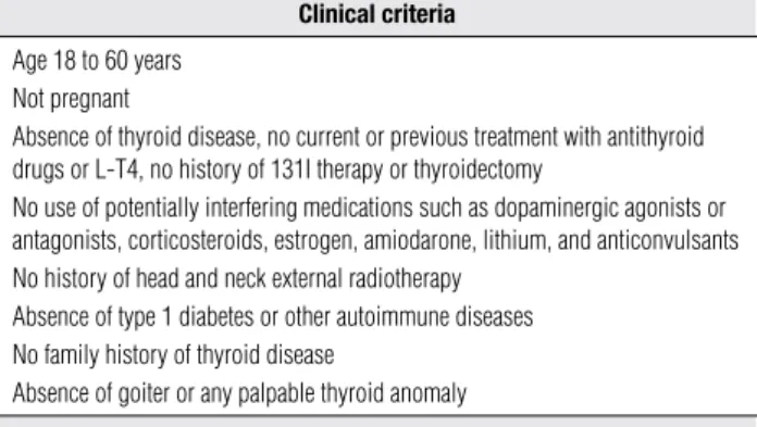
The population studied was from the metropolitan re-gion of Belo Horizonte (Minas Gerais, Brazil). Appa-rently healthy volunteers of both genders were initially interviewed and examined, and those fulilling the cli-nical criteria shown in table 1 were selected. Next, se-rum samples were obtained from the selected subjects in the morning (at about 8 a.m.) after an 8- to 10-h fast, and anti-thyroperoxidase antibodies (TPOAb), free T4 and TSH were measured. Subjects presenting positive TPOAb and/or altered free T4 were excluded. Ultrasonography (US) was not performed because of the following reasons: US is not required by the NACB (1); previous studies have shown no change in the re-ference limits of normal TSH when subjects with ul-trasonographic anomalies were excluded from the ini-tial sample (3,7-9), a inding also demonstrated in a previous study from our group (13); and no consensus exists regarding the ultrasonographic indings (3,6-9). A total of 1,802 volunteers were recruited; of these, 1,611 (89.4%) agreed to be interviewed and exami-ned and 641 were excluded because they did not fulill the selection criteria. The sample was considered to be complete after the inclusion of 480 participants of each gender [pre-deined and larger than that required by the NACB (1)]. The age distribution of the samples was: 18 to 30 years (n = 220); 30 to 45 years (n = 260) and 45 to 60 years (n = 480). The study was approved by the local Research Ethics Committee.
TSH was measured with a chemiluminescent assay (Immulite 2000, Diagnostic Products Corporation, Los Angeles, CA), with reference values of 0.4-4 mIU/L,
Table 1. Inclusion criteria
Clinical criteria
Age 18 to 60 years Not pregnant
Absence of thyroid disease, no current or previous treatment with antithyroid drugs or L-T4, no history of 131I therapy or thyroidectomy
No use of potentially interfering medications such as dopaminergic agonists or antagonists, corticosteroids, estrogen, amiodarone, lithium, and anticonvulsants No history of head and neck external radiotherapy
Absence of type 1 diabetes or other autoimmune diseases No family history of thyroid disease
Absence of goiter or any palpable thyroid anomaly
Complementary criteria
Anti-TPOAb negative Normal free T4
TPOAb: thyroperoxidase antibodies.
functional sensitivity of 0.02 mIU/L, and intra- and interassay coeficients of variation < 7% for values ran-ging from 0.1 to 40 mIU/L. TPOAb and free T4 were also measured by a chemiluminescent assay (Immulite 2000, Diagnostic Products Corporation, Los Angeles, CA), with reference values up to 35 IU/mL and from 0.8 to 2.0 ng/dL, respectively.
The reference limits of normal TSH were deined as follows: 1) 2.5th and 97.5th percentiles of the values obtained for the sample; 2) logarithmic transformation of the values obtained, calculation of the mean ± 1.96 SD of these values, and exponentiation to obtain the limits corresponding to the original scale (1).
RESULTS
The distribution of TSH values in the sample is shown in igure 1. The mean, median, minimum, maximum, and 2.5th and 97.5th percentiles of the values obtained were 1.52, 1.4, 0.35, 4.8, 0.43 and 3.24 mIU/L, res-pectively. TSH > 2.5 mIU/L was observed in 9.15% of the subjects and > 3 mIU/L in 3.11%.
Figure 1. Distribution of TSH in the cohort (n = 960). 0
5 10 15 20 25 30
Percent
< 0.5 0.5-1 1-1.5 1.5-2 2-2.5 2.5-3 3-3.5 3.5-4 > 4
Cop
yright
© ABE&M t
odos os dir
eit
os r
eser
vados
.
605 Arq Bras Endocrinol Metab. 2010;54/7
TSH reference values
The upper reference limit (mean + 1.96 SD) was estimated for the log-transformed data and then expo-nentiated back to the original scale. The resulting value (3.3 mIU/L) was very close to the 97.5th percentile of TSH values (3.24 mIU/L).
DISCUSSION
We irst would like to emphasize that the following dis-cussion, as well as our results, cannot be extrapolated to children, women in the irst trimester of gestation or subjects older than 70 years (11). Although the inluen-ce of other factors such as gender and rainluen-ce is possible, adjustment of TSH reference values for these factors is not recommended (1,14).
In determining the TSH reference range, it has been customaryto log-transform the data because of the skewed distribution (1).In fact, in the present stu-dy, TSH followed an approximatelognormal rather than Gaussian distribution. Nevertheless, the upper-re-ference limit based on “mean + 1.96 SD” of log-trans-formedTSH was very close to the 97.5th percentile.
Since occult thyroid disease cannot be excluded based only on clinical evaluation and antibody mea-surement and since the inadvertent inclusion of these cases may overestimate the upper limit of TSH (1,14), some studies have included ultrasonographic indings in the selection criteria of the population sample (3,6-10). Volzke and cols. (3), studying a previously iodi-ne-deicient population, found an upper limit of TSH (97.5th percentile) of 2.12 mIU/L. This result is in contrast to other studies that also excluded subjects with ultrasonographic anomalies and reported values of 3.6 mIU/L (5,6), 3.35 mIU/L (7), 3.77 mIU/L (8), 3.37 mIU/L (9), and 3.7 mIU/L (10). As can be seen, even in series including US, which is suppo-sed to minimize the risk of overestimating TSH, the upper limit was approximately 3.5 mIU/L, similar to the value found in the present study (3.24 mIU/L). Although the upper limits were similar, the comparison of the results of these different studies (5-10) is limited by population differences and the use of distinct TSH assays. Differences in the speciicitiesof the anti-TSH monoclonal antibodies used and in how TSH glycofor-ms are recognized by differentimmunoassays contribu-te to the need for assay-speciic values (15). Furthermo-re, differences related to the distribution of these TSH isoforms and to iodine ingestion contribute to the need for population-speciic TSH reference values.
Although patients without known or suspected thyroid disease and without antibodies may present ul-trasonographic anomalies, the need of this method for sample selection to establish TSH reference values is questionable (9,16). In the study of Kratsch and cols. (8), the upper limit of TSH changed from 3.63 to 3.77 mIU/L when subjects with a family history of thyroid disease, antibodies or ultrasonographic anomalies were excluded from the initial sample. Hamilton and cols. (9) showed that the exclusion of subjects with alte-red US did not modify the value corresponding to the 97.5th percentile (3.37 mIU/L). Zöphel and cols. (7) reported that the initial sample and the reference sam-ple (without antibodies and with normal US) yielded the same upper limit (97.5th percentile), 3.34 and 3.35 mIU/L, respectively. Even in the study of Volzke and cols. (3), no reduction in median TSH values (reported according to gender and age) was observed after the exclusion of subjects with TPOAb or ultrasonographic alterations. We also observed that the exclusion of 19 subjects presenting only an altered US from a sample of 169 patients without known or suspected thyroid disease, no family history and without TPOAb had no impact on the TSH upper limit (13). One current li-mitation of the use of US as a selection criterion is the discordance regarding the ultrasonographic indings. One study did not include glandular volume (9), ano-ther only considered nodules > 10 mm (3), two did not consider alterations in echogenicity (6,8), and one only included irregular hypoechogenicity (9). In addition, 641 (39%) of the 1,611 subjects initially evaluated in this study were excluded, a fact demonstrating that the selection criteria adopted were highly restrictive.
In view of these doubts regarding the need of US for sample selection, studies that do not perform US cannot be neglected. In the NHANES III involving 13,344 subjects older than 12 years (excluding preg-nant women) without known or laboratory evidence of thyroid disease (TSH < 0.1 mIU/L or > 4.5 mIU/L) and without antibodies (TPOAb or TgAb), the 97.5th percentile of TSH was 4.12 mIU/L (11). Jensen and cols. (12), studying 987 subjects aged 17 to 66 years (excluding pregnant women) without known thyroid disease or a family history and without antibodies, re-ported a value of 4.07 mIU/L.
in-Cop
yright
© ABE&M t
odos os dir
eit
os r
eser
vados
.
606 Arq Bras Endocrinol Metab. 2010;54/7
termediate values, TSH level is not the only parameter evaluated for the initiation of L-T4 therapy for sub-clinical hypothyroidism. It is essential that physicians consider various other patient-speciicfactors (17) be-foredeciding whether thyroid hormone replacement therapy is indicated inan individual patient. If L-T4 re-placement therapy is chosen, the upper limit of normal TSH should indeed be deined as the minimum value to indicate this therapy.
In a study also involving a Brazilian population (São Paulo), Duarte and cols. (10) evaluated 320 subjects aged 18 to 60 years (213 women) without known thy-roid disease who were submitted to clinical examination and were TPOAb and US negative, and reported an up-per limit of TSH of 3.7 mIU/L. These indings and the results of the present study suggest that the upper limit of normal TSH for the adult Brazilian population would be approximately 3.5 mIU/L, a limit lower than the tra-ditional value of 4.5 mIU/L but not as low as the limit suggested by some investigators (2.5 mIU/L) (1,2).
Acknowledgments: This study was partly supported by Sanoi-Aventis.
Disclosure: no potential conlict of interest relevant to this article was reported.
REFERENCES
1. Baloch Z, Carayon P, Conte-Devolx B, Demers LM, Feldt-Rasmus-sen U, Henry JF, et al. Laboratory support for the diagnosis and monitoring of thyroid disease. Thyroid. 2003;13:3-126.
2. Wartofsky L, Dickey RA. The evidence for a narrower thyrotro-pin reference range is compelling. J Clin Endocrinol Metab. 2005;90:5483-8.
3. Volzke H, Alte D, Kohlmann T, Ludemann J, Nauck M, John U, et al. Reference intervals of serum thyroid function tests in a pre-viously iodine-deicient area. Thyroid. 2005;15:279-85.
4. Surks MI, Goswami G, Daniels GH. The thyrotropin referen-ce range should remain unchanged. J Clin Endocrinol Metab. 2005;90:5489-96.
5. Brabant G, Beck-Peccoz P, Jarzab B, Laurberg P, Orgiazzi J, Sza-bolcs I, et al. Is there a need to redeine the upper normal limit of TSH? Eur J Endocrinol. 2006;154:633-7.
6. Knudsen N, Bulow I, Jorgensen T, Laurberg P, Ovesen L, Perrild H. Comparative study of thyroid function and types of thyroid dys-function in two areas in Denmark with slightly different iodine status. Eur J Endocrinol. 2000;143:485-91.
7. Zöphel K, Wunderlich G, Grüning T, Koch R, Döge H, Kotzerke J. Where does subclinical hypothyroidism start? Implications for the deinition of the upper reference limit for thyroid stimulating hormone (TSH). Nuklearmedizin. 2005;44:56-61.
8. Kratsch J, Fiedler GM, Leichtle A, Brugel M, Buchbinder S, Otto L, et al. New reference intervals for thyrotropin and thyroid hormones ba-sed on National Academy of Clinical Biochemistry criteria and re-gular ultrasonography of the thyroid. Clin Chem. 2005;51:1480-6. 9. Hamilton TE, Davis S, Onstad L, Kopecky KJ. Thyrotropin levels
in a population with no clinical, autoantibody, or ultrasono-graphic evidence of thyroid disease: implications for the diag-nosis of subclinical hypothyroidism. J Clin Endocrinol Metab. 2008;93:1224-30.
10. Duarte GC, Tomimori EK, Camargo RYA, Rubio IGS, Wajngarten M, Rodrigues AG, et al. The prevalence of thyroid dysfunction in el-derly cardiology patients with mild excessive iodine intake in the urban area of São Paulo. Clinics. 2009;64:135-42.
11. Surks MI, Hollowell JG. Age-speciic distribution of serum thyro-tropin and antithyroid antibodies in the U.S. population: impli-cations for the prevalence of subclinical hypothyroidism. J Clin Endocrinol Metab. 2007;92:4575-82.
12. Jensen E, Hyltoft Petersen P, Blaabjerg O, Hansen PS, Brix TH, He-gedüs L. Establishment of a serum thyroid stimulating hormone (TSH) reference interval in healthy adults. The importance of en-vironmental factors, including thyroid antibodies. Clin Chem Lab Med. 2004;42:824-32.
13. Borges MAR, Rosario PW, Purisch S. Normal serum TSH for our adult population. Congresso Mineiro de Endocrinologia 2007 (Abstract).
14. Spencer CA, Hollowell JG, Kazarosyan M, Braverman LE. National Health and Nutrition Examination Survey III thyroid-stimulating hormone (TSH)-thyroperoxidase antibody relationships demons-trate that TSH upper reference limits may be skewed by occult thyroid dysfunction. J Clin Endocrinol Metab. 2007;92:4236-40. 15. Rawlins ML, Roberts WL. Performance characteristics of six
third-generation assays for thyroid-stimulating hormone. Clin Chem. 2004;50:2338-44.
16. Zöphel K, Wunderlich G, Kotzerke J. Should we really determine a reference population for the deinition of thyroid-stimulating hormone reference interval? Clin Chem. 2006;52:329-30. 17. Biondi B, Cooper DS. The clinical signiicance of subclinical
thyroid dysfunction. Endocr Rev. 2008;29:76-131.